Mouse Anti-PDCD1LG2 antibody
PD L2; PD-L2;PD1L2_HUMAN; B7 dendritic cell molecule; B7-DC; B7DC; bA574F11.2; Btdc; Butyrophilin B7 DC; Butyrophilin B7-DC; Butyrophilin B7DC; CD 273; CD273; CD273 antigen; MGC142238; MGC142240; PD 1 ligand 2; PD-1 ligand 2; PD1 ligand 2; PDCD 1 ligand
View History [Clear]
Details
Product Name PDCD1LG2 Chinese Name 程序性死亡配体2(CD273)单克隆抗体 Alias PD L2; PD-L2; PD1L2_HUMAN; B7 dendritic cell molecule; B7-DC; B7DC; bA574F11.2; Btdc; Butyrophilin B7 DC; Butyrophilin B7-DC; Butyrophilin B7DC; CD 273; CD273; CD273 antigen; MGC142238; MGC142240; PD 1 ligand 2; PD-1 ligand 2; PD1 ligand 2; PDCD 1 ligand 2; PDCD1 ligand 2; PDL 2; PDL2; Programmed cell death 1 ligand 2; Programmed death ligand 2. Research Area immunology Immunogen Species Mouse Clonality Monoclonal Clone NO. G8F6 React Species Human Applications WB=1:1000-2000,IHC-P=1:100-400,IHC-F=1:100-400,IF=1:100-500(Paraffin sections need antigen repair)
not yet tested in other applications.
optimal dilutions/concentrations should be determined by the end user.Theoretical molecular weight 29kDa Cellular localization The cell membrane Secretory protein Form Liquid Concentration 1mg/ml immunogen KLH conjugated synthetic peptide derived from human PDCD1LG2: 31-130/273 Lsotype IgG1,κ Purification affinity purified by Protein G Buffer Solution 1M TBS(pH7.4) with 1% BSA, 3% Proclin300 and 50% Glycerol. Storage Shipped at 4℃. Store at -20 °C for one year. Avoid repeated freeze/thaw cycles. Attention This product as supplied is intended for research use only, not for use in human, therapeutic or diagnostic applications. PubMed PubMed Product Detail Involved in negative regulation of activated T cell proliferation; negative regulation of interferon-gamma production; and negative regulation of interleukin-10 production. Predicted to be located in plasma membrane. Predicted to be active in external side of plasma membrane. Biomarker of pulmonary tuberculosis. [provided by Alliance of Genome Resources, Nov 2021]
Function:
Involved in the costimulatory signal, essential for T-cell proliferation and IFNG production in a PDCD1-independent manner. Interaction with PDCD1 inhibits T-cell proliferation by blocking cell cycle progression and cytokine production.
Subunit:
Interacts with PDCD1.
Subcellular Location:
Isoform 3: Secreted (Probable).
Isoform 2: Endomembrane system; Single-pass type I membrane protein.
Isoform 1: Cell membrane; Single-pass type I membrane protein.
Tissue Specificity:
Highly expressed in heart, placenta, pancreas, lung and liver and weakly expressed in spleen, lymph nodes and thymus.
Post-translational modifications:
Phosphorylated by AKT1. Phosphorylation relieves inhibitory function on mTORC1.
Similarity:
Belongs to the immunoglobulin superfamily. BTN/MOG family.
Contains 1 Ig-like C2-type (immunoglobulin-like) domain.
Contains 1 Ig-like V-type (immunoglobulin-like) domain.
SWISS:
Q9BQ51
Gene ID:
80380
Database links:Entrez Gene: 80380 Human
Omim: 605723 Human
SwissProt: Q9BQ51 Human
Product Picture 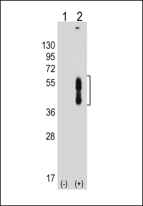 Sample:
Lane 1: Non-transfected 293T cell lysates
Lane 2: Transfected PD-L2-transfected 293T cell lysates
Primary: Anti-PDCD1LG2 (SLM-51708M) at 1/8000 dilution
Secondary: IRDye800CW Goat Anti-Mouse IgG at 1/20000 dilution
Predicted band size: 29 kD
Observed band size: 40-55 kD
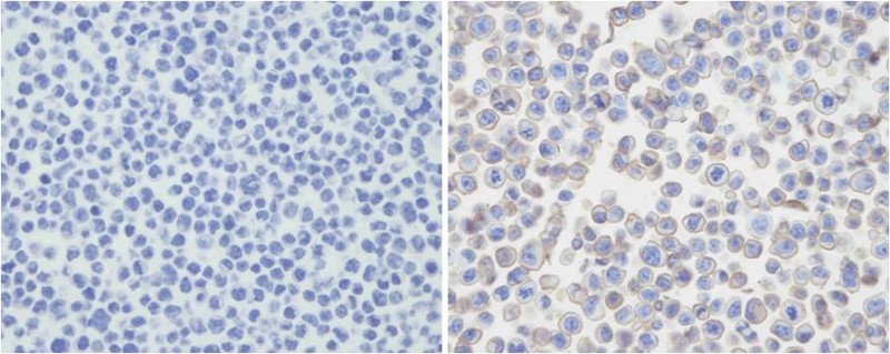 Immunohistochemical analysis of PDL-2 in untransfected(left) or transfected(right) with 293T cell sections. Cell was fixed with formaldehyde; antigen retrieval was by heat mediation with a EDTA buffer (pH9. 0). Samples were incubated with primary antibody (1:25) for 1 hours at room temperature. A undiluted biotinylated goat polyvalent antibody was used as the secondary antibody.
Cartpieces
Totalgoods,subtotals:¥Checkout
Partial purchase records(bought amounts latest0)
No one bought this product
User Comment(Total0User Comment Num)
- No comment


 +86 571 56623320
 SUNLONG BIOTECH

