Rabbit Anti-CD19 antibody
Antibody deficiency due to defect in CD19, included; AW495831; B lymphocyte antigen CD19; B lymphocyte surface antigen B4; B4; CD 19; CD19 antigen; CD19 molecule; Cd19 protein; Differentiation Antigen CD19; Leu 12; Leu12; Lymphocyte Surface Antigen; MGC10
View History [Clear]
Details
Product Name CD19 Chinese Name CD19Recombinant rabbit monoclonal anti Alias Antibody deficiency due to defect in CD19, included; AW495831; B lymphocyte antigen CD19; B lymphocyte surface antigen B4; B4; CD 19; CD19 antigen; CD19 molecule; Cd19 protein; Differentiation Antigen CD19; Leu 12; Leu12; Lymphocyte Surface Antigen; MGC109570; MGC12802; T-cell surface antigen Leu-12; CD19_HUMAN. Research Area Cardiovascular immunology Stem cells Immunogen Species Rabbit Clonality Monoclonal React Species Human Applications WB=1:500-2000,IHC-P=1:200-1000,IHC-F=1:200-1000,ICC/IF=1:50-200,IF=1:200-1000,Flow-Cyt=1:50-100 (Paraffin sections need antigen repair)
not yet tested in other applications.
optimal dilutions/concentrations should be determined by the end user.Theoretical molecular weight 59kDa Detection molecular weight 95 kDa Cellular localization The cell membrane Form Liquid Concentration 1mg/ml immunogen KLH conjugated synthetic peptide derived from human CD19 Lsotype IgG Purification affinity purified by Protein A Buffer Solution 1M TBS(pH7.4) with 1% BSA, 3% Proclin300 and 50% Glycerol. Storage Shipped at 4℃. Store at -20 °C for one year. Avoid repeated freeze/thaw cycles. Attention This product as supplied is intended for research use only, not for use in human, therapeutic or diagnostic applications. PubMed PubMed Product Detail This gene encodes a member of the immunoglobulin gene superfamily. Expression of this cell surface protein is restricted to B cell lymphocytes. This protein is a reliable marker for pre-B cells but its expression diminishes during terminal B cell differentiation in antibody secreting plasma cells. The protein has two N-terminal extracellular Ig-like domains separated by a non-Ig-like domain, a hydrophobic transmembrane domain, and a large C-terminal cytoplasmic domain. This protein forms a complex with several membrane proteins including complement receptor type 2 (CD21) and tetraspanin (CD81) and this complex reduces the threshold for antigen-initiated B cell activation. Activation of this B-cell antigen receptor complex activates the phosphatidylinositol 3-kinase signalling pathway and the subsequent release of intracellular stores of calcium ions. This protein is a target of chimeric antigen receptor (CAR) T-cells used in the treatment of lymphoblastic leukemia. Mutations in this gene are associated with the disease common variable immunodeficiency 3 (CVID3) which results in a failure of B-cell differentiation and impaired secretion of immunoglobulins. CVID3 is characterized by hypogammaglobulinemia, an inability to mount an antibody response to antigen, and recurrent bacterial infections. Alternative splicing results in multiple transcript variants encoding distinct isoforms. [provided by RefSeq, Jul 2020]
Function:
Assembles with the antigen receptor of B-lymphocytes in order to decrease the threshold for antigen receptor-dependent stimulation.
Subunit:
Forms a complex with CD21, CD81 and CD225 in the membrane of mature B-cells. Interacts with VAV. Interacts with GRB2 and SOS when phosphorylated on Tyr-348 and/or Tyr-378. Interacts with PLCG2 when phosphorylated on Tyr-409. Interacts with LYN.
Subcellular Location:
Membrane; Single-pass type I membrane protein.
Post-translational modifications:
Phosphorylated on serine and threonine upon DNA damage, probably by ATM or ATR. Phosphorylated on tyrosine following B-cell activation. Phosphorylated on tyrosine residues by LYN.
DISEASE:
Defects in CD19 are the cause of immunodeficiency common variable type 3 (CVID3) [MIM:613493]; also called antibody deficiency due to CD19 defect. CVID3 is a primary immunodeficiency characterized by antibody deficiency, hypogammaglobulinemia, recurrent bacterial infections and an inability to mount an antibody response to antigen. The defect results from a failure of B-cell differentiation and impaired secretion of immunoglobulins; the numbers of circulating B-cells is usually in the normal range, but can be low.
Similarity:
Belongs to the selectin/LECAM family.
Contains 2 Ig-like C2-type (immunoglobulin-like) domains.
SWISS:
P15391
Gene ID:
930
Product Picture 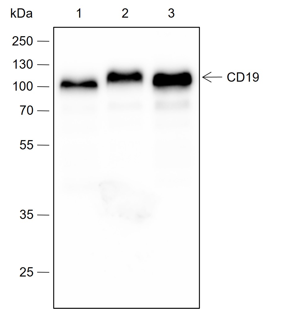 Blocking buffer: 5% NFDM/TBST
Primary ab dilution: 1:2000
Primary ab incubation condition: 2 hours at room temperature
Secondary ab: Goat Anti-Rabbit IgG H&L (HRP)
Lysate: 1: Ramos, 2: Daudi, 3: Raji
Protein loading quantity: 20 μg
Exposure time: 30 s
Predicted MW: 61 kDa
Observed MW: 95 kDa
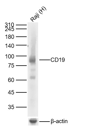 Sample:
Lane 1: Human Raji cell Lysates
Primary: Anti-CD19 (SLM-60605R) at 1/1000 dilution
Secondary: IRDye800CW Goat Anti-Rabbit IgG at 1/20000 dilution
Predicted band size: 59kDa
Observed band size: 90kDa
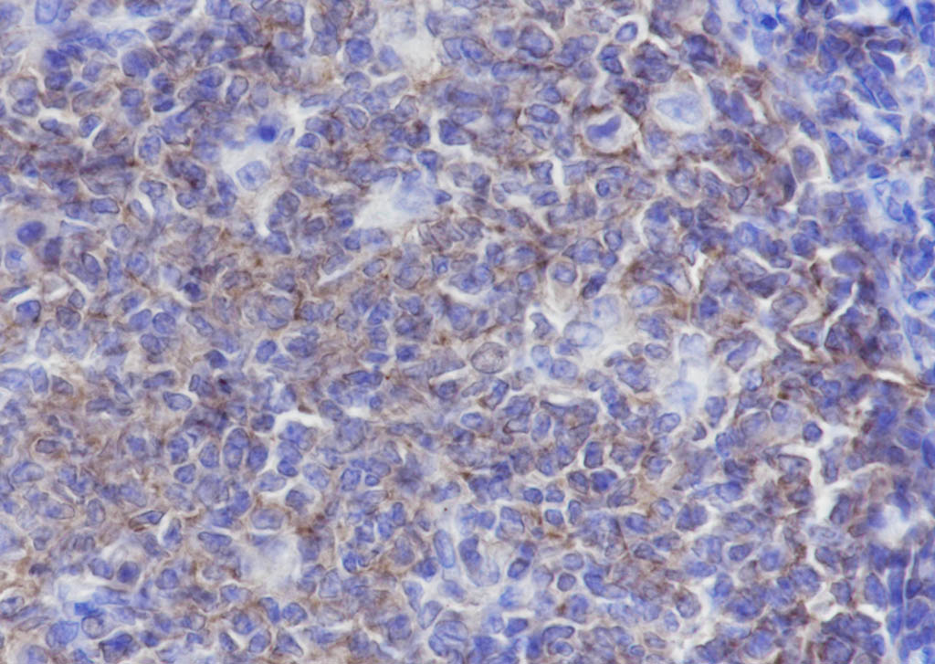 Tissue: Human tonsil Section type: Formalin fixed & Paraffin -embedded section Retrieval method: High temperature and high pressure Retrieval buffer: Tris/EDTA buffer, pH 9.0 Primary ab dilution: 1:1000 Primary ab incubation condition: 1 hour at room temperature Secondary ab: SP Kit(Rabbit)(sp-0023) Counter stain: Hematoxylin (Blue) Comment: Color brown is the positive signal for SLM-60605R
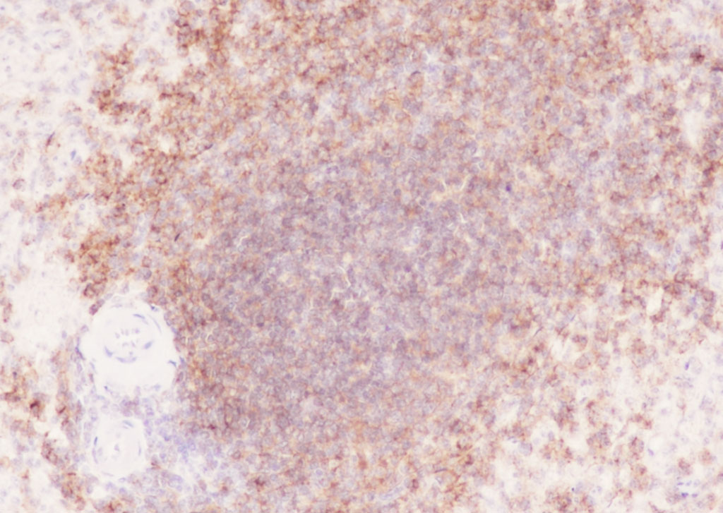 Tissue: Human spleen Section type: Formalin fixed & Paraffin -embedded section Retrieval method: High temperature and high pressure Retrieval buffer: Tris/EDTA buffer, pH 9.0 Primary ab dilution: 1:1000 Primary ab incubation condition: 1 hour at room temperature Secondary ab: SP Kit(Rabbit) Counter stain: Hematoxylin (Blue) Comment: Color brown is the positive signal for SLM-60605R
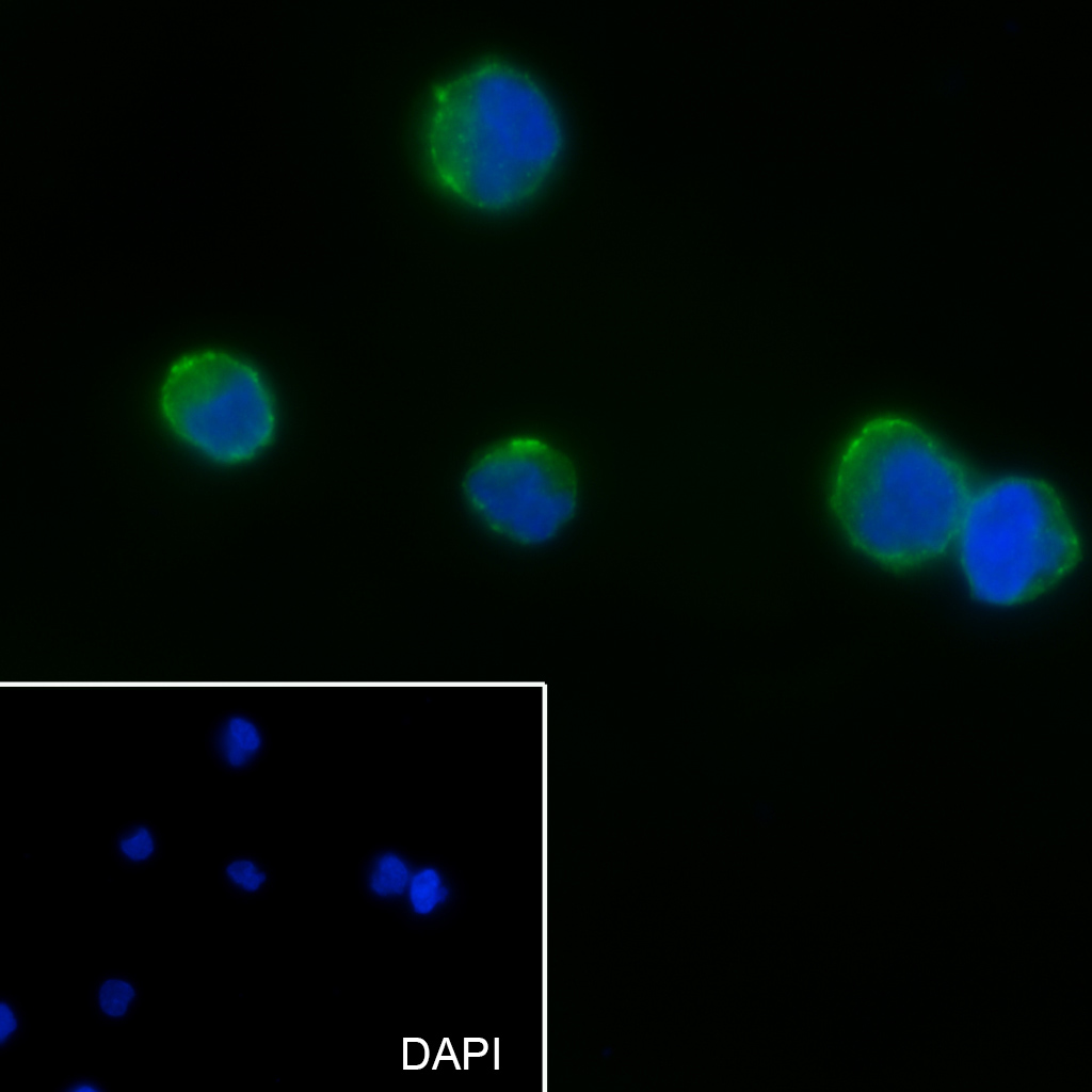 Cell line: Raji Fixative: 4% Paraformaldehyde Permeabilization: 0.1% TritonX-100 Primary ab dilution: 1:50 Primary incubation condition: 4°C overnight Secondary ab: Goat Anti-Rabbit IgG Nuclear counter stain: DAPI (Blue) Comment: Color green is the positive signal for SLM-60605R
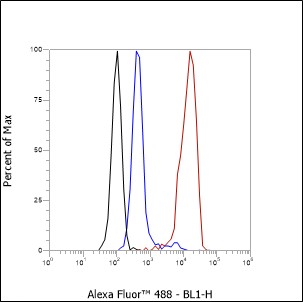 Cell line: Raji Fixative: 4% Paraformaldehyde Permeabilization: 90% Methanol Primary ab dilution: 1:100 Secondary ab: Goat anti Rabbit IgG Unlabelled control: The cell without incubation with primary antibody and secondary antibody (Black line). Isotype control: Rabbit monoclonal IgG (Blue line). Comment: Line red is the positive signal for SLM-60605R
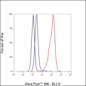 Cell line: Ramos Fixative: 4% Paraformaldehyde Permeabilization: 90% Methanol Primary ab dilution: 1:100 Secondary ab: Goat anti Rabbit IgG Unlabelled control: The cell without incubation with primary antibody and secondary antibody (Black line). Isotype control: Rabbit monoclonal IgG (Blue line). Comment: Line red is the positive signal for SLM-60605R
Cartpieces
Totalgoods,subtotals:¥Checkout
Partial purchase records(bought amounts latest0)
No one bought this product
User Comment(Total0User Comment Num)
- No comment


 +86 571 56623320
 SUNLONG BIOTECH

