Rabbit Anti-CD171/L1CAM antibody
L1CAM_HUMAN; Neural cell adhesion molecule L1; CAML1; MIC5; N-CAM-L1; NCAM-L1; CD171; L1 cell adhesion molecule; S10; HSAS; MASA; SPG1; HSAS1; N-CAM-L1;
View History [Clear]
Details
Product Name CD171/L1CAM Chinese Name 神经Cell adhesion molecule配体1Recombinant rabbit monoclonal anti Alias L1CAM_HUMAN; Neural cell adhesion molecule L1; CAML1; MIC5; N-CAM-L1; NCAM-L1; CD171; L1 cell adhesion molecule; S10; HSAS; MASA; SPG1; HSAS1; N-CAM-L1; Research Area immunology Neurobiology Signal transduction Immunogen Species Rabbit Clonality Monoclonal React Species Human Applications WB=1:1000-5000,IHC-P=1:200-800,IHC-F=1:200-800,IF=1:200-800 (Paraffin sections need antigen repair)
not yet tested in other applications.
optimal dilutions/concentrations should be determined by the end user.Theoretical molecular weight 138kDa Cellular localization The cell membrane Form Liquid Concentration 1mg/ml immunogen Recombinant human L1CAM protein Lsotype IgG Purification affinity purified by Protein A Buffer Solution 1*TBS (pH7.4), 5% BSA, 40% Glycerol. Preservative: 5% Sodium Azide Storage Shipped at 4℃. Store at -20 °C for one year. Avoid repeated freeze/thaw cycles. Attention This product as supplied is intended for research use only, not for use in human, therapeutic or diagnostic applications. PubMed PubMed Product Detail L1cam (L1 cell adhesion molecule isoform 1 precursor) is an axonal glycoprotein belonging to the immunoglobulin supergene family. The ectodomain, consisting of several immunoglobulin-like domains and fibronectin-like repeats (type III), is linked via a single transmembrane sequence to a conserved cytoplasmic domain. This cell adhesion molecule plays an important role in nervous system development, including neuronal migration and differentiation. Mutations in the gene cause three X-linked neurological syndromes known by the acronym CRASH (corpus callosum hypoplasia, retardation, aphasia, spastic paraplegia and hydrocephalus). Alternative splicing of a neuron-specific exon is thought to be functionally relevant. [provided by RefSeq].
SWISS:
P32004
Gene ID:
3897
Product Picture 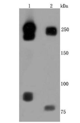 Western blot analysis of L1CAM on different lysates. Proteins were transferred to a PVDF membrane and blocked with 5% BSA in PBS for 1 hour at room
temperature. The primary antibody (ET1703-51, 1/500) was used in 5% BSA at room temperature for 2 hours. Goat Anti-Rabbit IgG - HRP Secondary Antibody at 1:50,000 dilution was used for 1 hour at room temperature.
Positive control:
Lane 1: Hela cell lysate
Lane 2: Human brain tissue lysate
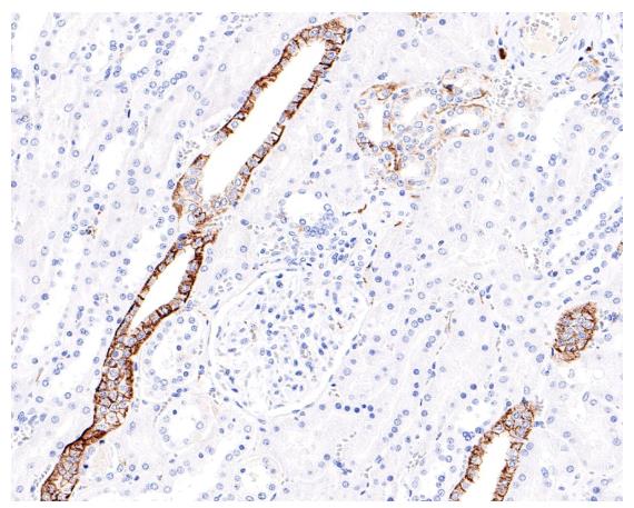 Immunohistochemical analysis of paraffin-embedded human kidney tissue with Rabbit anti-L1CAM antibody at 1/200 dilution. The section was pre-treated using heat mediated antigen retrieval with Tris-EDTA buffer (pH 9.0) for 20 minutes. The tissues were blocked in 1% BSA for 20 minutes at room temperature, washed with ddH2O and PBS, and then probed with the primary antibody at 1/200 dilution for 1 hour at room temperature. The detection was performed using an HRP conjugated compact polymer system. DAB was used as the chromogen. Tissues were counterstained with hematoxylin and mounted with DPX.
Cartpieces
Totalgoods,subtotals:¥Checkout
Partial purchase records(bought amounts latest0)
No one bought this product
User Comment(Total0User Comment Num)
- No comment


 +86 571 56623320
 SUNLONG BIOTECH

