
Details
Product Name Recombinant Rabbit Anti-CDKN2A Monoclonal Antibody Cat No. SLA2888M Chinese Name 抑癌基因p16重组兔单抗 Alias ARF; CAI2; CDK4I; CDKN2; CMM2; INK4; INK4A; MLM; MTS-1; MTS1; P14; P14ARF; P16; P16-INK4A; P16INK4; P16INK4A; P19; P19ARF; TP16; ARF-INK4a; INK4a-ARF; Ink4a/Arf; Pctr1; p16(INK4a); p19 ; p16Cdkn2a; CDN2A_HUMAN; CDKN2A; Cyclin-dependent kinase 4 inhibitor A (CDK4I); Multiple tumor suppressor 1 (MTS-1); p16-INK4a (p16-INK4 | p16INK4A); CDN2A_MOUSE; p16-INK4a (p16-INK4); CDN2A_RAT; p16-INK4a (p16 | p16-INK4); Host Species Rabbit Isotype IgG Clonality Monoclonal Clone No. 14E19 React Species Human Applications WB (1:500-2000)
IHC-P (1:100-500)
IHC-F (1:100-500)
IF (1:100-500)
ICC/IF (1:50-200)
Not yet tested in other applications.
Optimal working dilutions must be determined by the end user.Immunogen A synthesized peptide derived from human CDKN2A Purification affinity purified by Protein A Predicted MW. 17 kDa Observed MW. 16 kDa Subcellular Location Nucleus Gene ID 1029 Swiss-Prot P42771 Storage 0.01M TBS (pH7.4) with 1% BSA, 0.02% Proclin300 and 50% Glycerol.
Shipped at 4℃. Store at -20℃ for one year. Avoid repeated freeze/thaw cycles.Background This gene generates several transcript variants which differ in their first exons. At least three alternatively spliced variants encoding distinct proteins have been reported, two of which encode structurally related isoforms known to function as inhibitors of CDK4 kinase. The remaining transcript includes an alternate first exon located 20 Kb upstream of the remainder of the gene; this transcript contains an alternate open reading frame (ARF) that specifies a protein which is structurally unrelated to the products of the other variants. This ARF product functions as a stabilizer of the tumor suppressor protein p53 as it can interact with, and sequester, the E3 ubiquitin-protein ligase MDM2, a protein responsible for the degradation of p53. In spite of the structural and functional differences, the CDK inhibitor isoforms and the ARF product encoded by this gene, through the regulatory roles of CDK4 and p53 in cell cycle G1 progression, share a common functionality in cell cycle G1 control. This gene is frequently mutated or deleted in a wide variety of tumors, and is known to be an important tumor suppressor gene. [provided by RefSeq, Sep 2012]
Attention This product as supplied is intended for research use only, not for use in human, therapeutic or diagnostic applications.
Validation Images
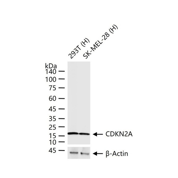
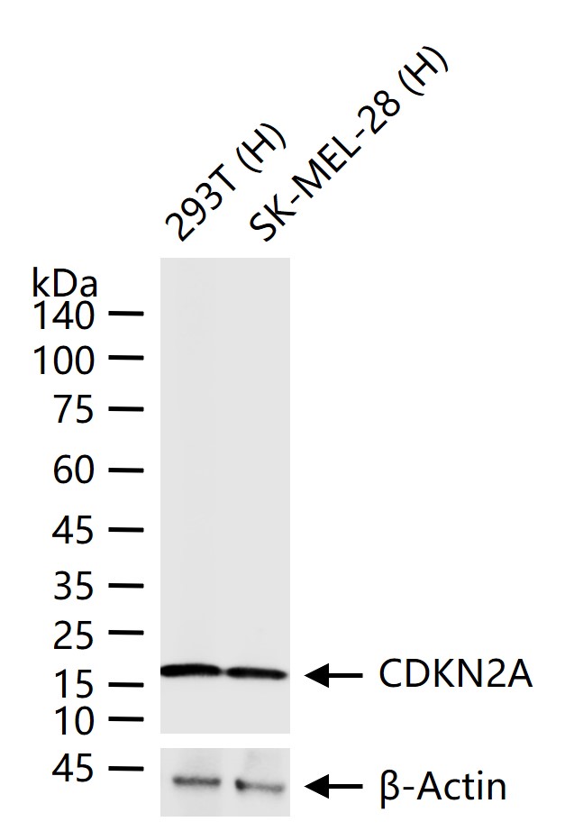
Figure 1. 25 ug total protein per lane of various lysates (see on figure) probed with CDKN2A monoclonal antibody, unconjugated (SLA2888M) at 1:1000 dilution and 4°C overnight incubation. Followed by conjugated secondary antibody incubation at r.t. for 60 min.
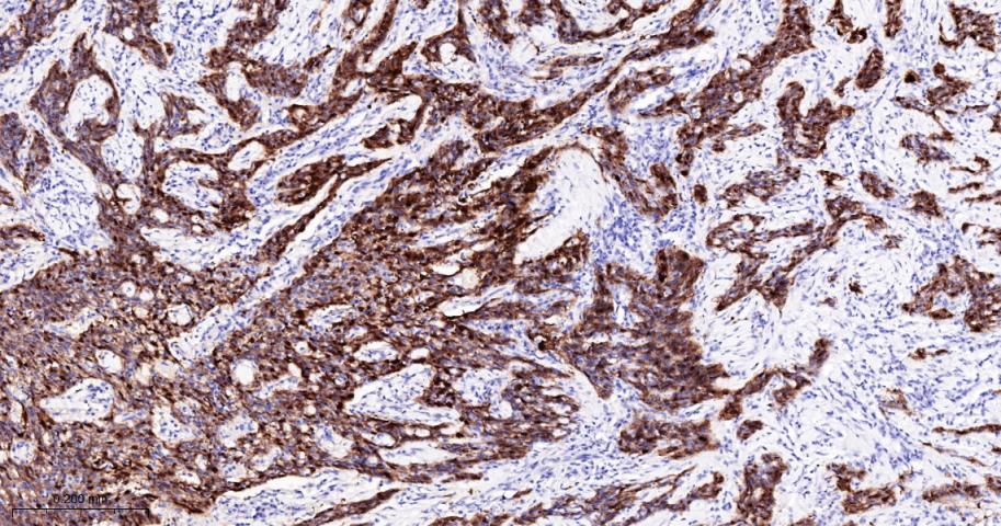
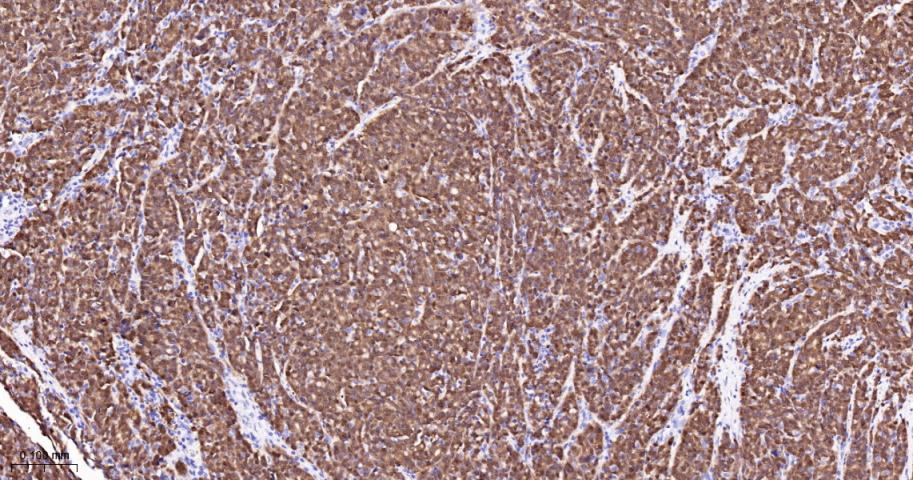
Figure 2. Paraformaldehyde-fixed, paraffin embedded Human Cervical Cancer; Antigen retrieval by boiling in sodium citrate buffer (pH6.0) for 15 min; The section was incubated with CDKN2A Monoclonal Antibody, Unconjugated (SLA2888M) at 1:200 overnight at 4°C, followed by conjugation to the SL0295G-HRP and DAB (C-0010) staining.
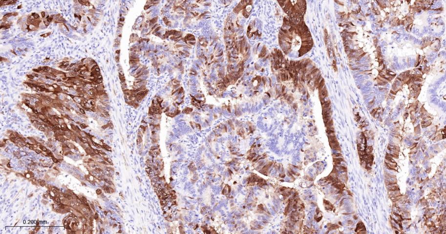
Figure 3. Paraformaldehyde-fixed, paraffin embedded Human Colon Cancer; Antigen retrieval by boiling in sodium citrate buffer (pH6.0) for 15 min; The section was incubated with CDKN2A Monoclonal Antibody, Unconjugated (SLA2888M) at 1:200 overnight at 4°C, followed by conjugation to the SL0295G-HRP and DAB (C-0010) staining.
Figure 4. Paraformaldehyde-fixed, paraffin embedded Human Ovarian mucinous carcinoma; Antigen retrieval by boiling in sodium citrate buffer (pH6.0) for 15 min; The section was incubated with CDKN2A Monoclonal Antibody, Unconjugated (SLA2888M) at 1:200 overnight at 4°C, followed by conjugation to the SL0295G-HRP and DAB (C-0010) staining.
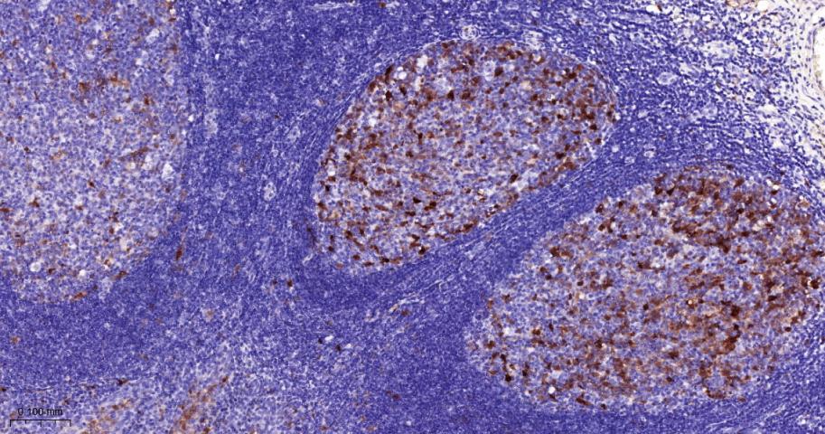
Figure 5. Paraformaldehyde-fixed, paraffin embedded Human Tonsil; Antigen retrieval by boiling in sodium citrate buffer (pH6.0) for 15 min; The section was incubated with CDKN2A Monoclonal Antibody, Unconjugated (SLA2888M) at 1:200 overnight at 4°C, followed by conjugation to the SL0295G-HRP and DAB (C-0010) staining.
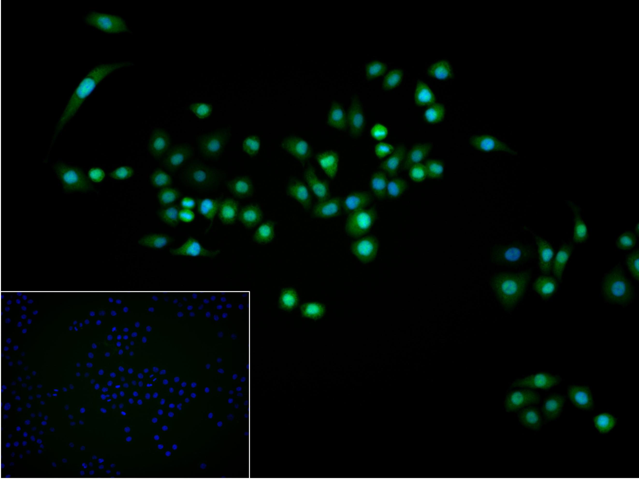
Figure 6. 4% Paraformaldehyde-fixed Hela (H) cell; Triton X-100 at r.t. for 20 min; Antibody incubation with (CDKN2A) monoclonal Antibody, unconjugated (SLA2888M) 1:100, 90 min at 37°C; followed by conjugated Goat Anti-Rabbit IgG antibody (green, SL60295G-BF488) at 37°C for 90 min, DAPI (blue, C02-04002) was used to stain the cell nuclei. PBS instead of the primary antibody was used as the blank control.
FOR RESEARCH USE ONLY. Not for use in diagnostic or therapeutic procedures.
Partial purchase records(bought amounts latest0)
User Comment(Total0User Comment Num)
- No comment


 +86 571 56623320

 SUNLONG BIOTECH


