
Details
Product Name BCL2 | Human Bcl-2 Mouse mAb Cat No. SLM-30235M Chinese Name Bcl-2单克隆抗体 Alias Bcl-2; PPP1R50; BCL2_HUMAN; BCL2; BCL2 apoptosis regulator; B-cell CLL/lymphoma 2; BCL2, apoptosis regulator; protein phosphatase 1, regulatory subunit 50 Host Species Mouse Isotype Mouse IgG2b,k Clonality Monoclonal Clone No. 8B4 React Species Human Applications Flow-Cyt (1ug/Test)
ICC/IF (1:100-500)
Not yet tested in other applications.
Optimal working dilutions must be determined by the end user.Immunogen Purification affinity purified by Protein G Concentration Predicted MW. 26 kDa Observed MW. 26 kDa Subcellular Location Nucleus Gene ID Swiss-Prot Storage 0.01M PBS (pH7.4).
Shipped at 4℃. Store at -20℃ for one year. Avoid repeated freeze/thaw cycles.Background The Bcl-2 gene was isolated at the chromosomal breakpoint of t(14;18)-bearing follicular B cell lymphomas(1,2).Bcl-2 blocks cell death following a variety of stimuli and confers a death-sparing effect to certain hematopoietic cell lines following growth factor withdrawal (3,5).Bcl-2 appears to function in several subcellular locations yet lacks any known motifs that would confer insight into its mechanism of action (6,7).A more recently identified protein,designated Bax p21(i.e., Bcl-associated X protein ),has extensive amino acid homology with Bcl-2 and both homodimerizes and forms heterodimers with Bcl-2(8). Overexpression of Bax accelerates apoptotic death induced by cytokine deprivation in an IL-3 dependent cell line and Bax also counters the death repressor activty of Bcl-2(8). Attention This product as supplied is intended for research use only, not for use in human, therapeutic or diagnostic applications.
Validation Images
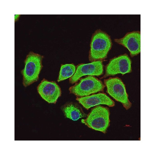
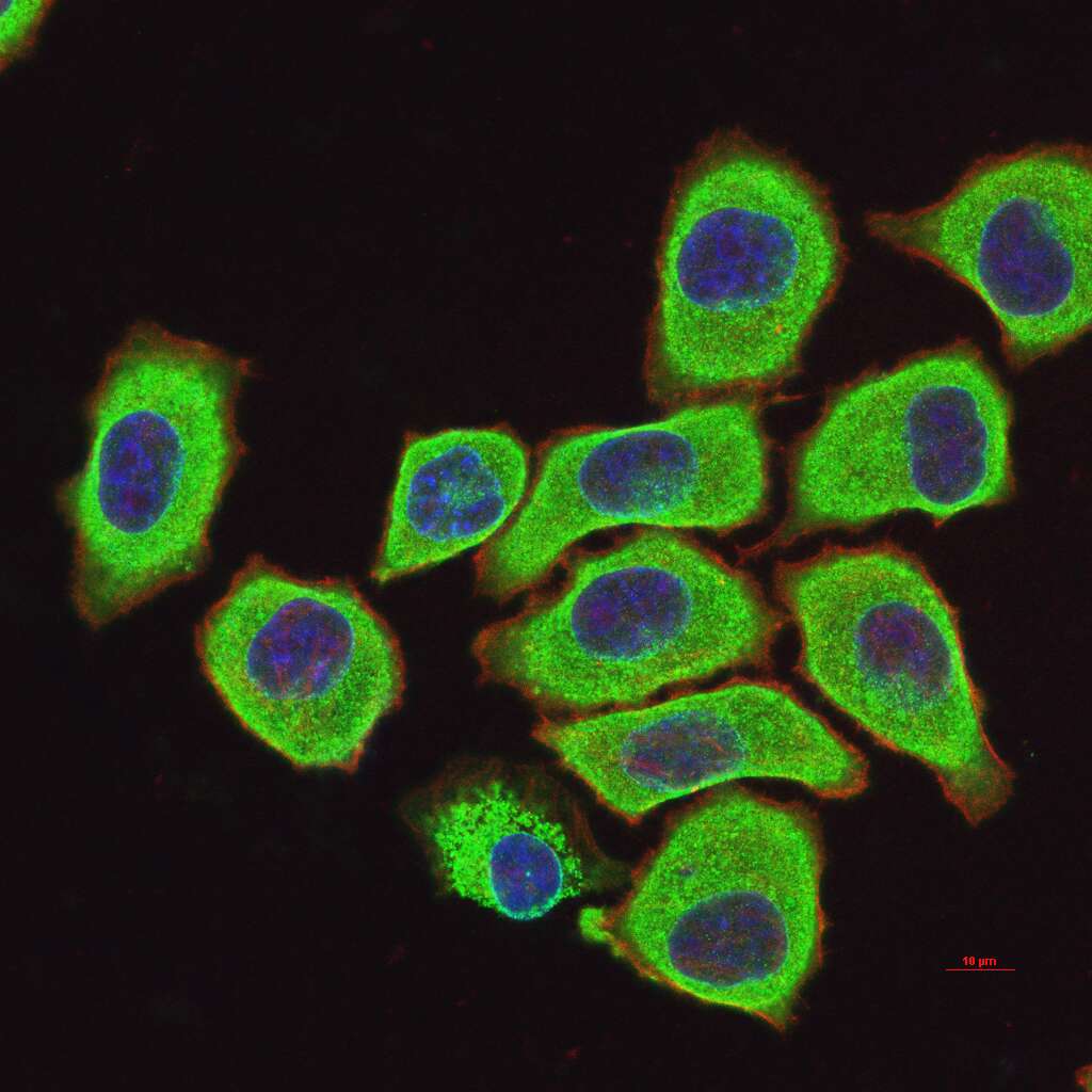
Figure 1. 4% Paraformaldehyde-fixed HeLa (H) cell; Triton X-100 at r.t. for 20 min; Antibody incubation with (Bcl-2) monoclonal Antibody, unconjugated (SLM-30235M) 1:100, 90 min at 37°C; followed by BF488 conjugated Goat Anti-Mouse IgG antibody (green, SL60296G-BF488) at 37°C for 90 min, then stained with Phalloidin/BF555 for 30 min at room temperature; DAPI (blue, C02-04002) was used to stain the cell nuclei.
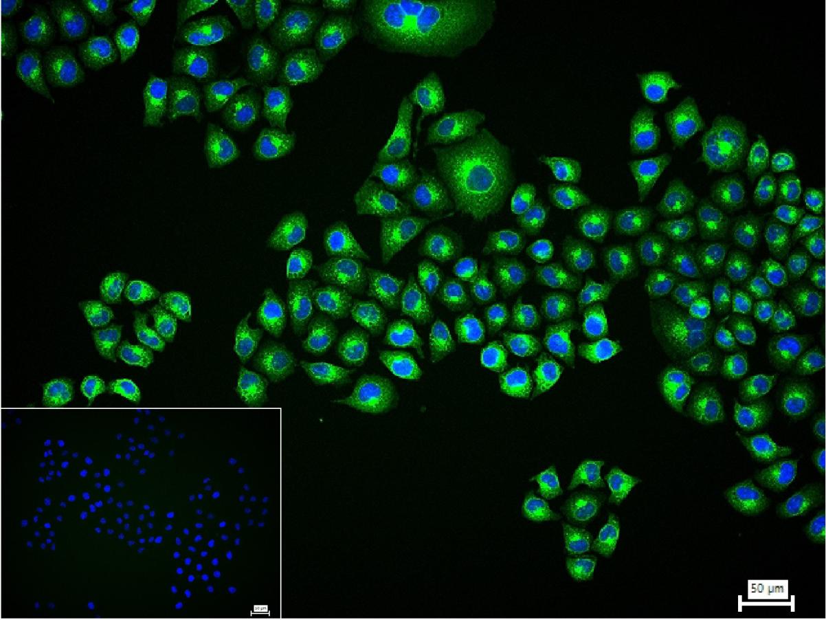
Figure 2. 4% Paraformaldehyde-fixed HeLa (H) cell; Triton X-100 at r.t. for 20 min; Antibody incubation with (Bcl-2) monoclonal Antibody, unconjugated (SLM-30235M) 1:100, 90 min at 37°C; followed by conjugated Goat Anti-Mouse IgG antibody (green, SL60296G-BF488) at 37°C for 90 min, DAPI (blue, C02-04002) was used to stain the cell nuclei. PBS instead of the primary antibody was used as the blank control.
FOR RESEARCH USE ONLY. Not for use in diagnostic or therapeutic procedures.
Partial purchase records(bought amounts latest0)
User Comment(Total0User Comment Num)
- No comment


 +86 571 56623320

 SUNLONG BIOTECH


