Details
Product Name PRTN3 | PR3 Mouse mAb Cat No. SLM-43164M Chinese Name 白细胞蛋白酶3单克隆抗体 Alias ACPA; AGP7; C-ANCA; CANCA; MBN; MBT; NP-4; NP4; P29; PR-3; PR3; PRTN3_HUMAN; PRTN3; C-ANCA antigen; Leukocyte proteinase 3 (PR-3 | PR3); Neutrophil proteinase 4 (NP-4); Wegener autoantigen; 3.4.21.76; proteinase 3; myeloblastin; serine proteinase, neutrophil; Wegener granulomatosis autoantigen Host Species Mouse Isotype IgG Clonality Monoclonal Clone No. 16B8 React Species Human Applications IHC-P (1:100-500)
IHC-F (1:100-500)
IF (1:100-500)
ICC/IF (1:100-500)
Not yet tested in other applications.
Optimal working dilutions must be determined by the end user.Immunogen Recombinant human PR3 Purification affinity purified by Protein A Predicted MW. 28 kDa Subcellular Location Membrane Storage 0.01M PBS (pH7.4).
Shipped at 4℃. Store at -20℃ for one year. Avoid repeated freeze/thaw cycles.Background The major features of Wegener granulomatosis are necrotizing granulomatous lesions, which most often affect the upper and lower airways and are associated with vasculitis, necrotizing glomerulonephritis and pulmonary capillaritis. The antigen responsible for this disease is Proteinase 3 (PR3, P29 or myeloblastin), which is one of the antibiotic proteins of neutrophilic granules belonging to the serine protease family. It is closely related to two others: Neutrophil Elastase and azurocidin. All three genes are expressed coordinately and their protein products are packaged together into azurophil granules during neutrophil differentiation. PR3 is a neutrophil protein which is able to cleave elastin and is involved in proliferation of human leukemia cells. PR3 is expressed specifically in immature myeloid cells and is a G-CSF-responsive protein critical to factor-independent growth. The genes for all three of the related serine protease family members are located in a cluster on the tip of the short arm of human chromosome 19. Attention This product as supplied is intended for research use only, not for use in human, therapeutic or diagnostic applications.
Validation Images
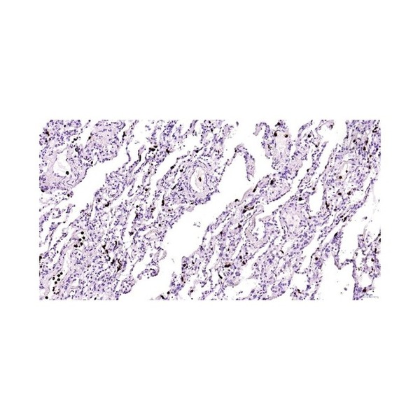
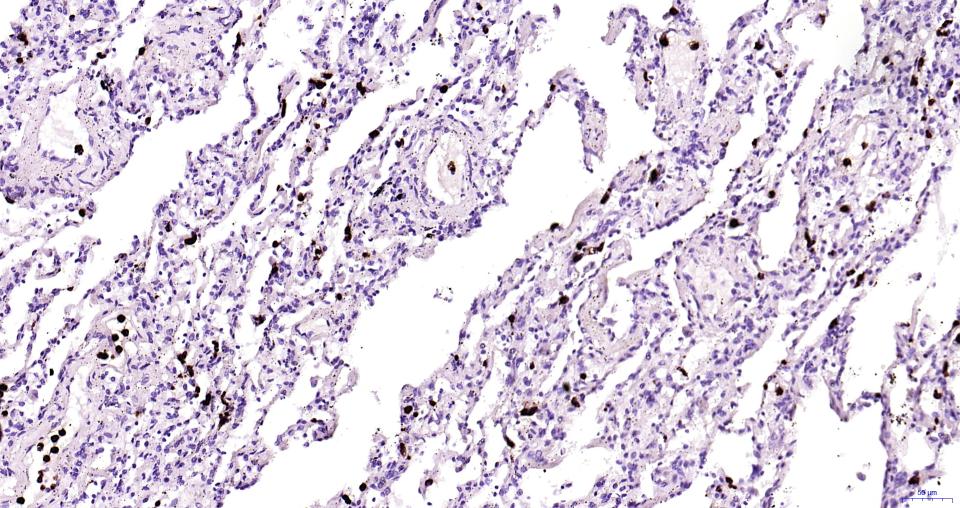
Figure 1. Paraformaldehyde-fixed, paraffin embedded Human lung; Antigen retrieval by boiling in sodium citrate buffer (pH6.0) for 15 min; Antibody incubation with PR3 Monoclonal Antibody, Unconjugated(SLM-43164M) at 1:200 overnight at 4°C, followed by conjugation to the SL40296G-HRP and DAB (C-0010) staining.
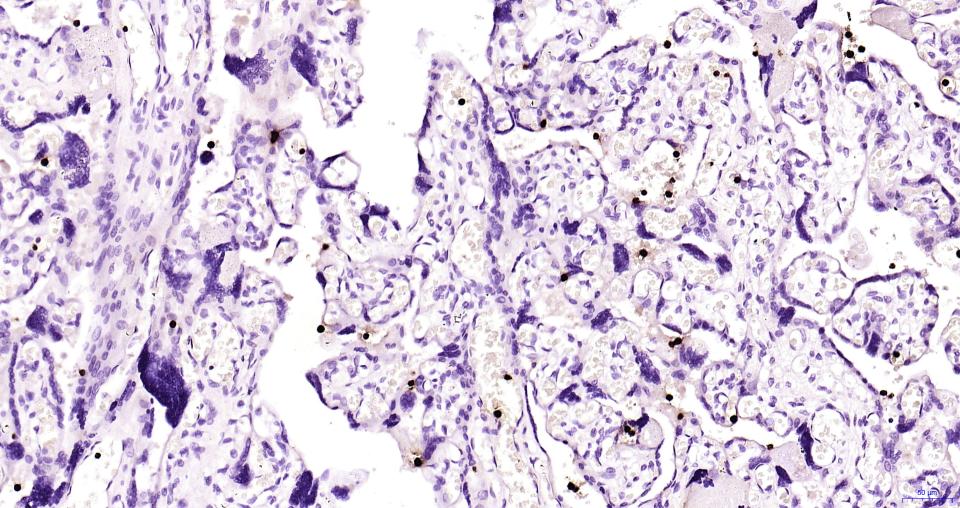
Figure 2. Paraformaldehyde-fixed, paraffin embedded Human placenta; Antigen retrieval by boiling in sodium citrate buffer (pH6.0) for 15 min; Antibody incubation with PR3 Monoclonal Antibody, Unconjugated(SLM-43164M) at 1:200 overnight at 4°C, followed by conjugation to the SL40296G-HRP and DAB (C-0010) staining.
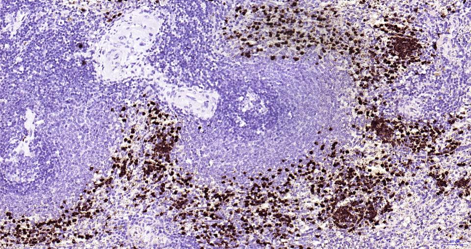
Figure 3. Paraformaldehyde-fixed, paraffin embedded Human spleen; Antigen retrieval by boiling in sodium citrate buffer (pH6.0) for 15 min; Antibody incubation with PR3 Monoclonal Antibody, Unconjugated(SLM-43164M) at 1:200 overnight at 4°C, followed by conjugation to the SL40296G-HRP and DAB (C-0010) staining.
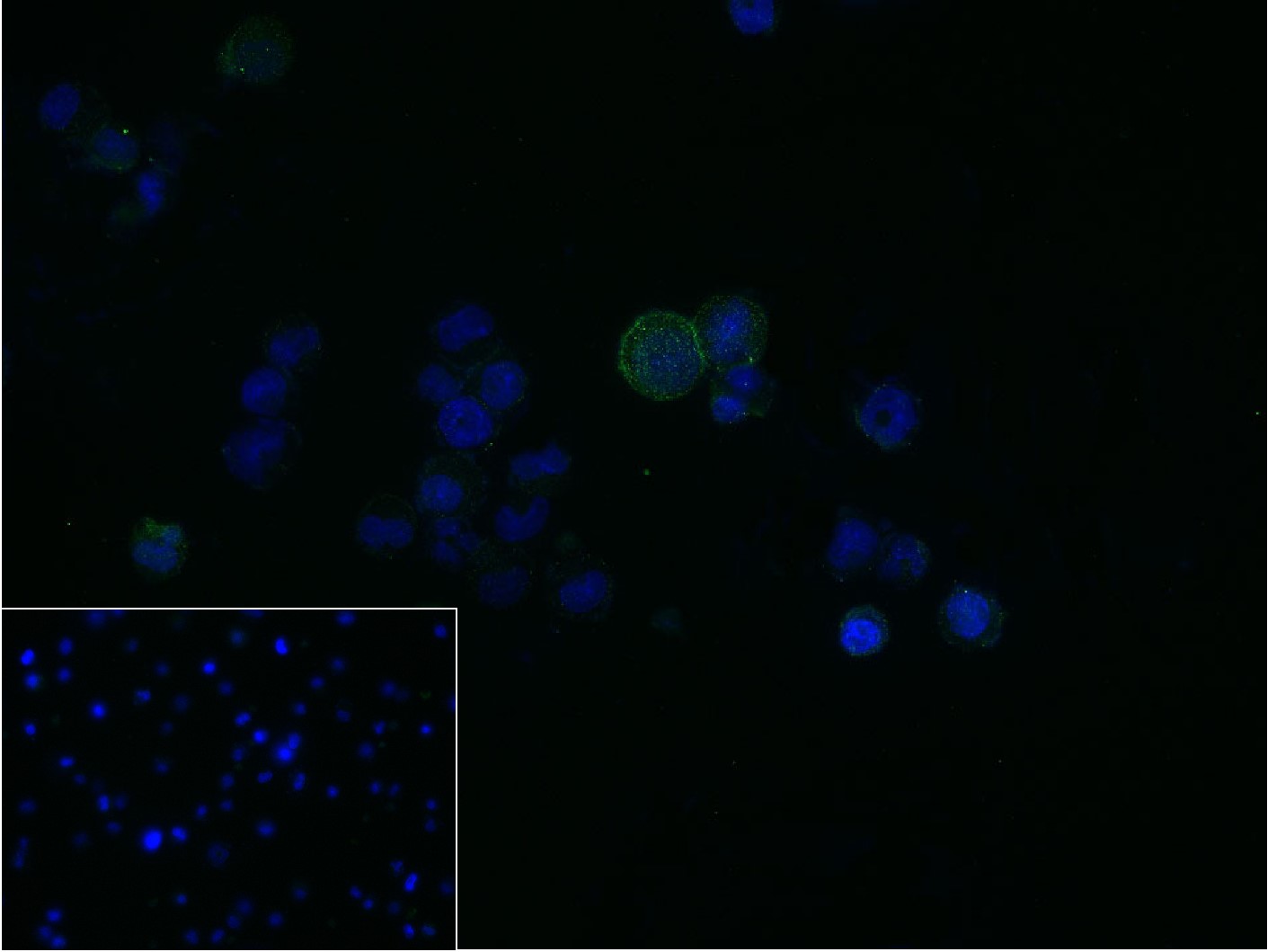
Figure 4. 4% Paraformaldehyde-fixed HL-60 (H) cell; Triton X-100 at r.t. for 20 min; Antibody incubation with (PR3) monoclonal Antibody, unconjugated (SLM-43164M) 1:100, 90 min at 37°C; followed by conjugated Goat Anti-Mouse IgG antibody (green, SL60296G-FITC) at 37°C for 90 min, DAPI (blue, C02-04002) was used to stain the cell nuclei. PBS instead of the primary antibody was used as the blank control.
FOR RESEARCH USE ONLY. Not for use in diagnostic or therapeutic procedures.
Partial purchase records(bought amounts latest0)
User Comment(Total0User Comment Num)
- No comment


 +86 571 56623320

 SUNLONG BIOTECH


