Rabbit Anti-CLDN1 antibody
CLD1_HUMAN; Claudin-1; Claudin 1; Claudin1; CLD1; SEMP1; Senescence-associated epithelial membrane protein; UNQ481/PRO944; ILVASC;
View History [Clear]
Details
Product Name CLDN1 Chinese Name 紧密连接蛋白1抗体 Alias CLD1_HUMAN; Claudin-1; Claudin 1; Claudin1; CLD1; SEMP1; Senescence-associated epithelial membrane protein; UNQ481/PRO944; ILVASC; literatures Research Area Signal transduction Cell adhesion molecule Cytoskeleton Immunogen Species Rabbit Clonality Polyclonal React Species Human,Mouse(predicted:Rat) Applications WB=1:500-2000 (Paraffin sections need antigen repair)
not yet tested in other applications.
optimal dilutions/concentrations should be determined by the end user.Theoretical molecular weight 23kDa Cellular localization The cell membrane Form Liquid Concentration 1mg/ml immunogen KLH conjugated synthetic peptide derived from mouse Claudin 1: 121-211/211 <Cytoplasmic> Lsotype IgG Purification affinity purified by Protein A Buffer Solution 1M TBS(pH7.4) with 1% BSA, 3% Proclin300 and 50% Glycerol. Storage Shipped at 4℃. Store at -20 °C for one year. Avoid repeated freeze/thaw cycles. Attention This product as supplied is intended for research use only, not for use in human, therapeutic or diagnostic applications. PubMed PubMed Product Detail Tight junctions represent one mode of cell-to-cell adhesion in epithelial or endothelial cell sheets, forming continuous seals around cells and serving as a physical barrier to prevent solutes and water from passing freely through the paracellular space. These junctions are comprised of sets of continuous networking strands in the outwardly facing cytoplasmic leaflet, with complementary grooves in the inwardly facing extracytoplasmic leaflet. The protein encoded by this gene, a member of the claudin family, is an integral membrane protein and a component of tight junction strands. Loss of function mutations result in neonatal ichthyosis-sclerosing cholangitis syndrome. [provided by RefSeq, Jul 2008]
Function:
Plays a major role in tight junction-specific obliteration of the intercellular space, through calcium-independent cell-adhesion activity.
Subunit:
Can form homo- and heteropolymers with other CLDN. Homopolymers interact with CLDN3, but not CLDN2, homopolymers. Directly interacts with TJP1/ZO-1, TJP2/ZO-2 and TJP3/ZO-3. Interacts with MPDZ and INADL.
Subcellular Location:
Cell junction, tight junction. Cell membrane; Multi-pass membrane protein.
Tissue Specificity:
Widely expressed, with highest levels in liver and kidney.
Similarity:
Belongs to the claudin family.
SWISS:
O88551
Gene ID:
12737
Database links:Entrez Gene: 9076 Human
Entrez Gene: 12737 Mouse
SwissProt: O95832 Human
SwissProt: O88551 Mouse
Claudin 1蛋白是紧密连接区相关的转膜蛋白Claudin的家族成员,在构成紧密连接的完整膜蛋白中发挥着一定作用,目前多用于Tumour方面的研究。Product Picture 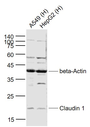 Sample:
Lane 1: A549 (Human) Cell Lysate at 30 ug
Lane 2: HepG2 (Human) Cell Lysate at 30 ug
Primary:
Anti- Claudin 1 (SL1428R) at 1/1000 dilution
Anti- beta-Actin (SL0061R) at 1/2000 dilution
Secondary: IRDye800CW Goat Anti-Rabbit IgG at 1/20000 dilution
Predicted band size: 21 kD
Observed band size: 21 kD
 Sample:
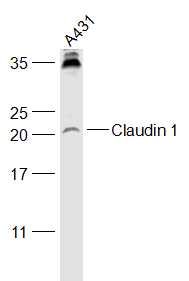
A431(Human) Cell Lysate at 30 ug
Primary: Anti-Claudin 1 (SL1428R) at 1/1000 dilution
Secondary: IRDye800CW Goat Anti-Rabbit IgG at 1/20000 dilution
Predicted band size: 23 kD
Observed band size: 23 kD
 Sample:
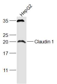
HepG2(Human) Cell Lysate at 30 ug
Primary: Anti-Claudin 1 (SL1428R) at 1/1000 dilution
Secondary: IRDye800CW Goat Anti-Rabbit IgG at 1/20000 dilution
Predicted band size: 23 kD
Observed band size: 23 kD
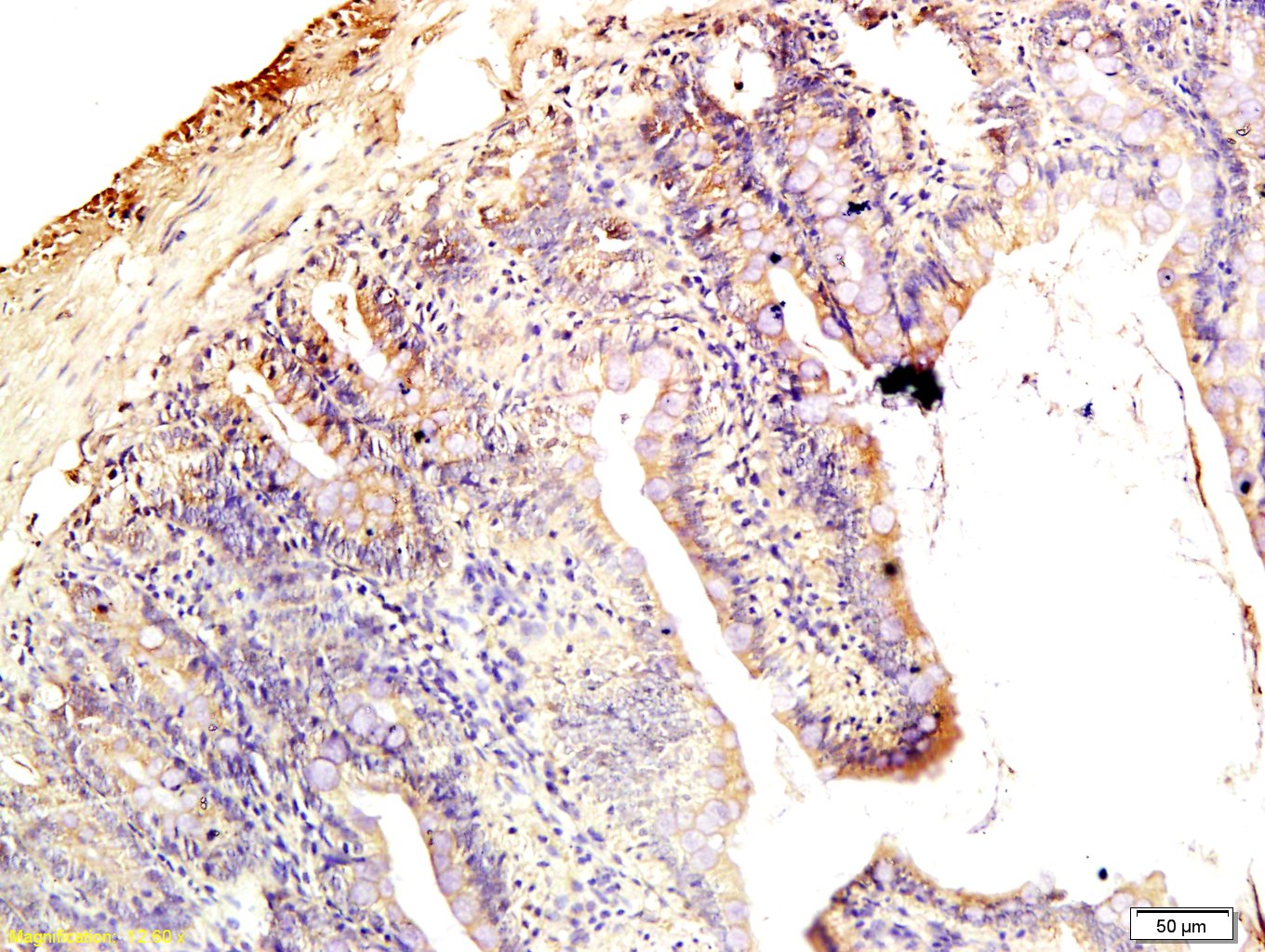 Tissue/cell: rat intestine tissue; 4% Paraformaldehyde-fixed and paraffin-embedded;
Antigen retrieval: citrate buffer ( 1M, pH 6.0 ), Boiling bathing for 15min; Block endogenous peroxidase by 3% Hydrogen peroxide for 30min; Blocking buffer (normal goat serum,C-0005) at 37℃ for 20 min;
Incubation: Anti-Claudin-1 Polyclonal Antibody, Unconjugated(SL1428R) 1:200, overnight at 4°C, followed by conjugation to the secondary antibody(SP-0023) and DAB(C-0010) staining
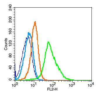 Blank control: Raji(blue).
Primary Antibody:Rabbit Anti-Claudin 1 antibody(SL1428R), Dilution: 1μg in 100 μL 1X PBS containing 0.5% BSA;
Isotype Control Antibody: Rabbit IgG(orange) ,used under the same conditions );
Secondary Antibody: Goat anti-rabbit IgG-PE(white blue), Dilution: 1:200 in 1 X PBS containing 0.5% BSA.
Protocol
The cells were fixed with 2% paraformaldehyde (10 min). Antibody (SL1428R, 1μg /1x10^6 cells) were incubated for 30 min on the ice, followed by 1 X PBS containing 0.5% BSA + 1 0% goat serum (15 min) to block non-specific protein-protein interactions. Then the Goat Anti-rabbit IgG/PE antibody was added into the blocking buffer mentioned above to react with the primary antibody of SL1428R at 1/200 dilution for 30 min on ice. Acquisition of 20,000 events was performed.
Cartpieces
Totalgoods,subtotals:¥Checkout
Partial purchase records(bought amounts latest0)
No one bought this product
User Comment(Total0User Comment Num)
- No comment


 +86 571 56623320
 SUNLONG BIOTECH

