Rabbit Anti-Collagen XVII antibody
Bullous Pemphigoid 180; 180 kDa bullous pemphigoid antigen 2; Alpha 1 type XVII collagen; BA16H23.2; BP 180; BP180; BPAG 2; BPAG2; Bullous pemphigoid antigen 2; COL17A1; Collagen 17; Collagen alpha 1 XVII chain; Collagen alpha 1(XVII) chain; Collagen alph
View History [Clear]
Details
Product Name Collagen XVII Chinese Name Collagen protein17Recombinant rabbit monoclonal anti Alias Bullous Pemphigoid 180; 180 kDa bullous pemphigoid antigen 2; Alpha 1 type XVII collagen; BA16H23.2; BP 180; BP180; BPAG 2; BPAG2; Bullous pemphigoid antigen 2; COL17A1; Collagen 17; Collagen alpha 1 XVII chain; Collagen alpha 1(XVII) chain; Collagen alpha1 XVII chain; Collagen type XVII alpha 1; Collagen XVII alpha 1 polypeptide; CollagenXVII; Epidermolysis bullosa junctional localisata variant; FLJ60881; KIAA0204; LAD 1; LAD. XVII型Collagen protein; 17型Collagen protein Research Area Cell biology Signal transduction Immunogen Species Rabbit Clonality Monoclonal Clone NO. 1C11 React Species Human,Mouse,Rat Applications WB=1:500-2000,IHC-P=1:50-200,IHC-F=1:50-200,IF=1:50-200 (Paraffin sections need antigen repair)
not yet tested in other applications.
optimal dilutions/concentrations should be determined by the end user.Theoretical molecular weight 150kDa Cellular localization The cell membrane Form Liquid Concentration 1mg/1ml immunogen Recombinant human Collagen XVII protein: 1300-1450/1497 <Extracellular> Lsotype IgG Purification affinity purified by Protein A Buffer Solution 1M TBS(pH7.4) with 1% BSA, 3% Proclin300 and 50% Glycerol. Storage Shipped at 4℃. Store at -20 °C for one year. Avoid repeated freeze/thaw cycles. Attention This product as supplied is intended for research use only, not for use in human, therapeutic or diagnostic applications. PubMed PubMed Product Detail This gene encodes the alpha chain of type XVII collagen. Unlike most collagens, collagen XVII is a transmembrane protein. Collagen XVII is a structural component of hemidesmosomes, multiprotein complexes at the dermal-epidermal basement membrane zone that mediate adhesion of keratinocytes to the underlying membrane. Mutations in this gene are associated with both generalized atrophic benign and junctional epidermolysis bullosa. Two homotrimeric forms of type XVII collagen exist. The full length form is the transmembrane protein. A soluble form, referred to as either ectodomain or LAD-1, is generated by proteolytic processing of the full length form. [provided by RefSeq, Jul 2008]
Function:
Unlike most collagens, collagen XVII is a transmembrane protein. Collagen XVII is a structural component of hemidesmosomes, multiprotein complexes at the dermal epidermal basement membrane zone that mediate adhesion of keratinocytes to the underlying membrane. Mutations in the gene coding for collagen XVII are associated with both generalized atrophic benign and junctional epidermolysis bullosa. Two homotrimeric forms of type XVII collagen exist. The full length form is the transmembrane protein. A soluble form, referred to as either ectodomain or LAD 1, is generated by proteolytic processing of the full length form. Two transcript variants, one resulting from alternative splicing in the 3' UTR, have been identified for this gene.
Subunit:
Homotrimers of alpha 1(XVII)chains. Interacts (via cytoplasmic region) with ITGB4 (via cytoplasmic region). Interacts (via cytoplasmic region) with DST isoform 3 (via N-terminus). Interacts (via N-terminus) with PLEC. Interacts (via cytoplasmic region) with DSP.
Subcellular Location:
Cell junction, hemidesmosome. Membrane; Single-pass type II membrane protein. Note=Localized along the plasma membrane of the hemidesmosome. 120 kDa linear IgA disease antigen and 97 kDa linear IgA disease antigen: Secreted, extracellular space, extracellular matrix, basement membrane.
Post-translational modifications:
The intracellular/endo domain is disulfide-linked.
DISEASE:
Generalized atrophic benign epidermolysis bullosa (GABEB) [MIM:226650]: A non-lethal, adult form of junctional epidermolysis bullosa characterized by life-long blistering of the skin, associated with hair and tooth abnormalities. Note=The disease is caused by mutations affecting the gene represented in this entry.
SWISS:
Q9UMD9
Gene ID:
1308
Database links:Entrez Gene: 1308 Human
Omim: 113811 Human
SwissProt: Q9UMD9 Human
Unigene: 117938 Human
Product Picture 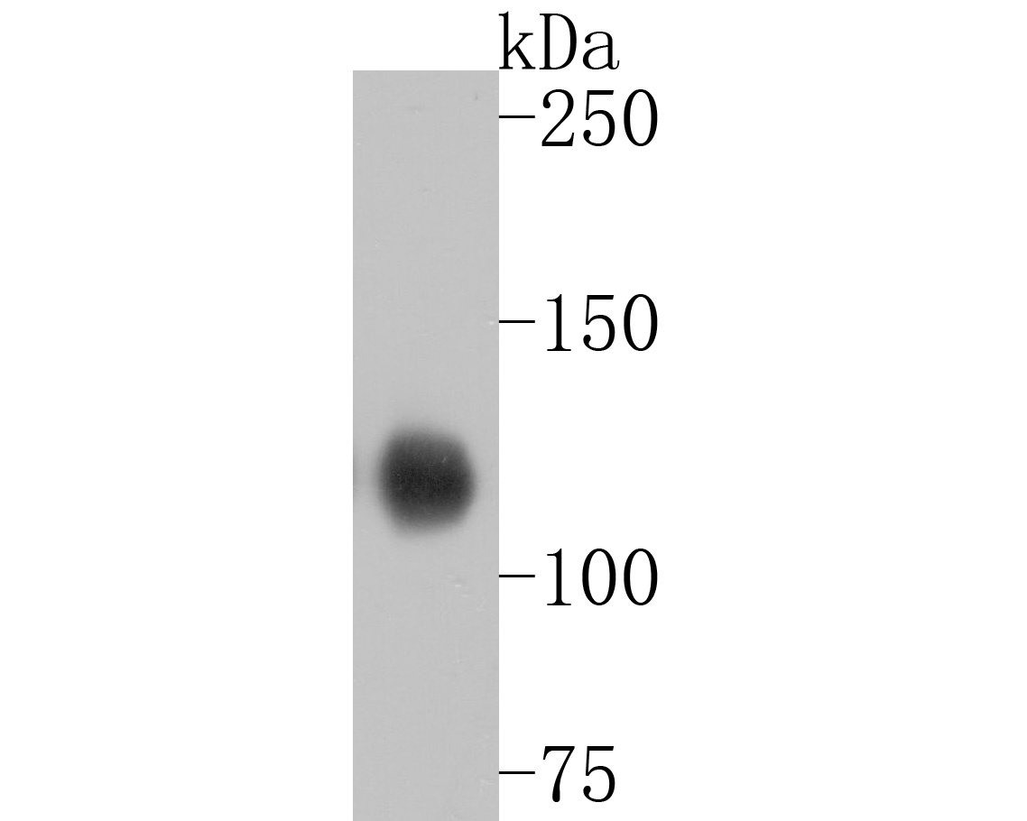 Sample:
Lane 1: Skin (Rat) Lysate at 40 ug
Primary: Anti-Collagen XVII (SLM-52041R) at 1/1000 dilution
Secondary: Goat Anti-Rabbit IgG - HRP at 1/5000 dilution
Predicted band size: 150 kD
Observed band size: 120 kD
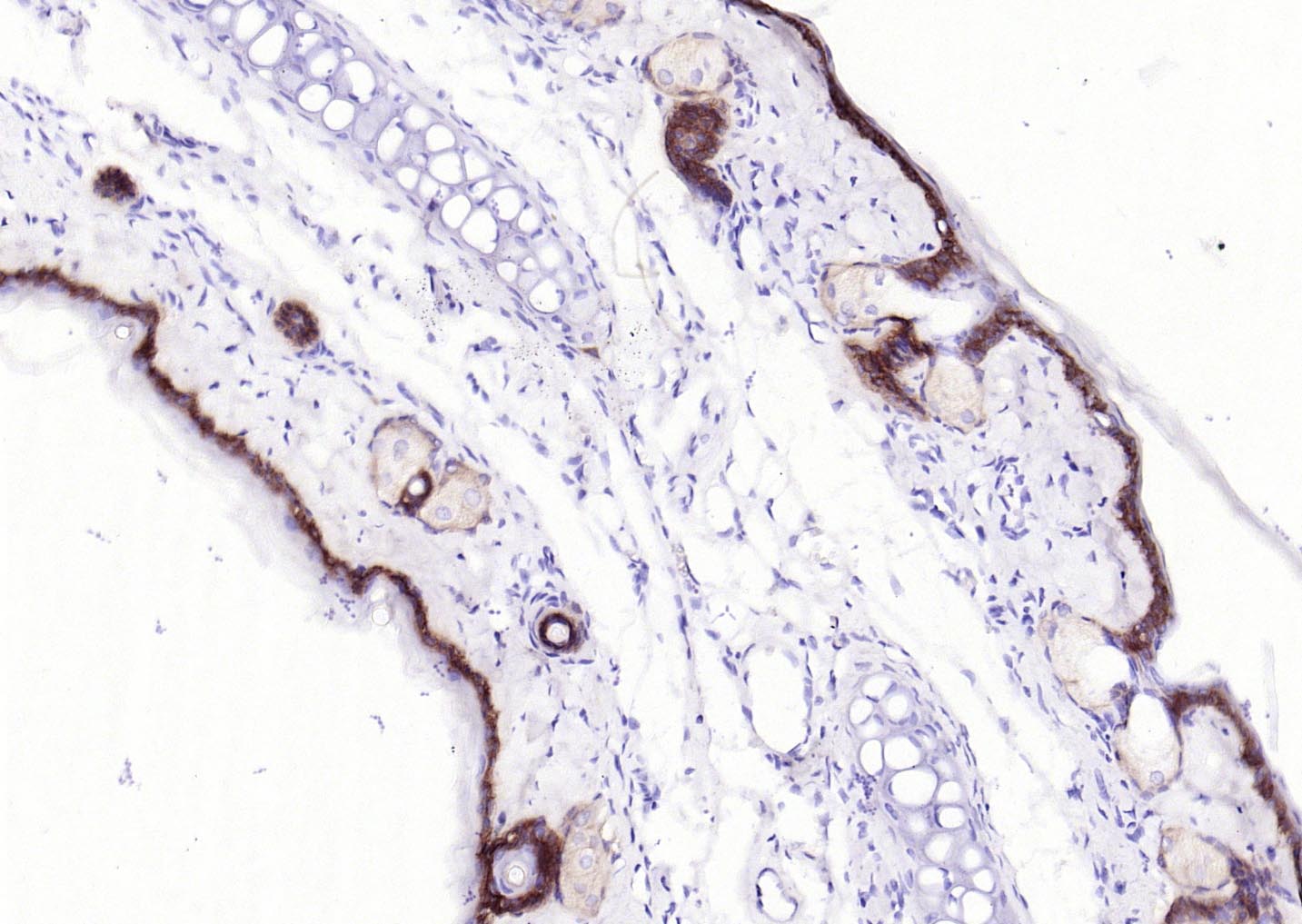 Paraformaldehyde-fixed, paraffin embedded (rat skin); Antigen retrieval by boiling in sodium citrate buffer (pH6.0) for 15min; Block endogenous peroxidase by 3% hydrogen peroxide for 20 minutes; Blocking buffer (normal goat serum) at 37°C for 30min; Antibody incubation with (Collagen XVII) Monoclonal Antibody, Unconjugated (SLM-52041R) at 1:200 overnight at 4°C, followed by operating according to SP Kit(Rabbit) (sp-0023) instructionsand DAB staining.
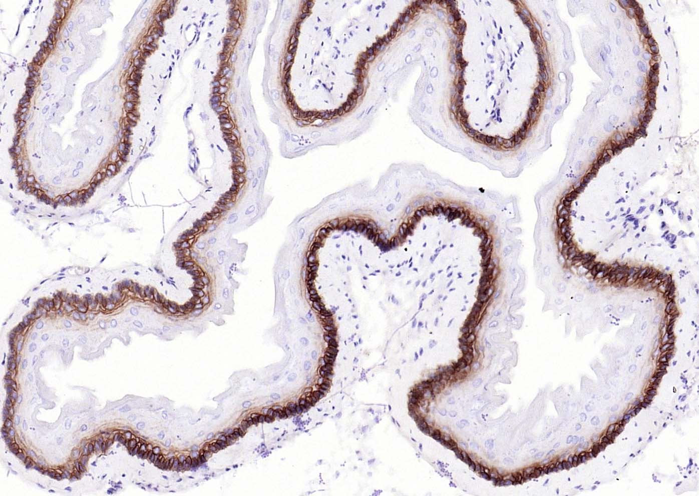 Paraformaldehyde-fixed, paraffin embedded (mouse esophageal); Antigen retrieval by boiling in sodium citrate buffer (pH6.0) for 15min; Block endogenous peroxidase by 3% hydrogen peroxide for 20 minutes; Blocking buffer (normal goat serum) at 37°C for 30min; Antibody incubation with (Collagen XVII) Monoclonal Antibody, Unconjugated (SLM-52041R) at 1:200 overnight at 4°C, followed by operating according to SP Kit(Rabbit) (sp-0023) instructionsand DAB staining.
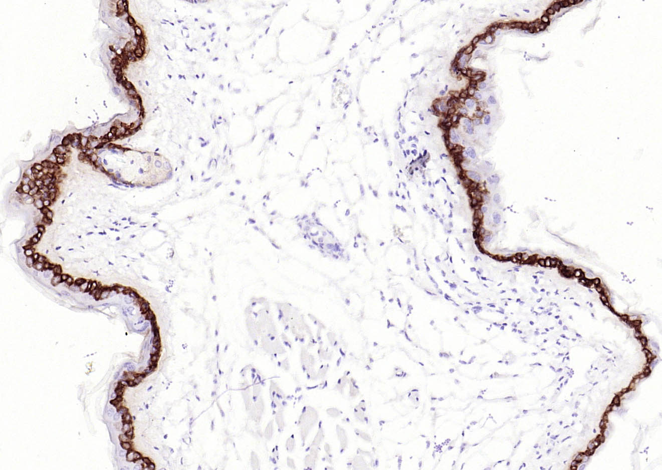 Paraformaldehyde-fixed, paraffin embedded (mouse skin); Antigen retrieval by boiling in sodium citrate buffer (pH6.0) for 15min; Block endogenous peroxidase by 3% hydrogen peroxide for 20 minutes; Blocking buffer (normal goat serum) at 37°C for 30min; Antibody incubation with (Collagen XVII) Monoclonal Antibody, Unconjugated (SLM-52041R) at 1:200 overnight at 4°C, followed by operating according to SP Kit(Rabbit) (sp-0023) instructionsand DAB staining.
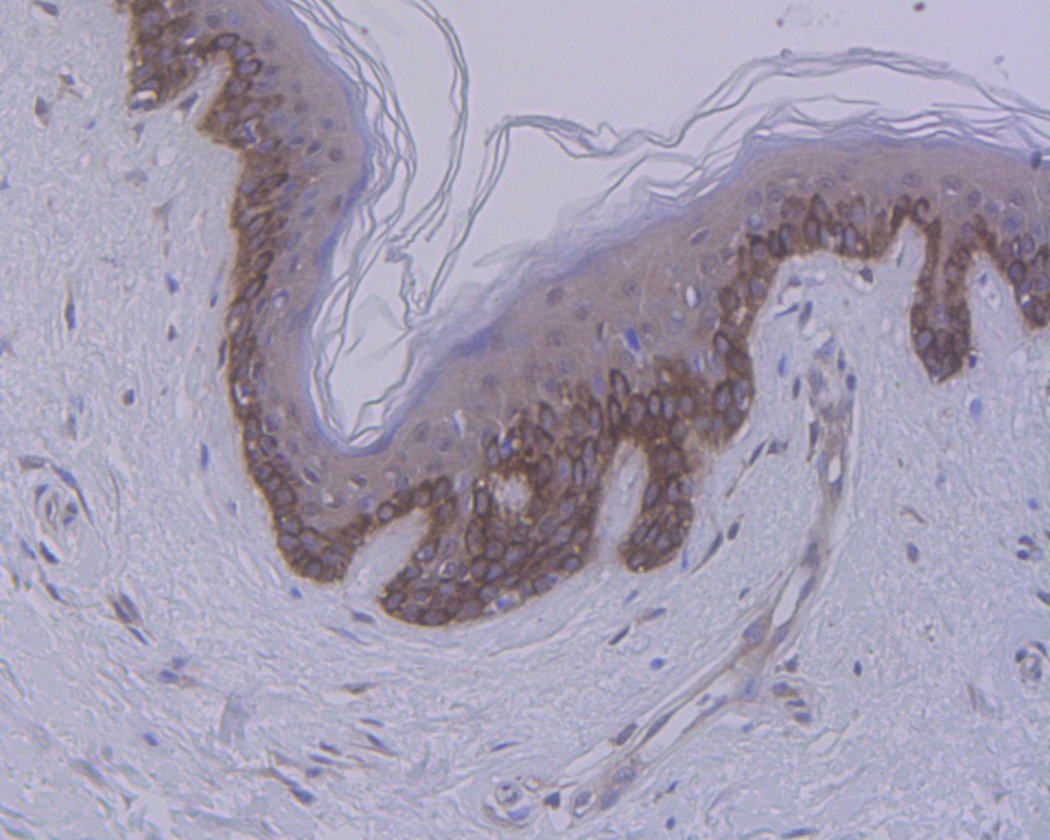 Paraformaldehyde-fixed, paraffin embedded (human skin); Antigen retrieval by boiling in sodium citrate buffer (pH6.0) for 15min; Block endogenous peroxidase by 3% hydrogen peroxide for 20 minutes; Blocking buffer (normal goat serum) at 37°C for 30min; Antibody incubation with (Collagen XVII) Monoclonal Antibody, Unconjugated (SLM-52041R) at 1:50 overnight at 4°C, followed by operating according to SP Kit(Rabbit) (sp-0023) instructionsand DAB staining.
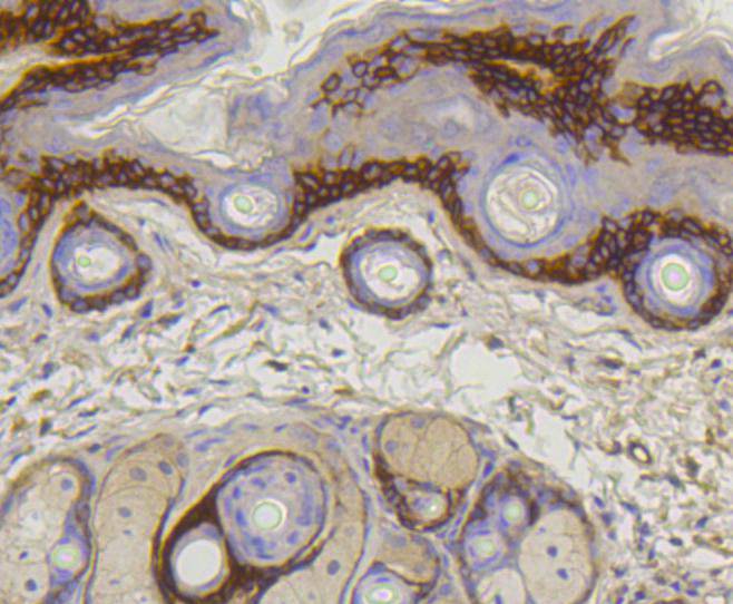 Paraformaldehyde-fixed, paraffin embedded (mouse skin); Antigen retrieval by boiling in sodium citrate buffer (pH6.0) for 15min; Block endogenous peroxidase by 3% hydrogen peroxide for 20 minutes; Blocking buffer (normal goat serum) at 37°C for 30min; Antibody incubation with (Collagen XVII) Monoclonal Antibody, Unconjugated (SLM-52041R) at 1:50 overnight at 4°C, followed by operating according to SP Kit(Rabbit) (sp-0023) instructionsand DAB staining.
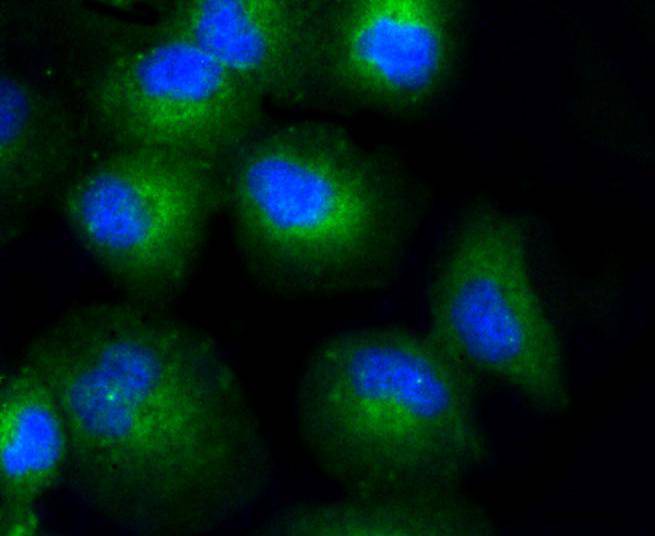 HUVEC cell; 4% Paraformaldehyde-fixed; Triton X-100 at room temperature for 20 min; Blocking buffer (normal goat serum, C-0005) at 37°C for 20 min; Antibody incubation with (Collagen XVII) monoclonal Antibody, Unconjugated (SLM-52041R) 1:50, 90 minutes at 37°C; followed by a conjugated Goat Anti-Rabbit IgG antibody at 37°C for 90 minutes, DAPI (blue, C02-04002) was used to stain the cell nuclei.
Cartpieces
Totalgoods,subtotals:¥Checkout
Partial purchase records(bought amounts latest0)
No one bought this product
User Comment(Total0User Comment Num)
- No comment


 +86 571 56623320
 SUNLONG BIOTECH

