Mouse Anti-PTPN2 antibody
PTN2_HUMAN; Tyrosine-protein phosphatase non-receptor type 2; PTPT; EC:3.1.3.48; T-cell protein-tyrosine phosphatase (TCPTP); protein tyrosine phosphatase non-receptor type 2; PTN2; TCPTP; TC-PTP; TCELLPTP;
View History [Clear]
Details
Product Name PTPN2 Chinese Name 酪氨酸蛋白磷酸酶非受体2型单克隆抗体 Alias PTN2_HUMAN; Tyrosine-protein phosphatase non-receptor type 2; PTPT; EC:3.1.3.48; T-cell protein-tyrosine phosphatase (TCPTP); protein tyrosine phosphatase non-receptor type 2; PTN2; TCPTP; TC-PTP; TCELLPTP; Research Area Signal transduction Kinases and Phosphatases Immunogen Species Mouse Clonality Monoclonal Clone NO. F3H2 React Species Human,Mouse,Rat Applications WB=1:500-2000,IHC-P=1:100-500,IHC-F=1:100-500,IF=1:100-500,Flow-Cyt=1:50-100,Flow-Cyt=1:50-100 (Paraffin sections need antigen repair)
not yet tested in other applications.
optimal dilutions/concentrations should be determined by the end user.Cellular localization The nucleus cytoplasmic The cell membrane Form Liquid Concentration 1mg/ml Lsotype IgG1/Kappa Purification Affinity purified by Protein G Buffer Solution 1M TBS(pH7.4) with 1% BSA, 3% Proclin300 and 50% Glycerol. Storage Shipped at 4℃. Store at -20 °C for one year. Avoid repeated freeze/thaw cycles. Attention This product as supplied is intended for research use only, not for use in human, therapeutic or diagnostic applications. PubMed PubMed Product Detail The protein encoded by this gene is a member of the protein tyrosine phosphatase (PTP) family. Members of the PTP family share a highly conserved catalytic motif, which is essential for the catalytic activity. PTPs are known to be signaling molecules that regulate a variety of cellular processes including cell growth, differentiation, mitotic cycle, and oncogenic transformation. Epidermal growth factor receptor and the adaptor protein Shc were reported to be substrates of this PTP, which suggested the roles in growth factor mediated cell signaling. Multiple alternatively spliced transcript variants encoding different isoforms have been found. Two highly related but distinctly processed pseudogenes that localize to chromosomes 1 and 13, respectively, have been reported. [provided by RefSeq, May 2011]
Subcellular Location:
[Isoform 2]: Nucleus. Cytoplasm. Cell membrane. Note=Predominantly localizes to chromatin (By similarity). Able to shuttle between the nucleus and the cytoplasm and to dephosphorylate plasma membrane receptors (PubMed:9488479). Recruited by activated ITGA1 at the plasma membrane. {ECO:0000250, ECO:0000269|PubMed:9488479}.
Tissue Specificity:
Ubiquitously expressed. Isoform 2 is probably the major isoform. Isoform 1 is expressed in T-cells and in placenta.
SWISS:
P17706
Gene ID:
5771
Database links:Entrez Gene: 5771 Human
Entrez Gene: 19255 Mouse
SwissProt: P17706 Human
SwissProt: Q06180 Mouse
去磷酸化受体蛋白酪氨酸激酶的非受体型酪氨酸特异性磷酸酶,包括INSR、EGFR、CSF1R、PDGFR。在The nucleus或细胞质中也去磷酸化非受体蛋白酪氨酸激酶,如JAK1、JAK2、JAK3、Src家族激酶、STAT1、STAT3和STAT6。能够负调控许多信号通路和生物过程,如造血,炎症反应,细胞增殖和分化,葡萄糖稳态。在免疫系统的发育中起着多方面的重要作用,通过FYN和LCK的去磷酸化来控制T细胞的分化和活化,在T细胞受体Signal transduction中发挥作用。去磷酸化CSF1R,负调控其下游信号和巨噬Cell differentiation。通过细胞质激酶JAK1、JAK3及其底物STAT1的去磷酸化负调控cell factor(IL2/白介素-2和Interferon)介导的信号传导,这些激酶在cell factor受体下游传播信号。Product Picture 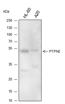 Blocking buffer: 5% NFDM/TBST
Primary ab dilution: 1:5000
Primary ab incubation condition: room temperature 2h
Secondary ab: Goat Anti-Mouse IgG H&L (HRP)
Lysate: HL-60, A20
Protein loading quantity: 20 μg
Exposure time: 10 s
Predicted MW: 45-50 kDa
Observed MW: 45-50 kDa
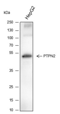 Blocking buffer: 5% NFDM/TBST
Primary ab dilution: 1:5000
Primary ab incubation condition: room temperature 2h
Secondary ab: Goat Anti-Mouse IgG H&L (HRP)
Lysate: HepG2
Protein loading quantity: 20 μg
Exposure time: 10 s
Predicted MW: 45-50 kDa
Observed MW: 45-50 kDa
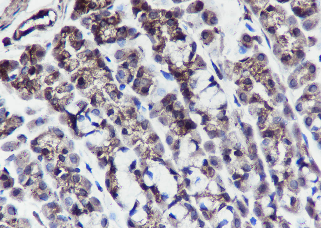
 Tissue: Human breast cancer
Section type: Formalin fixed & Paraffin -embedded section
Retrieval method: High temperature and high pressure
Retrieval buffer: Tris/EDTA buffer, pH 9.0 Primary ab dilution: 1:100
Primary ab incubation condition: 1 hour at room temperature
Secondary ab: SP Kit(Mouse)(sp-0024)
Counter stain: Hematoxylin (Blue)
Comment: Color brown is the positive signal for SLM-60446M
 Tissue: Rat stomach
Section type: Formalin fixed & Paraffin -embedded section
Retrieval method: High temperature and high pressure
Retrieval buffer: Tris/EDTA buffer, pH 9.0 Primary ab dilution: 1:100
Primary ab incubation condition: 1 hour at room temperature
Secondary ab: SP Kit(Mouse)(sp-0024)
Counter stain: Hematoxylin (Blue)
Comment: Color brown is the positive signal for SLM-60446M
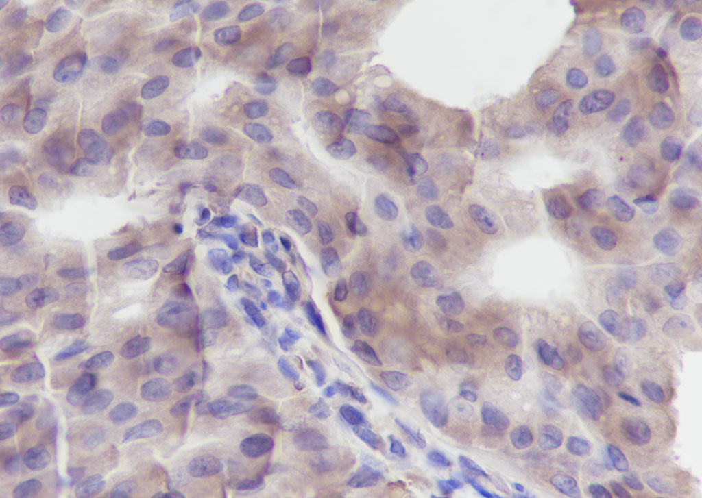
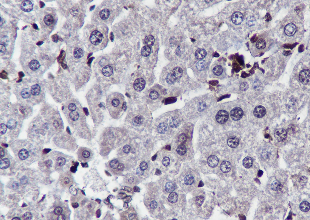 Tissue: Mouse liver
Section type: Formalin fixed & Paraffin -embedded section
Retrieval method: High temperature and high pressure
Retrieval buffer: Tris/EDTA buffer, pH 9.0 Primary ab dilution: 1:100
Primary ab incubation condition: 1 hour at room temperature
Secondary ab: SP Kit(Mouse)(sp-0024)
Counter stain: Hematoxylin (Blue)
Comment: Color brown is the positive signal for SLM-60446M
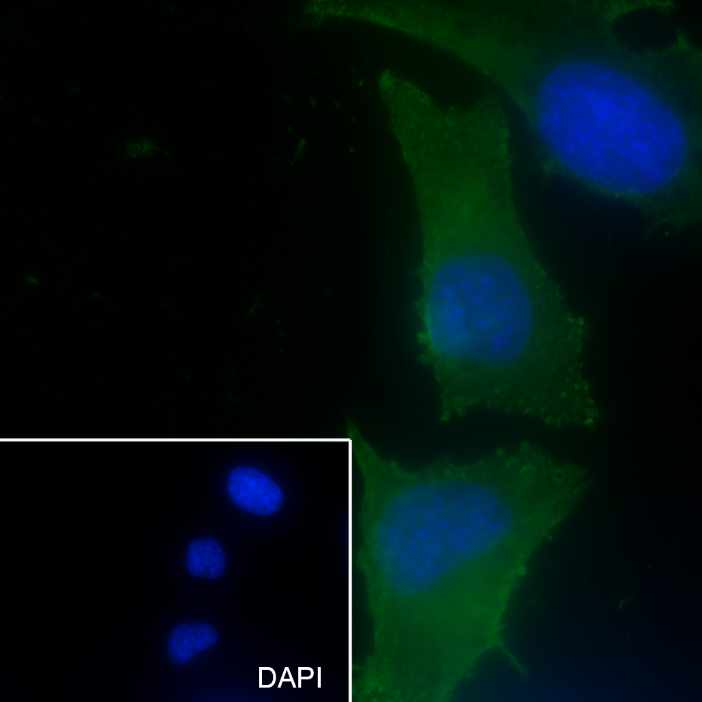 Cell line: HeLa
Fixative: 4% Paraformaldehyde
Permeabilization: 0.1% TritonX-100
Primary ab dilution: 1:50
Primary incubation condition: 4°C overnight
Secondary ab: Goat Anti-Mouse IgG
Nuclear counter stain: DAPI (Blue)
Comment: Color green is the positive signal for SLM-60446M
Cartpieces
Totalgoods,subtotals:¥Checkout
Partial purchase records(bought amounts latest0)
No one bought this product
User Comment(Total0User Comment Num)
- No comment


 +86 571 56623320
 SUNLONG BIOTECH

