Rabbit Anti-EDG1 antibody
CD363; CHEDG1; D1S3362; ECGF1; EDG1; EDG 1; Endothelial differentiation G-protein coupled receptor 1; Endothelial differentiation sphingolipid G protein coupled receptor 1; G protein coupled sphingolipid receptor; S1P receptor 1; S1P receptor Edg-1; S1P1;
View History [Clear]
Details
Product Name EDG1 Chinese Name endothelial cells分化鞘脂G protein-coupled receptor1Recombinant rabbit monoclonal anti Alias CD363; CHEDG1; D1S3362; ECGF1; EDG1; EDG 1; Endothelial differentiation G-protein coupled receptor 1; Endothelial differentiation sphingolipid G protein coupled receptor 1; G protein coupled sphingolipid receptor; S1P receptor 1; S1P receptor Edg-1; S1P1; S1PR1; S1PR1_HUMAN; Sphingosine 1 phosphate receptor Edg 1; Sphingosine 1 phosphate receptor EDG1; Sphingosine 1-phosphate receptor 1; Sphingosine 1-phosphate receptor Edg-1; Sphingosine 1phosphate receptor type 1 S1P1. Research Area Cell biology immunology Apoptosis Cell adhesion molecule G protein-coupled receptor endothelial cells Cytoskeleton Immunogen Species Rabbit Clonality Monoclonal React Species Human,Mouse,Rat Applications WB=1:500-2000 (Paraffin sections need antigen repair)
not yet tested in other applications.
optimal dilutions/concentrations should be determined by the end user.Theoretical molecular weight 44kDa Cellular localization The cell membrane Form Liquid Concentration 1mg/ml immunogen KLH conjugated synthetic peptide derived from human EDG1: 2-51/382 Lsotype IgG Purification affinity purified by Protein A Buffer Solution 1M TBS(pH7.4) with 1% BSA, 3% Proclin300 and 50% Glycerol. Storage Shipped at 4℃. Store at -20 °C for one year. Avoid repeated freeze/thaw cycles. Attention This product as supplied is intended for research use only, not for use in human, therapeutic or diagnostic applications. PubMed PubMed Product Detail Sphingosine-1-phosphate receptor 1 (S1P receptor 1 or S1P1), also known as endothelial differentiation gene 1 (EDG1) is a protein that in humans is encoded by the S1PR1 gene. S1PR1 is a G-protein-coupled receptor which binds the bioactive signaling molecule sphingosine 1-phosphate (S1P). S1PR1 belongs to a sphingosine-1-phosphate receptor subfamily comprising five members (S1PR1-5). S1PR1 was originally identified as an abundant transcript in endothelial cells and it has an important role in regulating endothelial cell cytoskeletal structure, migration, capillary-like network formation and vascular maturation. In addition, S1PR1 signaling is important in the regulation of lymphocyte maturation, migration and trafficking.
Function:
Receptor for the lysosphingolipid sphingosine 1-phosphate (S1P). S1P is a bioactive lysophospholipid that elicits diverse physiological effect on most types of cells and tissues. This inducible epithelial cell G-protein-coupled receptor may be involved in the processes that regulate the differentiation of endothelial cells. Seems to be coupled to the G(i) subclass of heteromeric G proteins.
Subcellular Location:
Cell membrane; Multi-pass membrane protein.
Tissue Specificity:
Endothelial cells, and to a lesser extent, in vascular smooth muscle cells, fibroblasts, melanocytes, and cells of epithelioid origin.
Post-translational modifications:
S1P-induced endothelial cell migration requires the PKB/AKT1-mediated phosphorylation of the third intracellular loop at the Thr-236 residue.
Similarity:
Belongs to the G-protein coupled receptor 1 family.
SWISS:
P21453
Gene ID:
1901
Database links:
Entrez Gene: 100050049 Horse
Entrez Gene: 1901 Human
Entrez Gene: 13609 Mouse
Omim: 601974 Human
SwissProt: P21453 Human
SwissProt: O08530 Mouse
Unigene: 154210 Human
Unigene: 982 Mouse
Unigene: 109455 Rat
研究发现,S1P1与VEGF肩并肩相互协作,从而促进血管生长。VEGF是很多不同抗癌药物的作用靶标。血管新生在很多疾病是异常的。靶向S1P1和VEGF可能要比只使用VEGF抑制剂更加有效地治疗疾病;S1P1是一种关键性的血管新生反应性基因。研究人员证实当新的血管网络形成时,由此产生的血液流动激活vascular endothelial cell表面上的S1P1,并把信号传递到这些细胞内部从而让新形成的血管网络稳定化。因此阻断S1P1可能有助于切断给癌性Tumour提供资源的血液供应。Product Picture 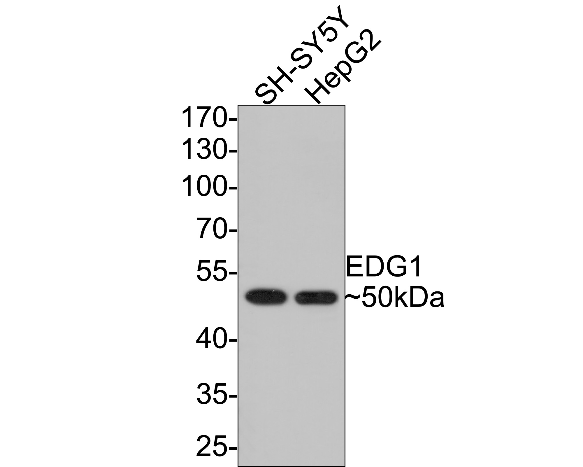 Western blot analysis of EDG1 on different lysates with Rabbit anti-EDG1 antibody (SLM-52902R) at 1/500 dilution.
Lane 1: SH-SY5Y cell lysate
Lane 2: HepG2 cell lysate
Lysates/proteins at 10 µg/Lane.
Predicted band size: 43 kDa
Observed band size: 50 kDa
Exposure time: 1 minute;
10% SDS-PAGE gel.
Proteins were transferred to a PVDF membrane and blocked with 5% NFDM/TBST for 1 hour at room temperature. The primary antibody (SLM-52902R) at 1/500 dilution was used in 5% NFDM/TBST at room temperature for 2 hours. Goat Anti-Rabbit IgG - HRP Secondary Antibody at 1:300,000 dilution was used for 1 hour at room temperature.
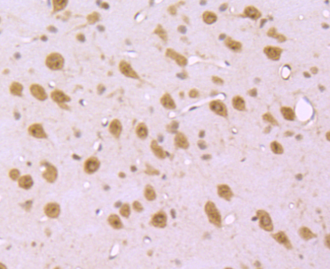 Immunohistochemical analysis of paraffin-embedded mouse brain tissue using anti-EDG1 antibody. The section was pre-treated using heat mediated antigen retrieval with Tris-EDTA buffer (pH 9.0) for 20 minutes.The tissues were blocked in 5% BSA for 30 minutes at room temperature, washed with ddH2O and PBS, and then probed with the primary antibody (SLM-52902R, 1/50) for 30 minutes at room temperature. The detection was performed using an HRP conjugated compact polymer system. DAB was used as the chromogen. Tissues were counterstained with hematoxylin and mounted with DPX
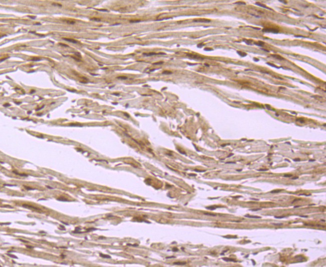 Immunohistochemical analysis of paraffin-embedded mouse heart tissue using anti-EDG1 antibody. The section was pre-treated using heat mediated antigen retrieval with Tris-EDTA buffer (pH 9.0) for 20 minutes.The tissues were blocked in 5% BSA for 30 minutes at room temperature, washed with ddH2O and PBS, and then probed with the primary antibody (SLM-52902R, 1/50) for 30 minutes at room temperature. The detection was performed using an HRP conjugated compact polymer system. DAB was used as the chromogen. Tissues were counterstained with hematoxylin and mounted with DPX.
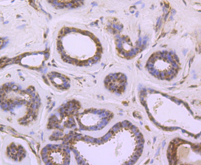 Immunohistochemical analysis of paraffin-embedded human breast carcinoma tissue using anti-EDG1 antibody. The section was pre-treated using heat mediated antigen retrieval with Tris-EDTA buffer (pH 9.0) for 20 minutes.The tissues were blocked in 5% BSA for 30 minutes at room temperature, washed with ddH2O and PBS, and then probed with the primary antibody (SLM-52902R, 1/50) for 30 minutes at room temperature. The detection was performed using an HRP conjugated compact polymer system. DAB was used as the chromogen. Tissues were counterstained with hematoxylin and mounted with DPX.
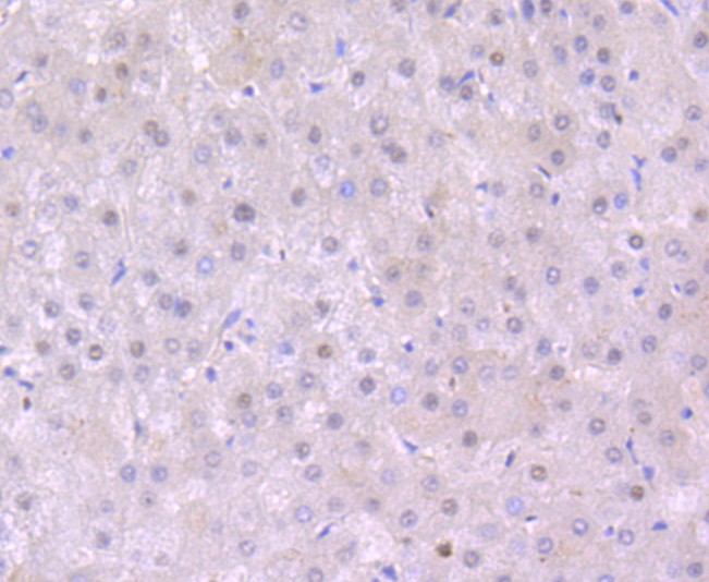 Immunohistochemical analysis of paraffin-embedded human liver tissue using anti-EDG1 antibody. The section was pre-treated using heat mediated antigen retrieval with Tris-EDTA buffer (pH 9.0) for 20 minutes.The tissues were blocked in 5% BSA for 30 minutes at room temperature, washed with ddH2O and PBS, and then probed with the primary antibody (SLM-52902R, 1/50) for 30 minutes at room temperature. The detection was performed using an HRP conjugated compact polymer system. DAB was used as the chromogen. Tissues were counterstained with hematoxylin and mounted with DPX.
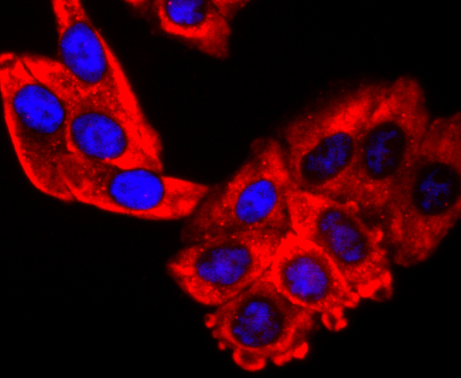 ICC staining of EDG1 in HepG2 cells (red). Formalin fixed cells were permeabilized with 0.1% Triton X-100 in TBS for 10 minutes at room temperature and blocked with 10% negative goat serum for 15 minutes at room temperature. Cells were probed with the primary antibody (SLM-52902R, 1/50) for 1 hour at room temperature, washed with PBS. Alexa Fluor®594 conjugate-Goat anti-Rabbit IgG was used as the secondary antibody at 1/1,000 dilution. The nuclear counter stain is DAPI (blue).
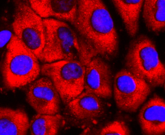 ICC staining of EDG1 in HUVEC cells (red). Formalin fixed cells were permeabilized with 0.1% Triton X-100 in TBS for 10 minutes at room temperature and blocked with 10% negative goat serum for 15 minutes at room temperature. Cells were probed with the primary antibody (SLM-52902R, 1/50) for 1 hour at room temperature, washed with PBS. Alexa Fluor®594 conjugate-Goat anti-Rabbit IgG was used as the secondary antibody at 1/1,000 dilution. The nuclear counter stain is DAPI (blue).
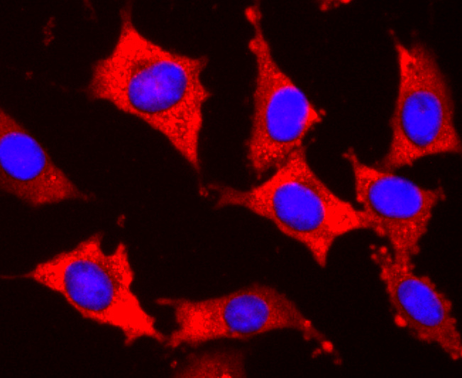 ICC staining of EDG1 in SH-SY5Y cells (red). Formalin fixed cells were permeabilized with 0.1% Triton X-100 in TBS for 10 minutes at room temperature and blocked with 10% negative goat serum for 15 minutes at room temperature. Cells were probed with the primary antibody (SLM-52902R, 1/50) for 1 hour at room temperature, washed with PBS. Alexa Fluor®594 conjugate-Goat anti-Rabbit IgG was used as the secondary antibody at 1/1,000 dilution. The nuclear counter stain is DAPI (blue).
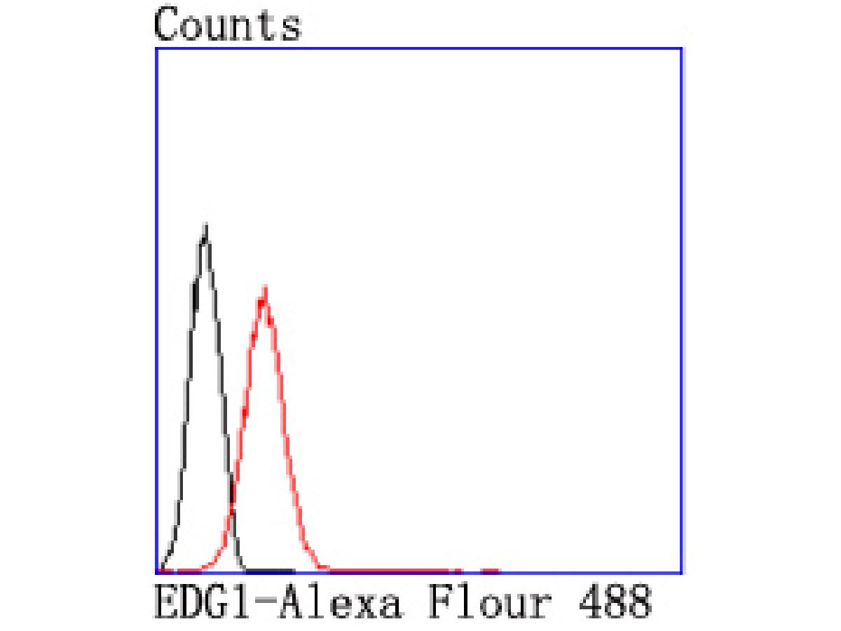 Flow cytometric analysis of EDG1 was done on Jurkat cells. The cells were fixed, permeabilized and stained with the primary antibody (SLM-52902R, 1/50) (red). After incubation of the primary antibody at room temperature for an hour, the cells were stained with a Alexa Fluor®488 conjugate-Goat anti-Rabbit IgG Secondary antibody at 1/1,000 dilution for 30 minutes.Unlabelled sample was used as a control (cells without incubation with primary antibody; black).
Cartpieces
Totalgoods,subtotals:¥Checkout
Partial purchase records(bought amounts latest0)
No one bought this product
User Comment(Total0User Comment Num)
- No comment


 +86 571 56623320
 SUNLONG BIOTECH

