Details
Product Name Recombinant Rabbit Anti-TG Monoclonal Antibody Cat No. SLA3109M Chinese Name 甲状腺球蛋白重组兔单抗 Alias AITD3; TGN; cog; THYG_HUMAN; TG; THYG_MOUSE; THYG_RAT; thyroglobulin Host Species Rabbit Isotype IgG Clonality Monoclonal Clone No. 6C9 React Species Human,Mouse,Rat Applications IHC-P (1:50-200)
IHC-F (1:50-200)
IF (1:50-200)
Flow-Cyt (1:50-100)
ICC/IF (1:50-200)
Not yet tested in other applications.
Optimal working dilutions must be determined by the end user.Immunogen A synthesized peptide derived from human Thyroglobulin Purification affinity purified by Protein A Concentration 1mg/ml Predicted MW. 305 kDa Observed MW. Subcellular Location Secreted Gene ID 7038 Swiss-Prot P01266 Storage 0.01M TBS (pH7.4) with 1% BSA, 0.02% Proclin300 and 50% Glycerol.
Shipped at 4℃. Store at -20℃ for one year. Avoid repeated freeze/thaw cycles.Background Thyroglobulin is the glycoprotein precursor of the iodinated thyroid hormones thyroxine (T4) and triiodothyronine (T3). Thyroglobulin is obtained from the thyroid gland and exhibits the general properties of the globulins. The human thyroglobulin (hTG) is a high molecular weight glycoprotein (605 kDa) found in the thyroid follicular cells. It plays a central role in the uptake, incorporation, and regulated biosynthesis of thyroid hormones, T4 and T3. Thyroid disorders are, in large part, due to autoimmune origin, and anti thyroglobulin autoantibodies were the first factor to be discovered. Anti hTG is found in all thyroid autoimmune diseases (Hashimoto's thyroiditis, Graves' diseases), with the highest level observed in Hashimoto's thyroiditis. Anti hTG is also characteristic of thyroid cancer, and its determination can be used for the follow up of cancer patients. Attention This product as supplied is intended for research use only, not for use in human, therapeutic or diagnostic applications.
Validation Images
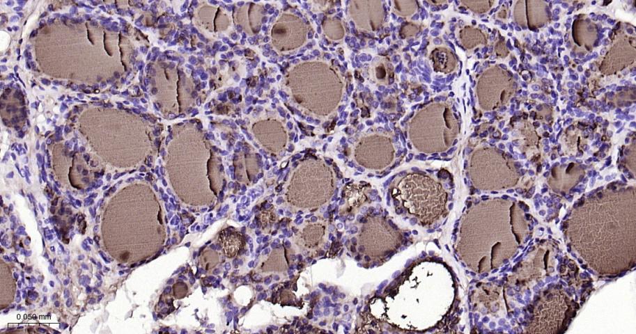
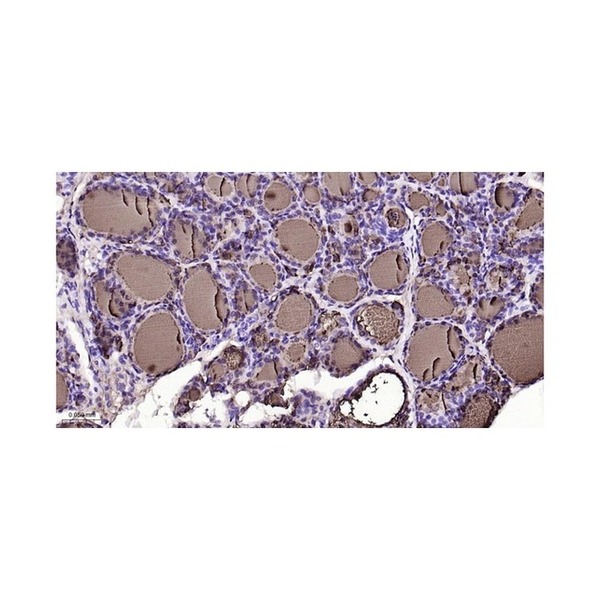
Figure 1. Paraformaldehyde-fixed, paraffin embedded Rat Thyroid Gland; Antigen retrieval by boiling in sodium citrate buffer (pH6.0) for 15 min; The section was incubated with TG Monoclonal Antibody, Unconjugated (SLA3109M) at 1:200 overnight at 4°C, followed by conjugation to the SL0295G-HRP and DAB (C-0010) staining
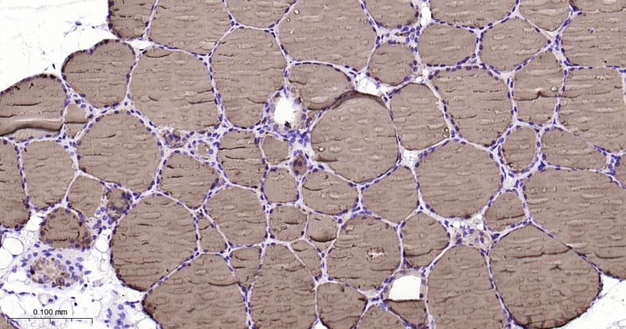
Figure 2. Paraformaldehyde-fixed, paraffin embedded Mouse Thyroid Gland; Antigen retrieval by boiling in sodium citrate buffer (pH6.0) for 15 min; The section was incubated with TG Monoclonal Antibody, Unconjugated (SLA3109M) at 1:200 overnight at 4°C, followed by conjugation to the SL0295G-HRP and DAB (C-0010) staining
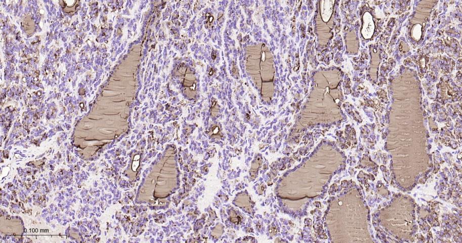
Figure 3. Paraformaldehyde-fixed, paraffin embedded Human Thyroid Cancer; Antigen retrieval by boiling in sodium citrate buffer (pH6.0) for 15 min; The section was incubated with TG Monoclonal Antibody, Unconjugated (SLA3109M) at 1:200 overnight at 4°C, followed by conjugation to the SL0295G-HRP and DAB (C-0010) staining
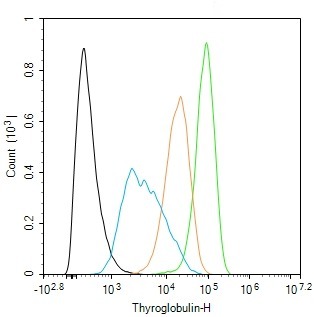
Figure 4. The TT (H) cells were fixed with 4% PFA (10 min at r.t.) and then permeabilized with 90% ice-cold methanol for 20 min at -20℃,the cells then were incubated in 5%BSA to block non-specific protein-protein interactions (30 min at r.t.).Primary Antibody (green):Rabbit Anti-Thyroglobulin antibody (SLA3109M,1:100); Secondary Antibody (white blue): Goat anti-Rabbit IgG-BF488 (SL60295G-BF488): 1 μg/test. Isotype Control (orange): Rabbit IgG (SL0295P). Blank control (black): PBS. Acquisition of 20,000 events was performed
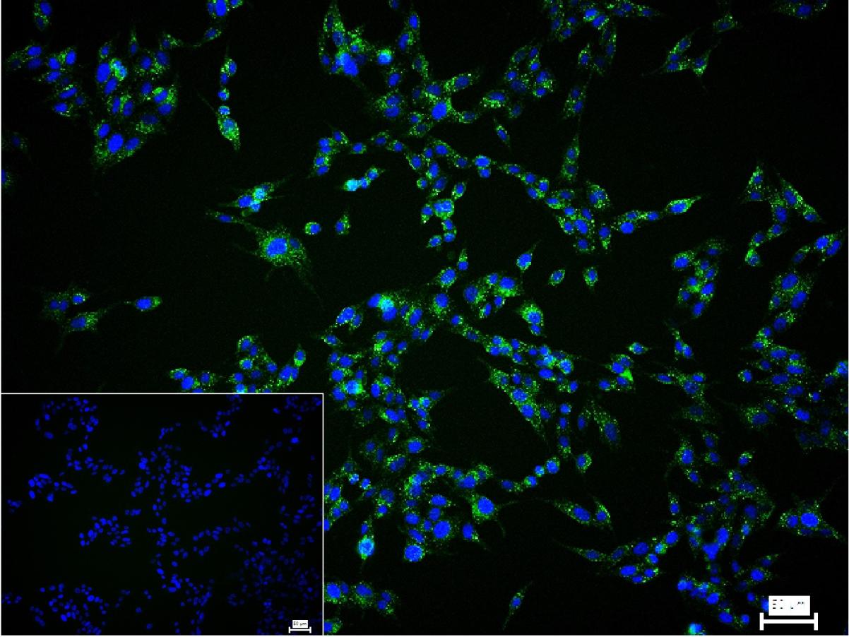
Figure 5. 4% Paraformaldehyde-fixed TT (H) cell; Triton X-100 at r.t. for 20 min; Antibody incubation with (Thyroglobulin) monoclonal Antibody, unconjugated (SLA3109M) 1:100, 90 min at 37°C; followed by conjugated Goat Anti-Rabbit IgG antibody (green, SL60295G-BF488) at 37°C for 90 min, DAPI (blue, C02-04002) was used to stain the cell nuclei. PBS instead of the primary antibody was used as the blank control
FOR RESEARCH USE ONLY. Not for use in diagnostic or therapeutic procedures.
Partial purchase records(bought amounts latest0)
User Comment(Total0User Comment Num)
- No comment


 +86 571 56623320

 SUNLONG BIOTECH


