
Details
Product Name Recombinant Rabbit Anti-NEFH Monoclonal Antibody Cat No. SLA1118M Chinese Name 高分子量神经丝蛋白重组兔单抗 Alias CMT2CC; NFH; NF-H; NF200; mKIAA0845; NFH_HUMAN; NEFH; 200 kDa neurofilament protein; Neurofilament triplet H protein; KIAA0845; NFH_MOUSE; NFH_RAT; neurofilament heavy chain; neurofilament, heavy polypeptide 200kDa; neurofilament heavy polypeptide; neurofilament heavy Host Species Rabbit Isotype IgG Clonality Monoclonal Clone No. 19F20 React Species Human,Mouse,Rat Applications IHC-P (1:200-500)
IHC-F (1:200-500)
IF (1:200-500)
Not yet tested in other applications.
Optimal working dilutions must be determined by the end user.Immunogen KLH conjugated synthetic peptide derived from human NF-H Purification affinity purified by Protein A Concentration 1mg/ml Predicted MW. 112 kDa Subcellular Location Cytoplasm Gene ID 4744 Swiss-Prot P12036 Storage 0.01M TBS (pH7.4) with 1% BSA, 0.02% Proclin300 and 50% Glycerol.
Shipped at 4℃. Store at -20℃ for one year. Avoid repeated freeze/thaw cycles.Background Neurofilaments can be defined as the intermediate or 10nm filaments found in specifically in neuronal cells. When visualised using an electron microscope, neurofilaments appear as 10nm diameter fibres of indeterminate length that generally have fine wispy protrusions from their sides. They are particularly abundant in axons of large projection neurons. They probably function to provide structural support for neurons and their synapses and to support the large axon diameters required for rapid conduction of impulses down axons. Neurofilaments are composed of a mixture of subunits, which usually includes the three neurofilament triplet proteins neurofilament light (NFL), neurofilament medium (NFM) and neurofilament heavy (NFH). Neurofilaments may also include smaller amounts of peripherin, alpha internexin, nestin and in some cases vimentin. Antibodies to the various neurofilament subunits are very useful cell type markers since the proteins are among the most abundant of the nervous system, are expressed only in neurons, and are biochemically very stable. Some studies have shown that levels of neurofilament heavy and neurofilament light are elevated in patients with Alzheimer's disease, frontotemporal lobe dementia, and vascular dementia. Attention This product as supplied is intended for research use only, not for use in human, therapeutic or diagnostic applications.
Validation Images
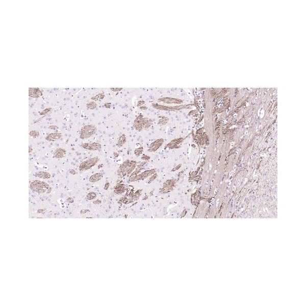
Figure 1. Paraformaldehyde-fixed, paraffin embedded Mouse Cerebrum; Antigen retrieval by boiling in sodium citrate buffer (pH6.0) for 15 min; Antibody incubation with NEFH Monoclonal Antibody, Unconjugated(SLA1118M) at 1:200 overnight at 4°C, followed by conjugation to the SP Kit (Rabbit, SP-0023) and DAB (C-0010) staining.
Figure 2. Paraformaldehyde-fixed, paraffin embedded Rat Cerebrum; Antigen retrieval by boiling in sodium citrate buffer (pH6.0) for 15 min; Antibody incubation with NEFH Monoclonal Antibody, Unconjugated(SLA1118M) at 1:200 overnight at 4°C, followed by conjugation to the SP Kit (Rabbit, SP-0023) and DAB (C-0010) staining.
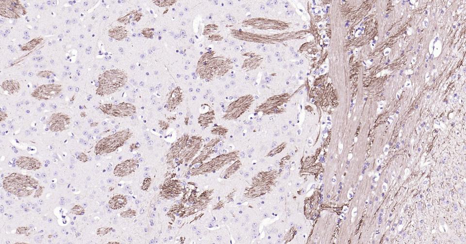
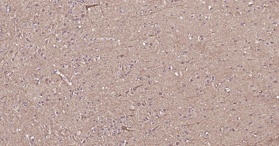
Figure 3. Paraformaldehyde-fixed, paraffin embedded Human Cerebrum; Antigen retrieval by boiling in sodium citrate buffer (pH6.0) for 15 min; Antibody incubation with NEFH Monoclonal Antibody, Unconjugated(SLA1118M) at 1:200 overnight at 4°C, followed by conjugation to the SP Kit (Rabbit, SP-0023) and DAB (C-0010) staining.
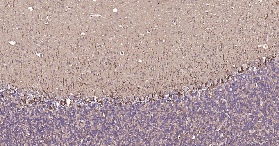
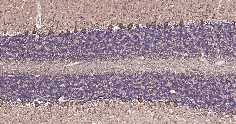
Figure 4. Paraformaldehyde-fixed, paraffin embedded Mouse Cerebellum; Antigen retrieval by boiling in sodium citrate buffer (pH6.0) for 15 min; Antibody incubation with NEFH Monoclonal Antibody, Unconjugated(SLA1118M) at 1:200 overnight at 4°C, followed by conjugation to the SP Kit (Rabbit, SP-0023) and DAB (C-0010) staining.
Figure 5. Paraformaldehyde-fixed, paraffin embedded Rat Cerebellum; Antigen retrieval by boiling in sodium citrate buffer (pH6.0) for 15 min; Antibody incubation with NEFH Monoclonal Antibody, Unconjugated(SLA1118M) at 1:200 overnight at 4°C, followed by conjugation to the SP Kit (Rabbit, SP-0023) and DAB (C-0010) staining.
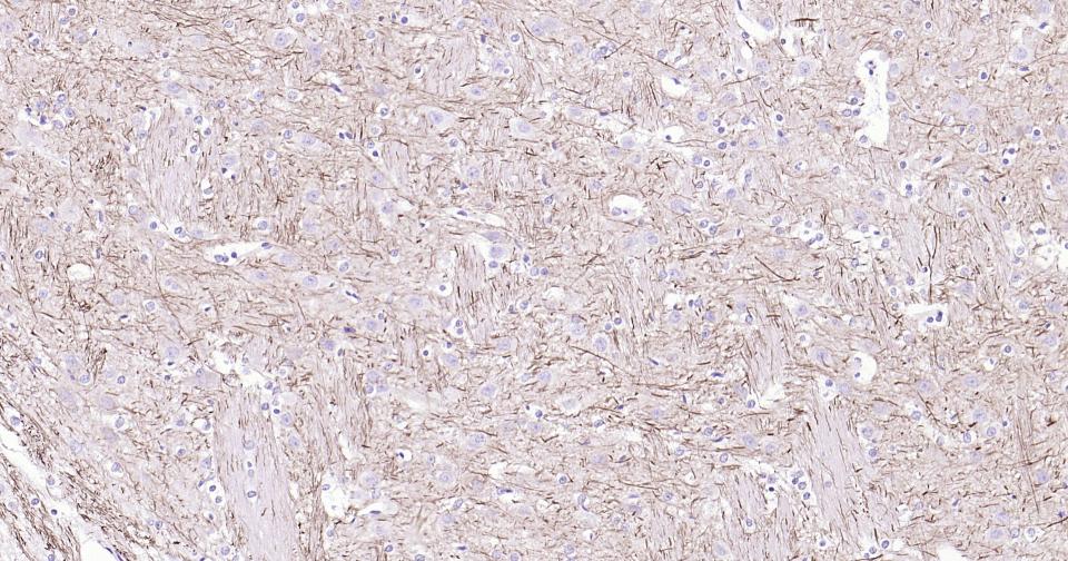
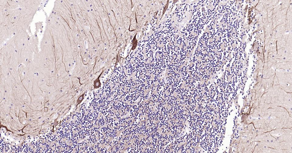
Figure 6. Paraformaldehyde-fixed, paraffin embedded Human Cerebellum; Antigen retrieval by boiling in sodium citrate buffer (pH6.0) for 15 min; Antibody incubation with NEFH Monoclonal Antibody, Unconjugated(SLA1118M) at 1:200 overnight at 4°C, followed by conjugation to the SP Kit (Rabbit, SP-0023) and DAB (C-0010) staining.
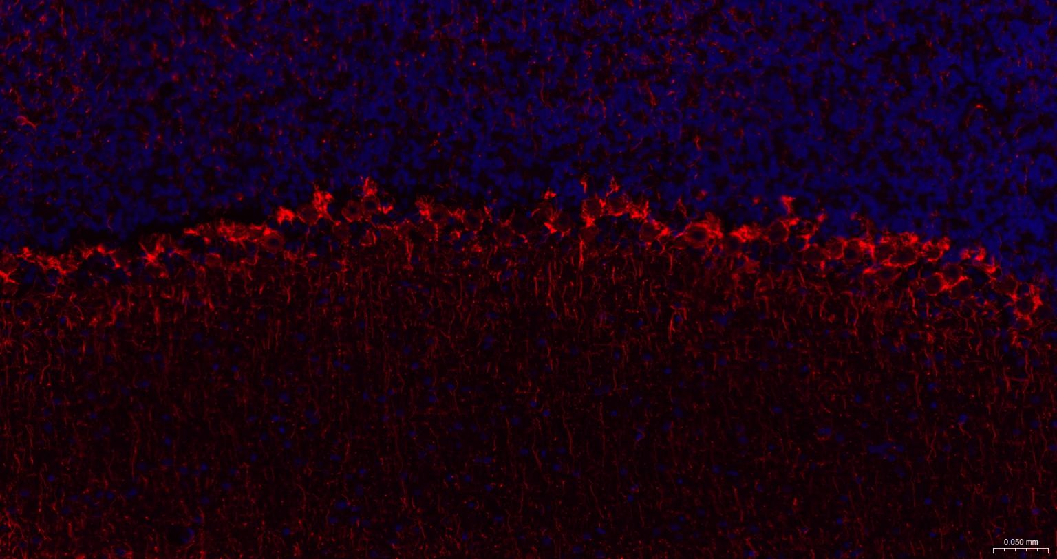
Figure 7. Paraformaldehyde-fixed, paraffin embedded Rat Cerebellum; Antigen retrieval by boiling in sodium citrate buffer (pH6.0) for 15 min; The section was incubated with NF-H Monoclonal Antibody, Unconjugated (SLA1118M) at 1:200 overnight at 4°C. Followed by conjugated Goat Anti-Rabbit IgG antibody (Red, SL0295G-BF594), DAPI (blue, C02-04002) was used to stain the cell nuclei.
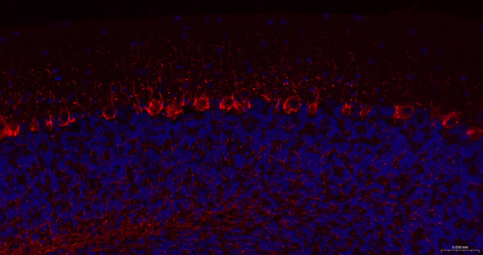
Figure 8. Paraformaldehyde-fixed, paraffin embedded Mouse Cerebellum; Antigen retrieval by boiling in sodium citrate buffer (pH6.0) for 15 min; The section was incubated with NF-H Monoclonal Antibody, Unconjugated (SLA1118M) at 1:200 overnight at 4°C. Followed by conjugated Goat Anti-Rabbit IgG antibody (Red, SL0295G-BF594), DAPI (blue, C02-04002) was used to stain the cell nuclei.
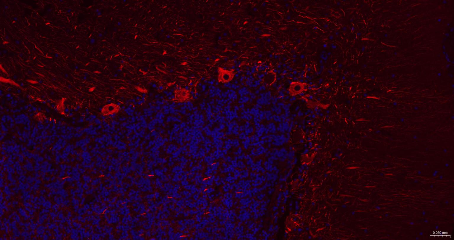
Figure 9. Paraformaldehyde-fixed, paraffin embedded Human Cerebellum; Antigen retrieval by boiling in sodium citrate buffer (pH6.0) for 15 min; The section was incubated with NF-H Monoclonal Antibody, Unconjugated (SLA1118M) at 1:200 overnight at 4°C. Followed by conjugated Goat Anti-Rabbit IgG antibody (Red, SL0295G-BF594), DAPI (blue, C02-04002) was used to stain the cell nuclei.
FOR RESEARCH USE ONLY. Not for use in diagnostic or therapeutic procedures.
Partial purchase records(bought amounts latest0)
User Comment(Total0User Comment Num)
- No comment


 +86 571 56623320

 SUNLONG BIOTECH


