Details
Product Name Recombinant Rabbit Anti-TGM2 Monoclonal Antibody Cat No. SLA0923M Chinese Name 谷氨酰胺转胺酶2重组鼠单抗 Alias G(h); TG(C); TGC; hTG2; tTG; G[a]h; TG2; TGase2; tTGas; TGM2_HUMAN; TGM2; Erythrocyte transglutaminase; Heart G alpha(h) (hhG alpha(h)); Isopeptidase TGM2; Protein G alpha(h) (G(h)); Protein-glutamine deamidase TGM2; Protein-glutamine dopaminyltransferase TGM2; Protein-glutamine histaminyltransferase TGM2; Protein-glutamine noradrenalinyltransferase TGM2; Protein-glutamine serotonyltransferase TGM2; Tissue transglutaminase (tTG | tTgase); Transglutaminase C (TG(C) | TGC | TGase C); Transglutaminase H (TGase H); Transglutaminase II (TGase II); Transglutaminase-2 (TG2 | TGase-2 | hTG2); 2.3.2.13; TGM2_MOUSE; Tissue transglutaminase (tTG); Transglutaminase-2 (TG2 | TGase-2 | TGase2); transglutaminase 2; transglutaminase 2 (C polypeptide, protein-glutamine-gamma-glutamyltransferase); C polypeptide, protein-glutamine-gamma-glutamyltransferase Host Species Mouse Isotype IgG1 Clonality Monoclonal Clone No. SB103 React Species Human Applications WB (1:500-2000)
IHC-P (1:200-800)
IHC-F (1:200-800)
IF (1:200-800)
ICC/IF (1:50-200)
Not yet tested in other applications.
Optimal working dilutions must be determined by the end user.Immunogen Recombinant Human TGM2 protein Purification Affinity Purification Predicted MW. 78 kDa Observed MW. 77 kDa Subcellular Location Cytoplasm Gene ID 7052 Swiss-Prot P21980 Storage 0.01M TBS (pH7.4) with 1% BSA, 0.02% Proclin300 and 50% Glycerol.
Shipped at 4℃. Store at -20℃ for one year. Avoid repeated freeze/thaw cycles.Background Calcium-dependent acyltransferase that catalyzes the formation of covalent bonds between peptide-bound glutamine and various primary amines, such as gamma-amino group of peptide-bound lysine, or mono- and polyamines, thereby producing cross-linked or aminated proteins, respectively. Involved in many biological processes, such as bone development, angiogenesis, wound healing, cellular differentiation, chromatin modification and apoptosis. Acts as a protein-glutamine gamma-glutamyltransferase by mediating the cross-linking of proteins, such as ACO2, HSPB6, FN1, HMGB1, RAP1GDS1, SLC25A4/ANT1, SPP1 and WDR54. Under physiological conditions, the protein cross-linking activity is inhibited by GTP; inhibition is relieved by Ca(2+) in response to various stresses. When secreted, catalyzes cross-linking of proteins of the extracellular matrix, such as FN1 and SPP1 resulting in the formation of scaffolds. Plays a key role during apoptosis, both by (1) promoting the cross-linking of cytoskeletal proteins resulting in condensation of the cytoplasm, and by (2) mediating cross-linking proteins of the extracellular matrix, resulting in the irreversible formation of scaffolds that stabilize the integrity of the dying cells before their clearance by phagocytosis, thereby preventing the leakage of harmful intracellular components. Attention This product as supplied is intended for research use only, not for use in human, therapeutic or diagnostic applications.
Validation Images
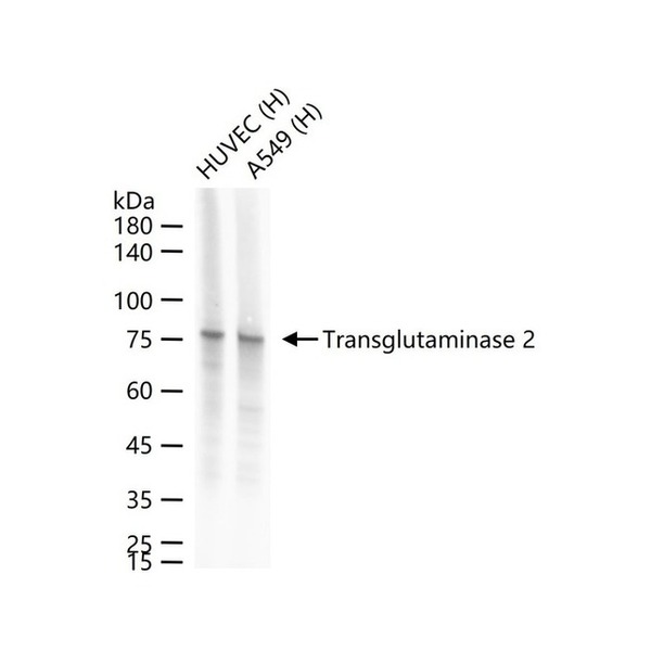
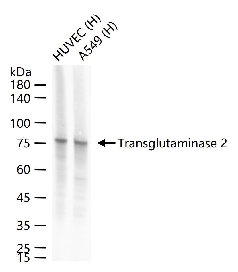
Figure 1. 25 ug total protein per lane of various lysates (see on figure) probed with Transglutaminase 2 monoclonal antibody, unconjugated (SLA0923M) at 1:1000 dilution and 4°C overnight incubation. Followed by conjugated secondary antibody incubation at r.t. for 60 min.
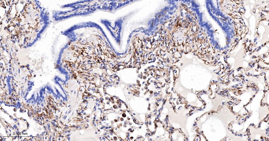
Figure 2. Paraformaldehyde-fixed, paraffin embedded Human Lung; Antigen retrieval by boiling in sodium citrate buffer (pH6.0) for 15 min; The section was incubated with Transglutaminase 2 Monoclonal Antibody, Unconjugated (SLA0923M) at 1:200 overnight at 4°C, followed by conjugation to the SL40296G-HRP and DAB (C-0010) staining.
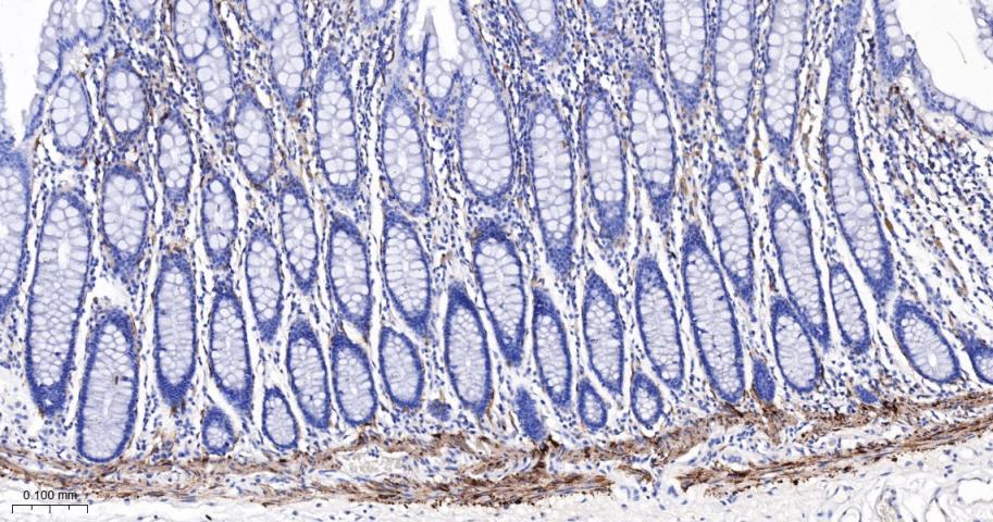
Figure 3. Paraformaldehyde-fixed, paraffin embedded Human Colon; Antigen retrieval by boiling in sodium citrate buffer (pH6.0) for 15 min; The section was incubated with Transglutaminase 2 Monoclonal Antibody, Unconjugated (SLA0923M) at 1:200 overnight at 4°C, followed by conjugation to the SL40296G-HRP and DAB (C-0010) staining.
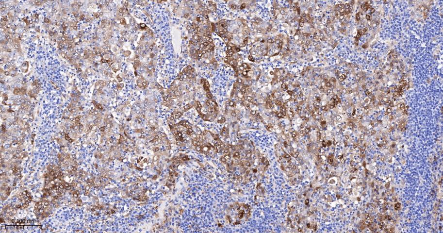
Figure 4. Paraformaldehyde-fixed, paraffin embedded Human Gastric Cancer; Antigen retrieval by boiling in sodium citrate buffer (pH6.0) for 15 min; The section was incubated with Transglutaminase 2 Monoclonal Antibody, Unconjugated (SLA0923M) at 1:200 overnight at 4°C, followed by conjugation to the SL40296G-HRP and DAB (C-0010) staining.
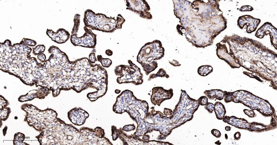
Figure 5. Paraformaldehyde-fixed, paraffin embedded Human Placenta; Antigen retrieval by boiling in sodium citrate buffer (pH6.0) for 15 min; The section was incubated with Transglutaminase 2 Monoclonal Antibody, Unconjugated (SLA0923M) at 1:200 overnight at 4°C, followed by conjugation to the SL40296G-HRP and DAB (C-0010) staining.
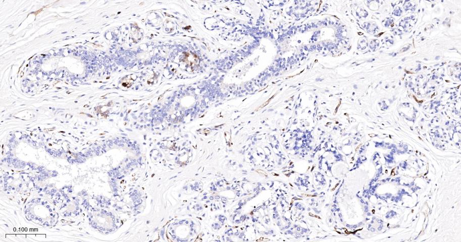
Figure 6. Paraformaldehyde-fixed, paraffin embedded Human Breast Cancer; Antigen retrieval by boiling in sodium citrate buffer (pH6.0) for 15 min; The section was incubated with Transglutaminase 2 Monoclonal Antibody, Unconjugated (SLA0923M) at 1:200 overnight at 4°C, followed by conjugation to the SL40296G-HRP and DAB (C-0010) staining.
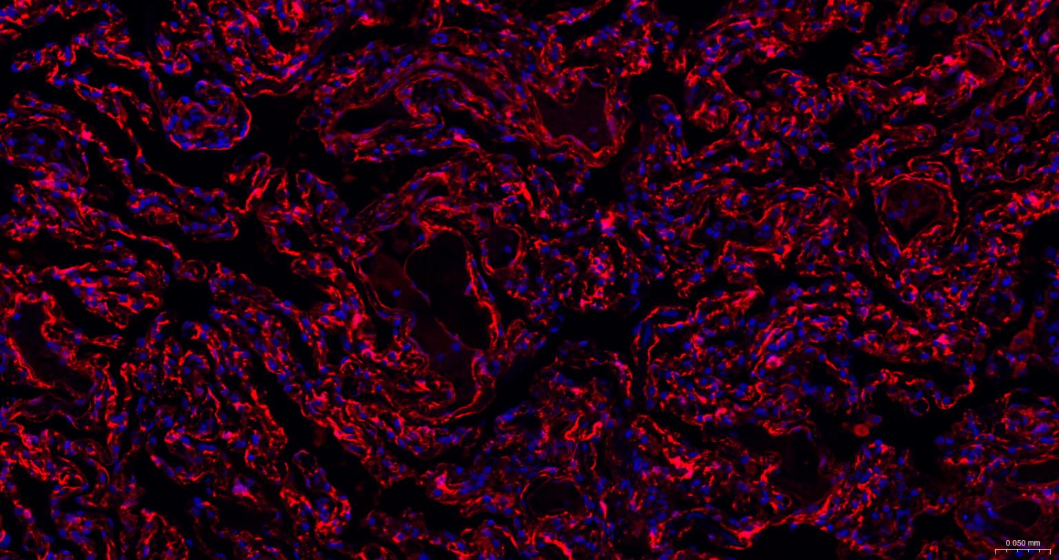
Figure 7. Paraformaldehyde-fixed, paraffin embedded Human Lung; Antigen retrieval by boiling in sodium citrate buffer (pH6.0) for 15 min; The section was incubated with Transglutaminase 2 Monoclonal Antibody, Unconjugated (SLA0923M) at 1:200 overnight at 4°C. Followed by conjugated Goat Anti-Mouse IgG antibody (Red, SL0296G-BF594), DAPI (blue, C02-04002) was used to stain the cell nuclei.
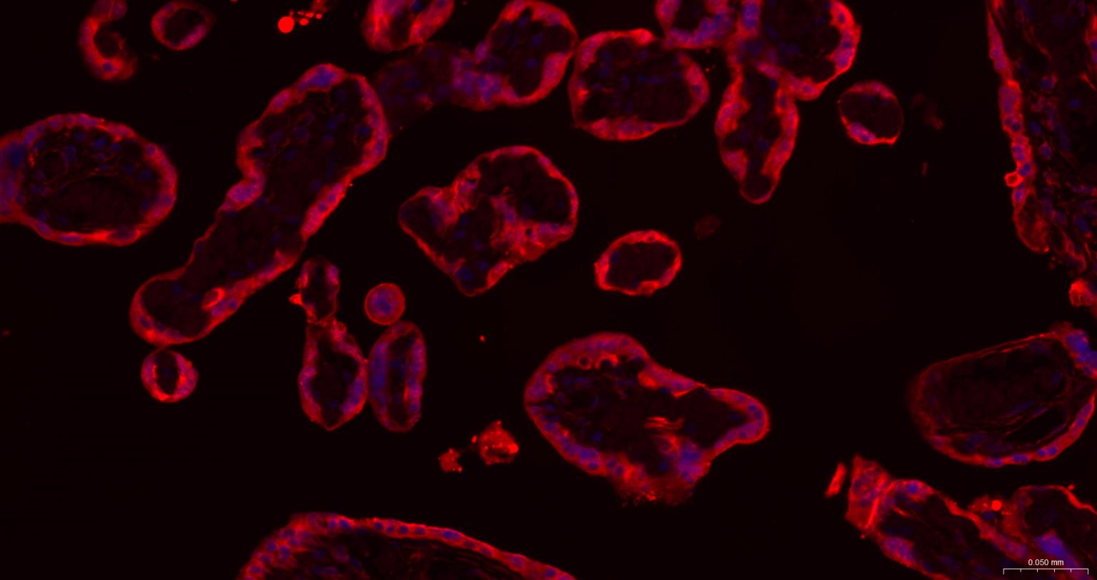
Figure 8. Paraformaldehyde-fixed, paraffin embedded Human Placenta; Antigen retrieval by boiling in sodium citrate buffer (pH6.0) for 15 min; The section was incubated with Transglutaminase 2 Monoclonal Antibody, Unconjugated (SLA0923M) at 1:200 overnight at 4°C. Followed by conjugated Goat Anti-Mouse IgG antibody (Red, SL0296G-BF594), DAPI (blue, C02-04002) was used to stain the cell nuclei.
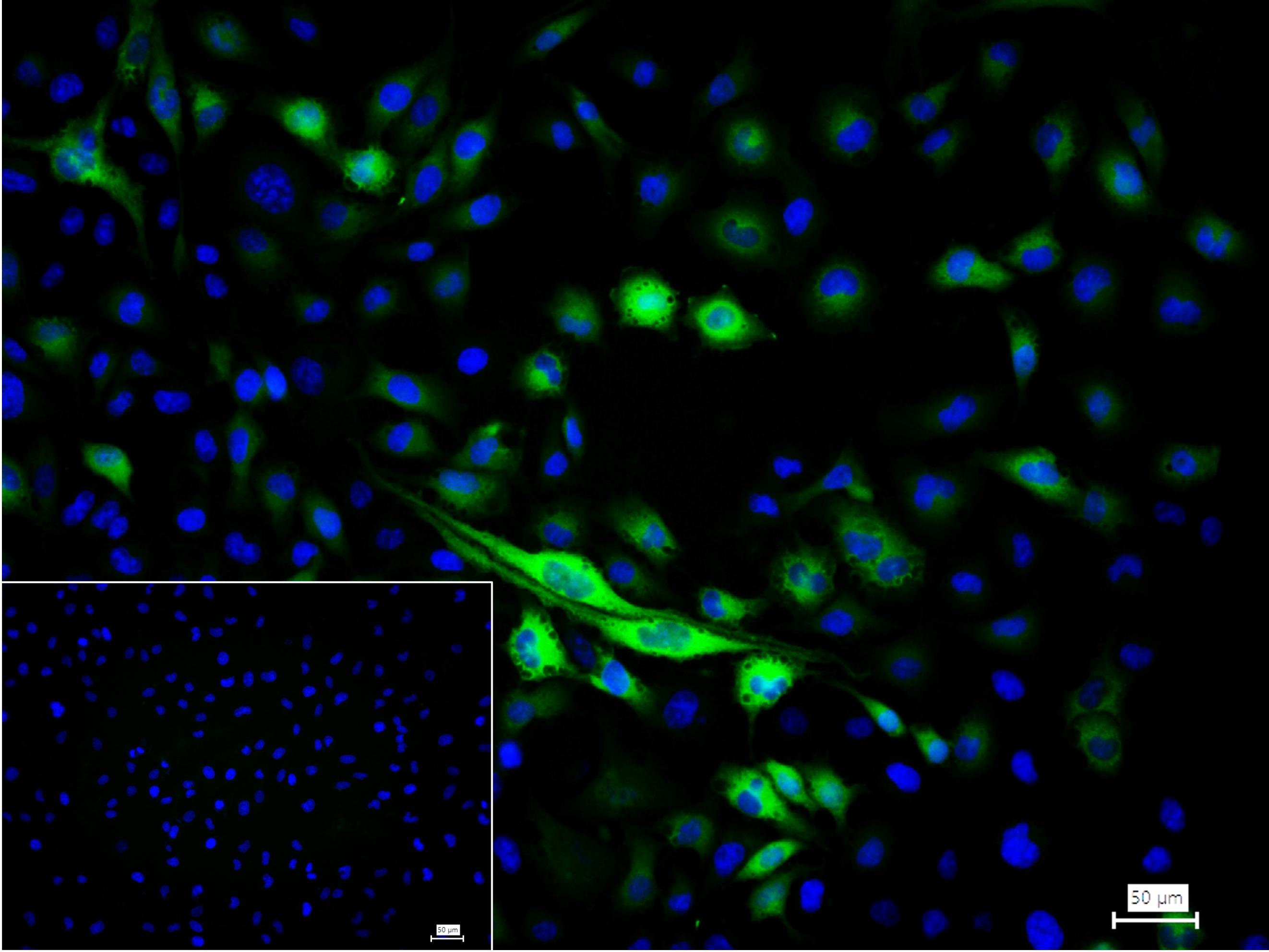
Figure 9. 4% Paraformaldehyde-fixed HUVEC (H) cell; Triton X-100 at r.t. for 20 min; Antibody incubation with (Transglutaminase 2) monoclonal Antibody, unconjugated (SLM-55526M) 1:100, 90 min at 37°C; followed by BF488 conjugated Goat Anti-Mouse IgG antibody (green, SL60296G-BF488) at 37°C for 90 min, DAPI (blue, C02-04002) was used to stain the cell nuclei. PBS instead of the primary antibody was used as the blank control.
FOR RESEARCH USE ONLY. Not for use in diagnostic or therapeutic procedures.
Partial purchase records(bought amounts latest0)
User Comment(Total0User Comment Num)
- No comment


 +86 571 56623320

 SUNLONG BIOTECH


