
Details
Product Name Recombinant Rabbit Anti-pan CK Monoclonal Antibody Cat No. SLA3120M Chinese Name 泛细胞角蛋白重组兔单抗 Alias CK13; K13; WSN2; K1C16_HUMAN; KRT16; Cytokeratin-16 (CK-16); Keratin-16 (K16); KRT16A; CK14; EBS1; EBS1A; EBS1B; EBS1C; EBS1D; EBS3; EBS4; K14; NFJ; K1C14_HUMAN; KRT14; Cytokeratin-14 (CK-14); Keratin-14 (K14); CK16; FNEPPK; K16; K1CP; NEPPK; PC1; 39.1; CK-17; K17; PC; PC2; PCHC1; K1C17_HUMAN; KRT17; Cytokeratin-17 (CK-17); Keratin-17 (K17); CK-24; K24; KA24; K1C24_HUMAN; KRT24; Cytokeratin-24 (CK-24); Keratin-24 (K24); Type I keratin-24; keratin 24; keratin 24, type I Host Species Rabbit Isotype IgG Clonality Monoclonal React Species Human Applications WB (1:500-2000)
IHC-P (1:100-500)
IHC-F (1:100-500)
IF (1:100-500)
Not yet tested in other applications.
Optimal working dilutions must be determined by the end user.Purification affinity purified by Protein A Concentration 1mg/ml Observed MW. 42-64 kDa Subcellular Location Cytoplasm Storage 0.01M TBS (pH7.4) with 1% BSA, 0.02% Proclin300 and 50% Glycerol.
Shipped at 4℃. Store at -20℃ for one year. Avoid repeated freeze/thaw cycles.Background Keratins are a large family of proteins that form the intermediate filament cytoskeleton of epithelial cells, which are classified into two major sequence types. Type I keratins are a group of acidic intermediate filament proteins, including K9–K23, and the hair keratins Ha1–Ha8. Type II keratins are the basic or neutral courterparts to the acidic type I keratins, including K1–K8, and the hair keratins, Hb1–Hb6. In the cytoplasm, the keratin filaments conform a complex network which extends from the surface of the nucleus to the cell membrane. Apart from the relatively static functions provided in terms of supporting the nucleus and providing tensile strength to the cell, the cytokeratin networks undergo rapid phosphate exchanges mediated depolymerization, with important implications in the more dynamic cellular processes such as mitosis and post-mitotic period, cell movement and differentiation. Cytokeratin pan is part of a subfamily of intermediate filament proteins that are characterized by remarkable biochemical diversity, and represented in human epithelial tissues by at least 20 different polypeptides. Cytokeratins range in molecular weight between 40 kDa- 68 kDa, and an isoelectric pH between 4.9-7.8. The individual human cytokeratins are numbered 1 to 20. The various epithelia in the human body usually express cytokeratins which are not only characteristic of the type of epithelium, but also related to the degree of maturation or differentiation within an epithelium. Cytokeratin subtype expression patterns are used to an increasing extent in the distinction of different types of epithelial malignancies. The cytokeratin antibodies are not only of assistance in the differential diagnosis of tumors using immunohistochemistry on tissue sections, but are also a useful tool in cytopathology and flow cytometric assays. The composition of cytokeratin pairs vary with the epithelial cell type, stage of differentiation, cellular growth environment, and disease state. Many studies have shown the usefulness of keratins as markers in cancer research and tumor diagnosis. Attention This product as supplied is intended for research use only, not for use in human, therapeutic or diagnostic applications.
Validation Images
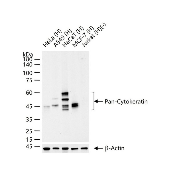
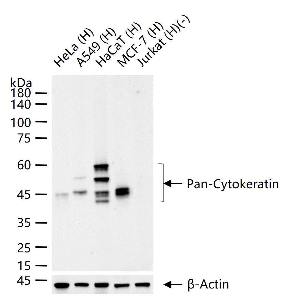
Figure 1. 25 ug total protein per lane of various lysates (see on figure) probed with Pan-Cytokeratin monoclonal antibody, unconjugated (SLA3120M) at 1:1000 dilution and 4°C overnight incubation. Followed by conjugated secondary antibody incubation at r.t. for 60 min.
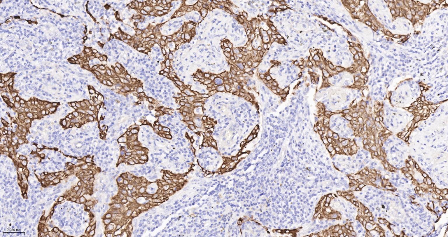
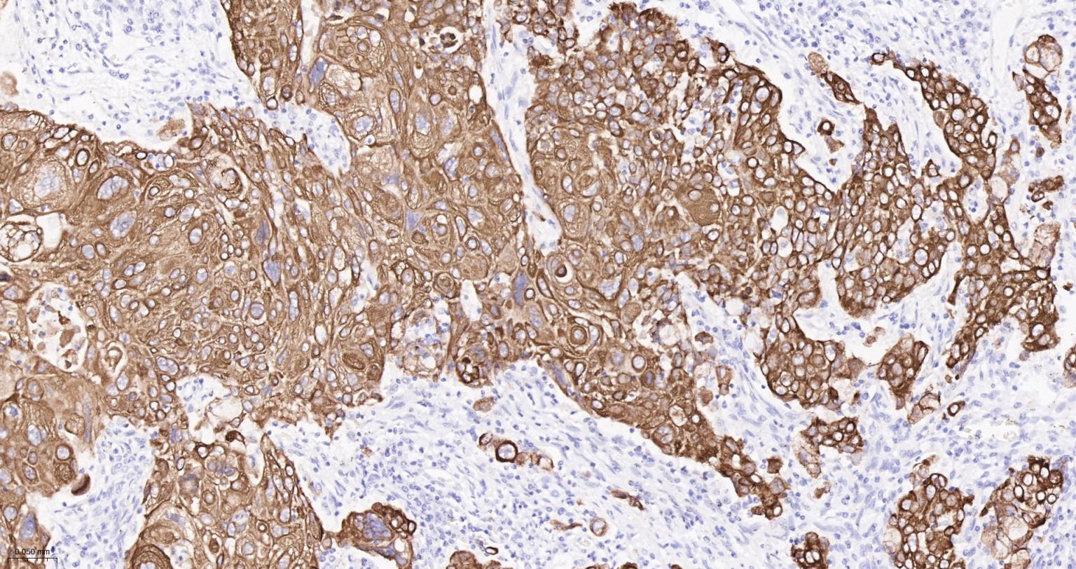
Figure 2. Paraformaldehyde-fixed, paraffin embedded Human Lung squamous cell carcinoma; Antigen retrieval by boiling in sodium citrate buffer (pH6.0) for 15 min; The section was incubated with Pan-Cytokeratin Monoclonal Antibody, Unconjugated (SLA3120M) at 1:200 overnight at 4°C, followed by conjugation to the SL0295G-HRP and DAB (C-0010) staining.
Figure 3. Paraformaldehyde-fixed, paraffin embedded Human Cervical Cancer; Antigen retrieval by boiling in sodium citrate buffer (pH6.0) for 15 min; The section was incubated with Pan-Cytokeratin Monoclonal Antibody, Unconjugated (SLA3120M) at 1:200 overnight at 4°C, followed by conjugation to the SL0295G-HRP and DAB (C-0010) staining.
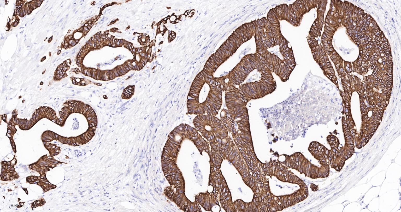
Figure 4. Paraformaldehyde-fixed, paraffin embedded Human Colon Cancer; Antigen retrieval by boiling in sodium citrate buffer (pH6.0) for 15 min; The section was incubated with Pan-Cytokeratin Monoclonal Antibody, Unconjugated (SLA3120M) at 1:200 overnight at 4°C, followed by conjugation to the SL0295G-HRP and DAB (C-0010) staining.
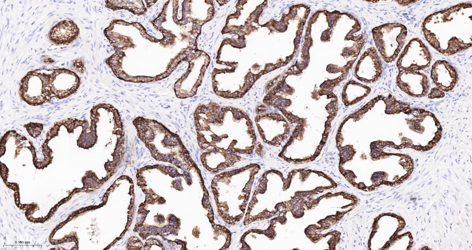
Figure 5. Paraformaldehyde-fixed, paraffin embedded Human Prostate Cancer; Antigen retrieval by boiling in sodium citrate buffer (pH6.0) for 15 min; The section was incubated with Pan-Cytokeratin Monoclonal Antibody, Unconjugated (SLA3120M) at 1:200 overnight at 4°C, followed by conjugation to the SL0295G-HRP and DAB (C-0010) staining.
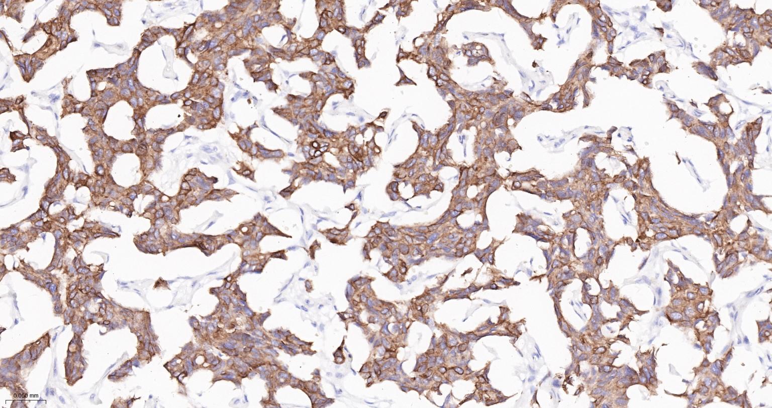
Figure 6. Paraformaldehyde-fixed, paraffin embedded Human Breast Cancer; Antigen retrieval by boiling in sodium citrate buffer (pH6.0) for 15 min; The section was incubated with Pan-Cytokeratin Monoclonal Antibody, Unconjugated (SLA3120M) at 1:200 overnight at 4°C, followed by conjugation to the SL0295G-HRP and DAB (C-0010) staining.
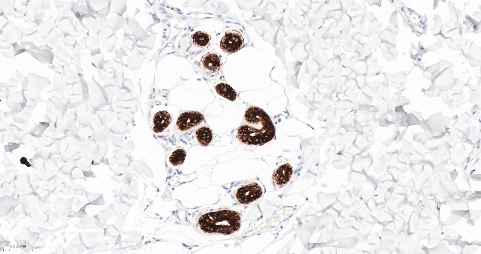
Figure 7. Paraformaldehyde-fixed, paraffin embedded Human abdominal skin; Antigen retrieval by boiling in sodium citrate buffer (pH6.0) for 15 min; The section was incubated with Pan-Cytokeratin Monoclonal Antibody, Unconjugated (SLA3120M) at 1:200 overnight at 4°C, followed by conjugation to the SL0295G-HRP and DAB (C-0010) staining.
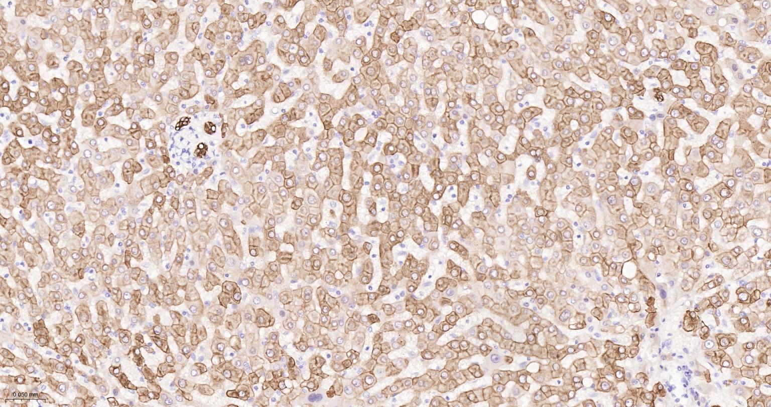
Figure 8. Paraformaldehyde-fixed, paraffin embedded Human Liver; Antigen retrieval by boiling in sodium citrate buffer (pH6.0) for 15 min; The section was incubated with Pan-Cytokeratin Monoclonal Antibody, Unconjugated (SLA3120M) at 1:200 overnight at 4°C, followed by conjugation to the SL0295G-HRP and DAB (C-0010) staining.
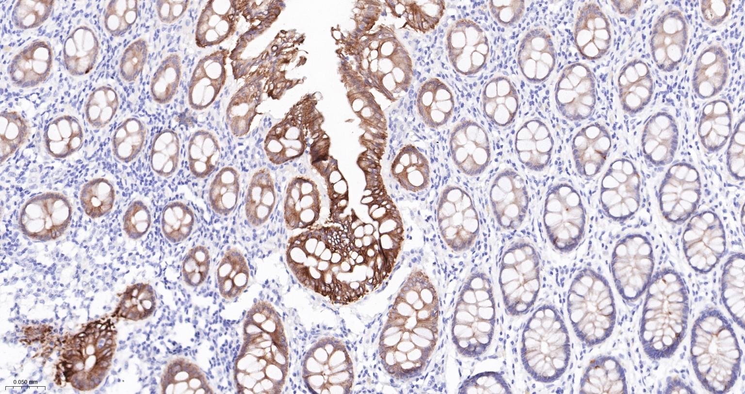
Figure 9. Paraformaldehyde-fixed, paraffin embedded Human Colon; Antigen retrieval by boiling in sodium citrate buffer (pH6.0) for 15 min; The section was incubated with Pan-Cytokeratin Monoclonal Antibody, Unconjugated (SLA3120M) at 1:200 overnight at 4°C, followed by conjugation to the SL0295G-HRP and DAB (C-0010) staining.
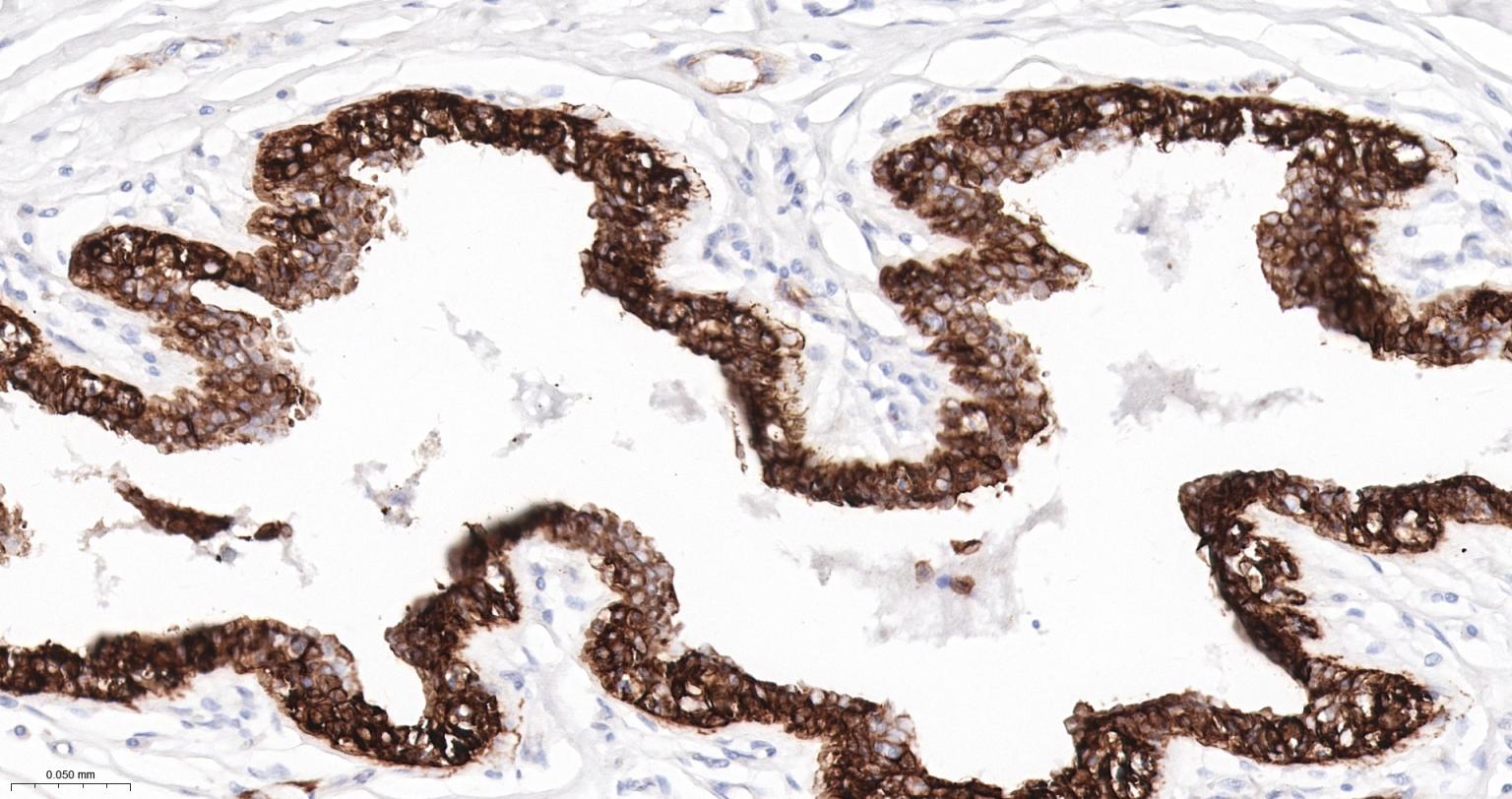
Figure 10. Paraformaldehyde-fixed, paraffin embedded Human Breast; Antigen retrieval by boiling in sodium citrate buffer (pH6.0) for 15 min; The section was incubated with Pan-Cytokeratin Monoclonal Antibody, Unconjugated (SLA3120M) at 1:200 overnight at 4°C, followed by conjugation to the SL0295G-HRP and DAB (C-0010) staining.
FOR RESEARCH USE ONLY. Not for use in diagnostic or therapeutic procedures.
Partial purchase records(bought amounts latest0)
User Comment(Total0User Comment Num)
- No comment


 +86 571 56623320

 SUNLONG BIOTECH


