Details
Product Name Recombinant Rabbit Anti-MyD88 Monoclonal Antibody Cat No. SLA1372M Chinese Name 髓样分化蛋白重组兔单抗 Alias IMD68; MYD88D; WM1; MYD88_HUMAN; MYD88; MYD88_MOUSE; MYD88_RAT; MYD88 innate immune signal transduction adaptor; myeloid differentiation primary response gene (88); myeloid differentiation primary response 88; TLR adaptor MYD88 Host Species Rabbit Isotype IgG Clonality Monoclonal Clone No. 3C9 React Species Human Applications WB (1:500-2000)
IHC-P (1:100-200)
IHC-F (1:100-200)
IF (1:100-200)
ICC/IF (1:50-200)
Not yet tested in other applications.
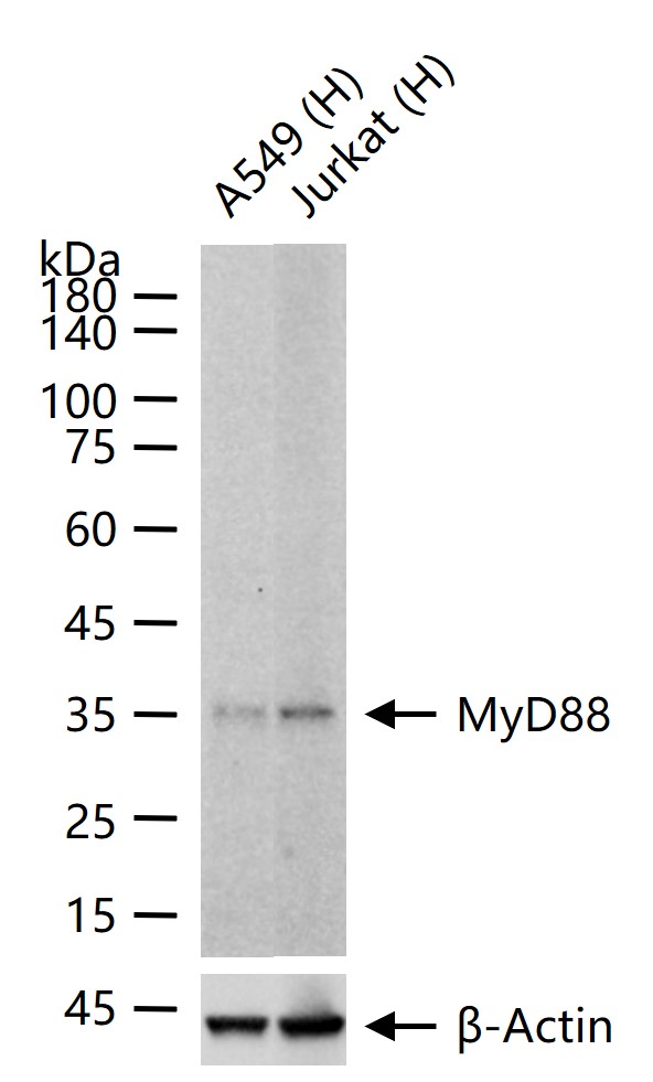
Optimal working dilutions must be determined by the end user.Immunogen A synthesized peptide derived from human MyD88 Purification affinity purified by Protein A Predicted MW. 33 kDa Observed MW. 33 kDa Gene ID 4615 Swiss-Prot Q99836 Storage 0.01M TBS (pH7.4) with 1% BSA, 0.02% Proclin300 and 50% Glycerol.
Shipped at 4℃. Store at -20℃ for one year. Avoid repeated freeze/thaw cycles.Background Adapter protein involved in the Toll-like receptor and IL-1 receptor signaling pathway in the innate immune response.Acts via IRAK1, IRAK2, IRF7 and TRAF6, leading to NF-kappa-B activation, cytokine secretion and the inflammatory response. Attention This product as supplied is intended for research use only, not for use in human, therapeutic or diagnostic applications.
Validation Images
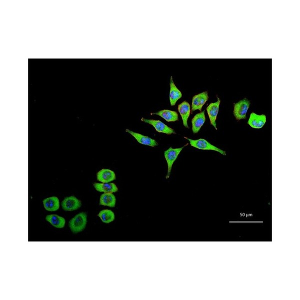
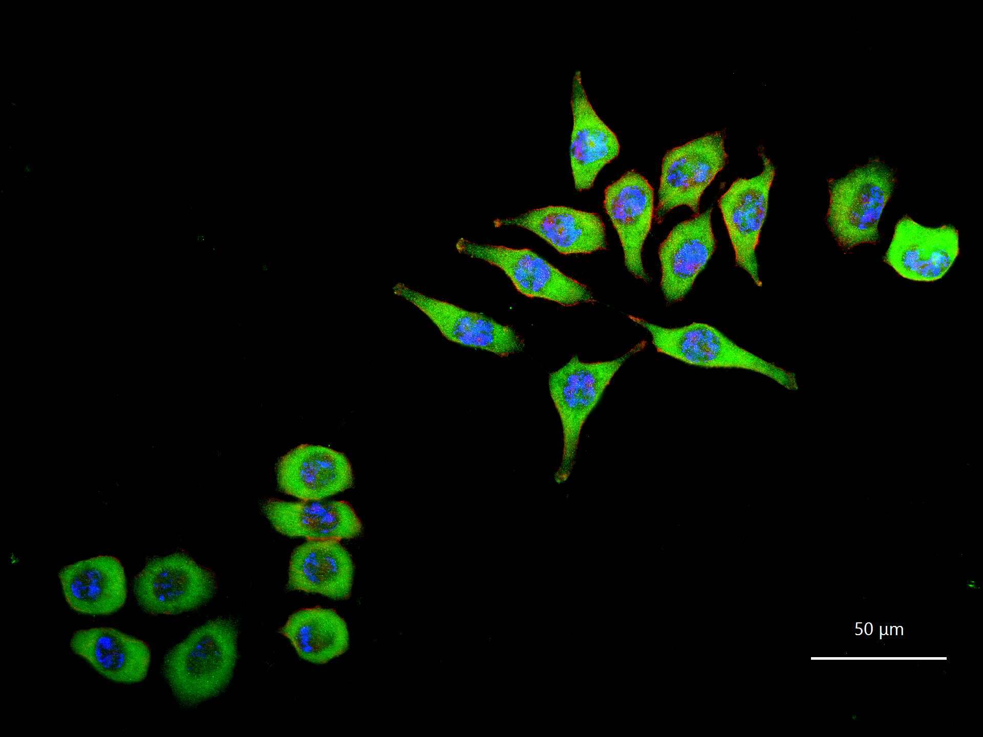
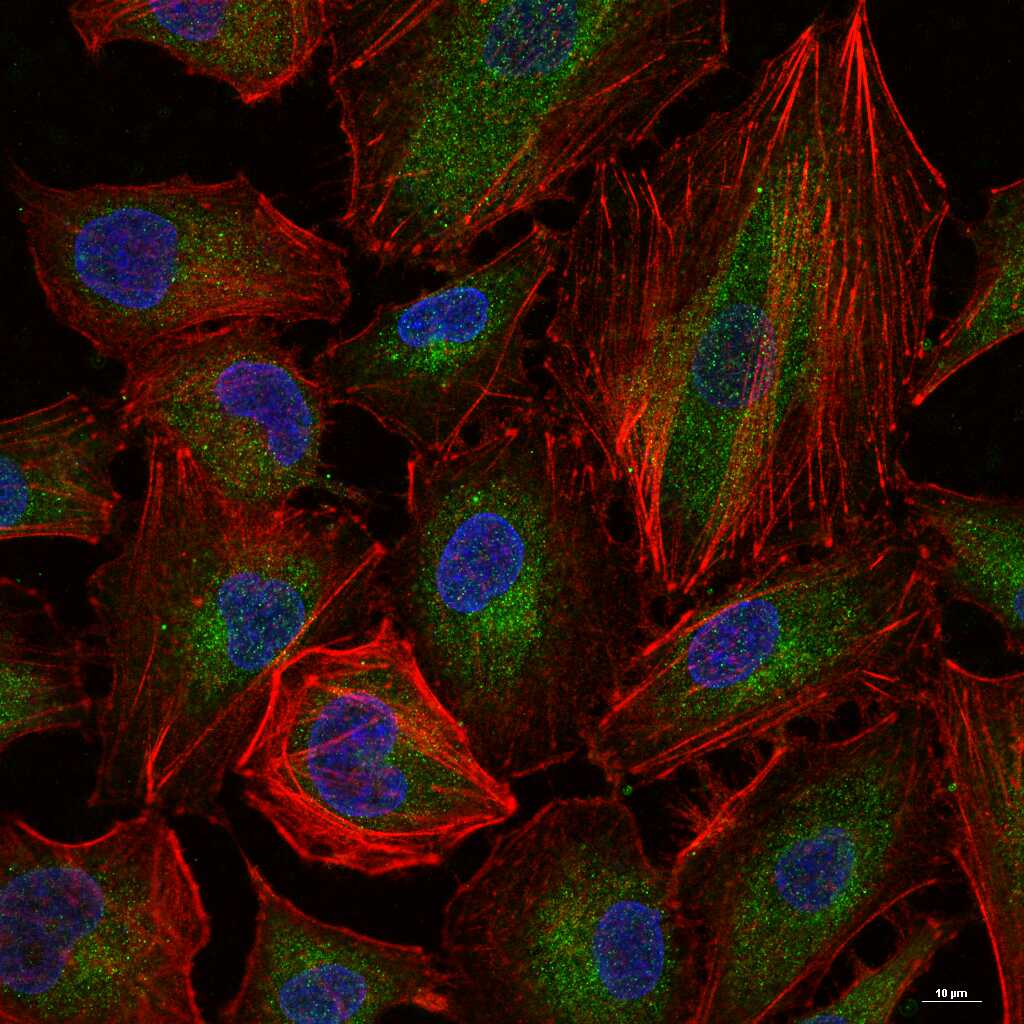
Figure 1. 4% Paraformaldehyde-fixed HeLa (H) cell; Triton X-100 at r.t. for 20 min; Antibody incubation with (MyD88) monoclonal Antibody, unconjugated (SLA1372M) 1:100, 90 min at 37°C; followed by BF488 conjugated Goat Anti-Rabbit IgG antibody (green, SL60295G-BF488) at 37°C for 90 min, then stained with Phalloidin/BF555 for 30 min at room temperature; DAPI (blue, C02-04002) was used to stain the cell nuclei.
Figure 2. 4% Paraformaldehyde-fixed A549 (H) cell; Triton X-100 at r.t. for 20 min; Antibody incubation with (MyD88) monoclonal Antibody, unconjugated (SLA1372M) 1:100, 90 min at 37°C; followed by BF488 conjugated Goat Anti-Rabbit IgG antibody (green, SL60295G-BF488) at 37°C for 90 min, then stained with Phalloidin/BF555 for 30 min at room temperature; DAPI (blue, C02-04002) was used to stain the cell nuclei.
Figure 3. 25 ug total protein per lane of various lysates (see on figure) probed with MyD88 monoclonal antibody, unconjugated (SLA1372M) at 1:2000 dilution and 4°C overnight incubation. Followed by conjugated secondary antibody incubation at r.t. for 60 min.
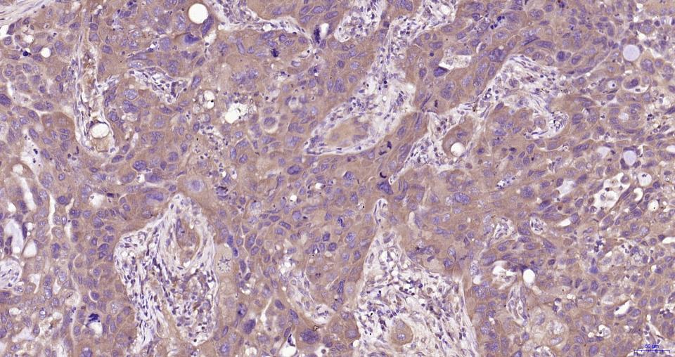
Figure 4. Paraformaldehyde-fixed, paraffin embedded Human Lung Cancer; Antigen retrieval by boiling in sodium citrate buffer (pH6.0) for 15 min; Antibody incubation with MyD88 Monoclonal Antibody, Unconjugated(SLA1372M) at 1:200 overnight at 4°C, followed by conjugation to the SP Kit(Rabbit, SP-0023) and DAB (C-0010) staining.
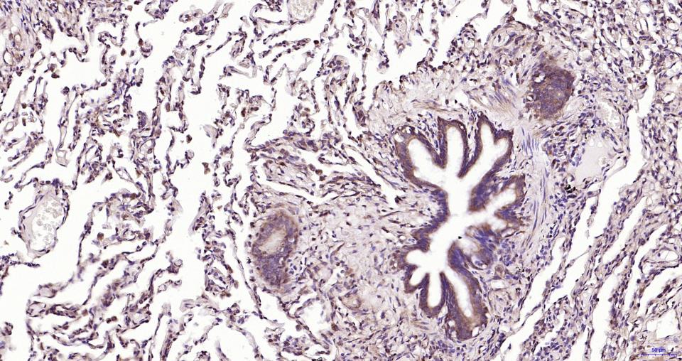
Figure 5. Paraformaldehyde-fixed, paraffin embedded Human Lung ; Antigen retrieval by boiling in sodium citrate buffer (pH6.0) for 15 min; Antibody incubation with MyD88 Monoclonal Antibody, Unconjugated(SLA1372M) at 1:200 overnight at 4°C, followed by conjugation to the SP Kit(Rabbit, SP-0023) and DAB (C-0010) staining.
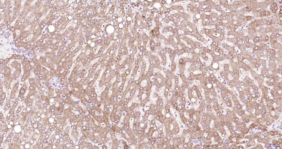
Figure 6. Paraformaldehyde-fixed, paraffin embedded Human Liver; Antigen retrieval by boiling in sodium citrate buffer (pH6.0) for 15 min; Antibody incubation with MyD88 Monoclonal Antibody, Unconjugated(SLA1372M) at 1:200 overnight at 4°C, followed by conjugation to the SP Kit(Rabbit, SP-0023) and DAB (C-0010) staining.
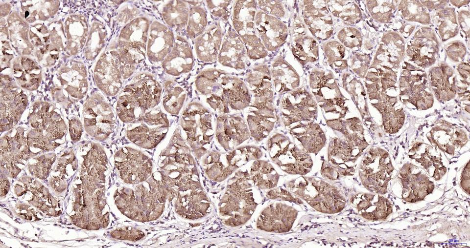
Figure 7. Paraformaldehyde-fixed, paraffin embedded Human Stomach; Antigen retrieval by boiling in sodium citrate buffer (pH6.0) for 15 min; Antibody incubation with MyD88 Monoclonal Antibody, Unconjugated(SLA1372M) at 1:200 overnight at 4°C, followed by conjugation to the SP Kit(Rabbit, SP-0023) and DAB (C-0010) staining.
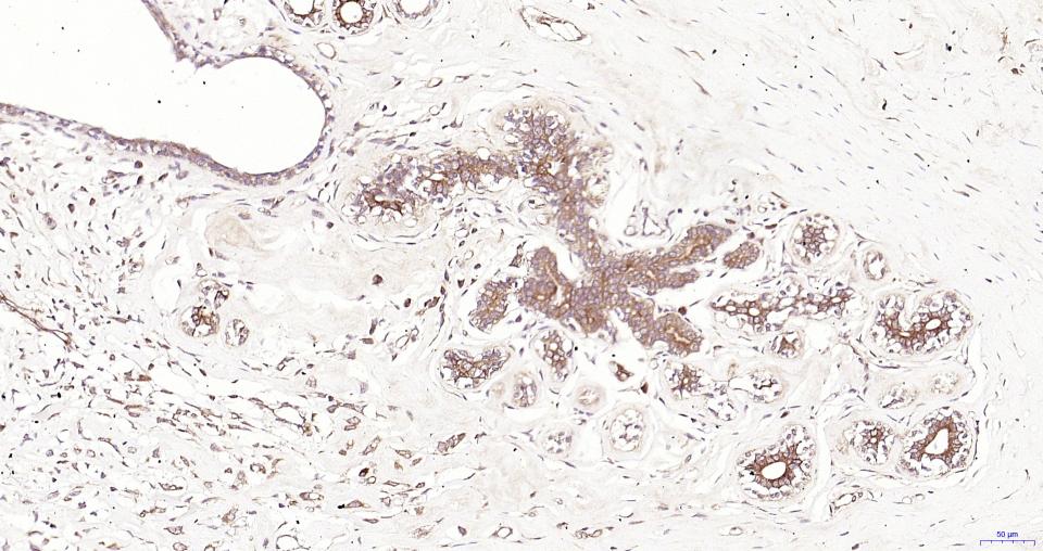
Figure 8. Paraformaldehyde-fixed, paraffin embedded Human Breast Cancer; Antigen retrieval by boiling in sodium citrate buffer (pH6.0) for 15 min; Antibody incubation with MyD88 Monoclonal Antibody, Unconjugated(SLA1372M) at 1:200 overnight at 4°C, followed by conjugation to the SP Kit(Rabbit, SP-0023) and DAB (C-0010) staining.
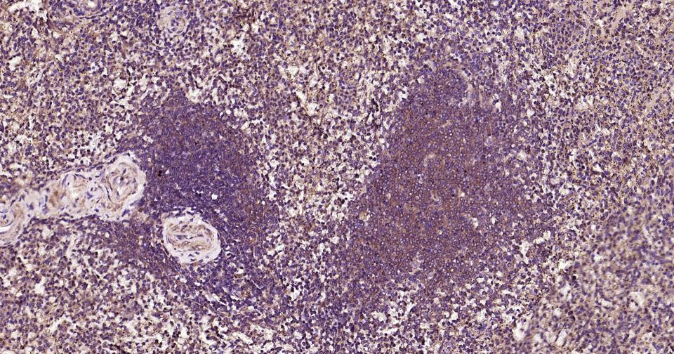
Figure 9. Paraformaldehyde-fixed, paraffin embedded Human Spleen; Antigen retrieval by boiling in sodium citrate buffer (pH6.0) for 15 min; Antibody incubation with MyD88 Monoclonal Antibody, Unconjugated(SLA1372M) at 1:200 overnight at 4°C, followed by conjugation to the SL0295G-HRP and DAB (C-0010) staining.
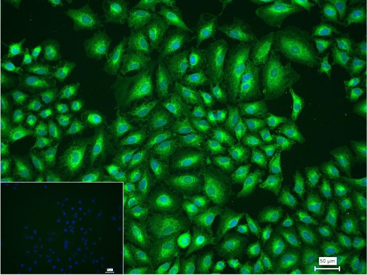
Figure 10. 4% Paraformaldehyde-fixed A549 (H) cell; Triton X-100 at r.t. for 20 min; Antibody incubation with (MyD88) monoclonal Antibody, unconjugated (SLA1372M) 1:100, 90 min at 37°C; followed by conjugated Goat Anti-Rabbit IgG antibody (green, SL40295G-FITC) at 37°C for 90 min, DAPI (blue, C02-04002) was used to stain the cell nuclei. PBS instead of the primary antibody was used as the blank control.
FOR RESEARCH USE ONLY. Not for use in diagnostic or therapeutic procedures.
Partial purchase records(bought amounts latest0)
User Comment(Total0User Comment Num)
- No comment


 +86 571 56623320

 SUNLONG BIOTECH


