Details
Product Name Rabbit Anti-HIF1A Polyclonal Antibody Cat No. SLA0194 Chinese Name 缺氧诱导因子1α /低氧诱导因子-1抗体 Alias HIF-1-alpha; HIF-1A; HIF-1alpha; HIF1; HIF1-ALPHA; MOP1; PASD8; bHLHe78; HIF1alpha; HIF1A_HUMAN; HIF1A; ARNT-interacting protein; Basic-helix-loop-helix-PAS protein MOP1; Class E basic helix-loop-helix protein 78 (bHLHe78); Member of PAS protein 1; PAS domain-containing protein 8; HIF1A_MOUSE; hypoxia inducible factor 1 subunit alpha; hypoxia inducible factor 1, alpha subunit (basic helix-loop-helix transcription factor); Class E basic helix-loop-helix protein 78 Host Species Rabbit Isotype IgG Clonality Polyclonal React Species Human Applications ICC/IF (1:50-200)
Not yet tested in other applications.
Optimal working dilutions must be determined by the end user.Immunogen Recombinant human HIF-1 Alpha protein Purification affinity purified by Protein A Concentration 1mg, ml Predicted MW. 92 kDa Subcellular Location Nucleus Gene ID 3091 Swiss-Prot Q16665 Storage 0.01M TBS (pH7.4) with 1% BSA, 0.02% Proclin300 and 50% Glycerol.
Shipped at 4℃. Store at -20℃ for one year. Avoid repeated freeze/thaw cycles.Background Hypoxia-inducible factor 1 alpha is not only of particular significance for the body to maintain normal physiological functions under hypoxic conditions,and plays an important role in pathological processes such as tumor growth and neuronal apoptosis. HIF1 alpha can regulate the expression levels of many downstream genes. HIF appears in mammalian cells under hypoxic stress conditions. HIF is a transcription factor, stabilizes cells under hypoxia. Attention This product as supplied is intended for research use only, not for use in human, therapeutic or diagnostic applications.
Validation Images
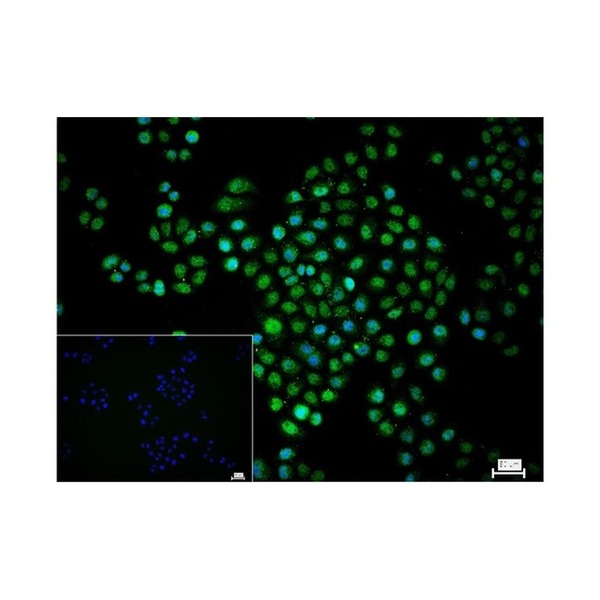
Figure 1. 4% Paraformaldehyde-fixed Hela (treated with 500uM CoCl2 for 6 hours) (H) cell; Triton X-100 at r.t. for 20 min; Antibody incubation with (HIF-1 Alpha) polyclonal Antibody, unconjugated (SLA0194) 1:100, 90 min at 37°C; followed by conjugated Goat Anti-Rabbit IgG antibody (green, SL60295G-BF488) at 37°C for 90 min, DAPI (blue, C02-04002) was used to stain the cell nuclei. PBS instead of the primary antibody was used as the blank control.
Figure 2. 4% Paraformaldehyde-fixed HepG2 (treated with 500uM CoCl2 for 6 hours) (H) cell; Triton X-100 at r.t. for 20 min; Antibody incubation with (HIF-1 Alpha) polyclonal Antibody, unconjugated (SLA0194) 1:100, 90 min at 37°C; followed by conjugated Goat Anti-Rabbit IgG antibody (green, SL60295G-BF488) at 37°C for 90 min, DAPI (blue, C02-04002) was used to stain the cell nuclei. PBS instead of the primary antibody was used as the blank control.
FOR RESEARCH USE ONLY. Not for use in diagnostic or therapeutic procedures.


Partial purchase records(bought amounts latest0)
User Comment(Total0User Comment Num)
- No comment


 +86 571 56623320

 SUNLONG BIOTECH


