Details
Product Name Recombinant Rabbit Anti-CST3 Monoclonal Antibody Cat No. SLA0423M Chinese Name 胱抑素C/半胱氨酸蛋白酶抑制剂C单克隆抗体 Alias ADLDWA; ARMD11; HEL-S-2; CysC; CYTC_HUMAN; CST3; Cystatin-3; Gamma-trace; Neuroendocrine basic polypeptide; Post-gamma-globulin; CYTC_MOUSE; CYTC_RAT; A0A8C0SEH9_CANLF; LOC607874; D5MTH1_FELCA; Host Species Mouse Isotype IgG Clonality Monoclonal Clone No. 7B2 React Species Human Applications WB (1:500-1000)
IHC-P (1:200-800)
IHC-F (1:200-800)
IF (1:200-800)
Not yet tested in other applications.
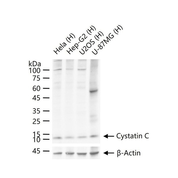
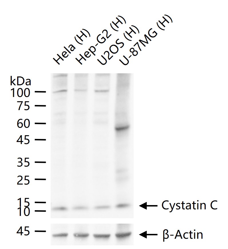
Optimal working dilutions must be determined by the end user.Immunogen Recombinant human Cystatin C Protein Purification affinity purified by Protein G Concentration 1mg/ml Predicted MW. 14 kDa Observed MW. 13 kDa Subcellular Location Secreted Gene ID 1471 Swiss-Prot P01034 Storage 0.01M TBS (pH7.4) with 1% BSA, 0.02% Proclin300 and 50% Glycerol.
Shipped at 4℃. Store at -20℃ for one year. Avoid repeated freeze/thaw cycles.Background The cystatin superfamily encompasses proteins that contain multiple cystatin-like sequences. Some of the members are active cysteine protease inhibitors, while others have lost or perhaps never acquired this inhibitory activity. There are three inhibitory families in the superfamily, including the type 1 cystatins(stefins), type 2 cystatins and the kininogens. The type 2 cystatin proteins are a class of cysteine proteinase inhibitors found in a variety of human fluids and secretions, where they appear to provide protective functions. The cystatin locus on chromosome 20 contains the majority of the type 2 cystatin genes and pseudogenes. This gene is located in the cystatin locus and encodes the most abundant extracellular inhibitor of cysteine proteases, which is found in high concentrations in biological fluids and is expressed in virtually all organs of the body. A mutation in this gene has been associated with amyloid angiopathy. Expression of this protein in vascular wall smooth muscle cells is severely reduced in both atherosclerotic and aneurysmal aortic lesions, establishing its role in vascular disease. [provided by RefSeq]. Attention This product as supplied is intended for research use only, not for use in human, therapeutic or diagnostic applications.
Validation Images
Figure 1. 25 ug total protein per lane of various lysates (see on figure) probed with Cystatin C monoclonal antibody, unconjugated (SLA0423M) at 1:500 dilution and 4°C overnight incubation. Followed by conjugated secondary antibody incubation at r.t. for 60 min.
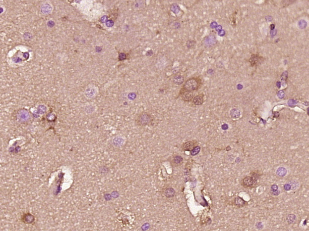
Figure 2. Paraformaldehyde-fixed, paraffin embedded (Human glioma); Antigen retrieval by boiling in sodium citrate buffer (pH6.0) for 15min; Block endogenous peroxidase by 3% hydrogen peroxide for 20 minutes; Blocking buffer (normal goat serum) at 37°C for 30min; Antibody incubation with (Cystatin C) Monoclonal Antibody, Unconjugated (SLA0423M) at 1:400 overnight at 4°C, followed by operating according to SP Kit(Mouse) (sp-0024) instructionsand DAB staining.
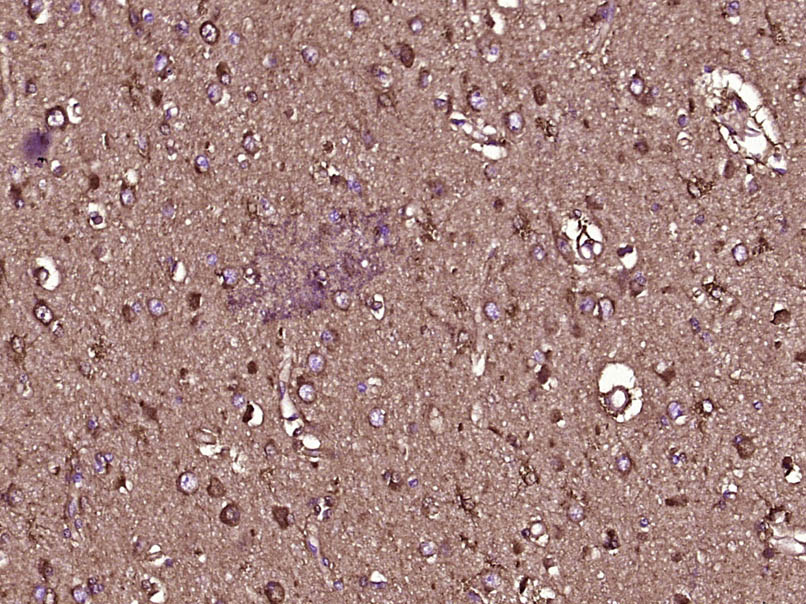
Figure 3. Paraformaldehyde-fixed, paraffin embedded (Human brain glioma); Antigen retrieval by boiling in sodium citrate buffer (pH6.0) for 15min; Block endogenous peroxidase by 3% hydrogen peroxide for 20 minutes; Blocking buffer (normal goat serum) at 37°C for 30min; Antibody incubation with (Cystatin C) Monoclonal Antibody, Unconjugated (ascites of SLA0423M 7B2) at 1:2000 overnight at 4°C, followed by a conjugated secondary (sp-0023) for 20 minutes and DAB staining.
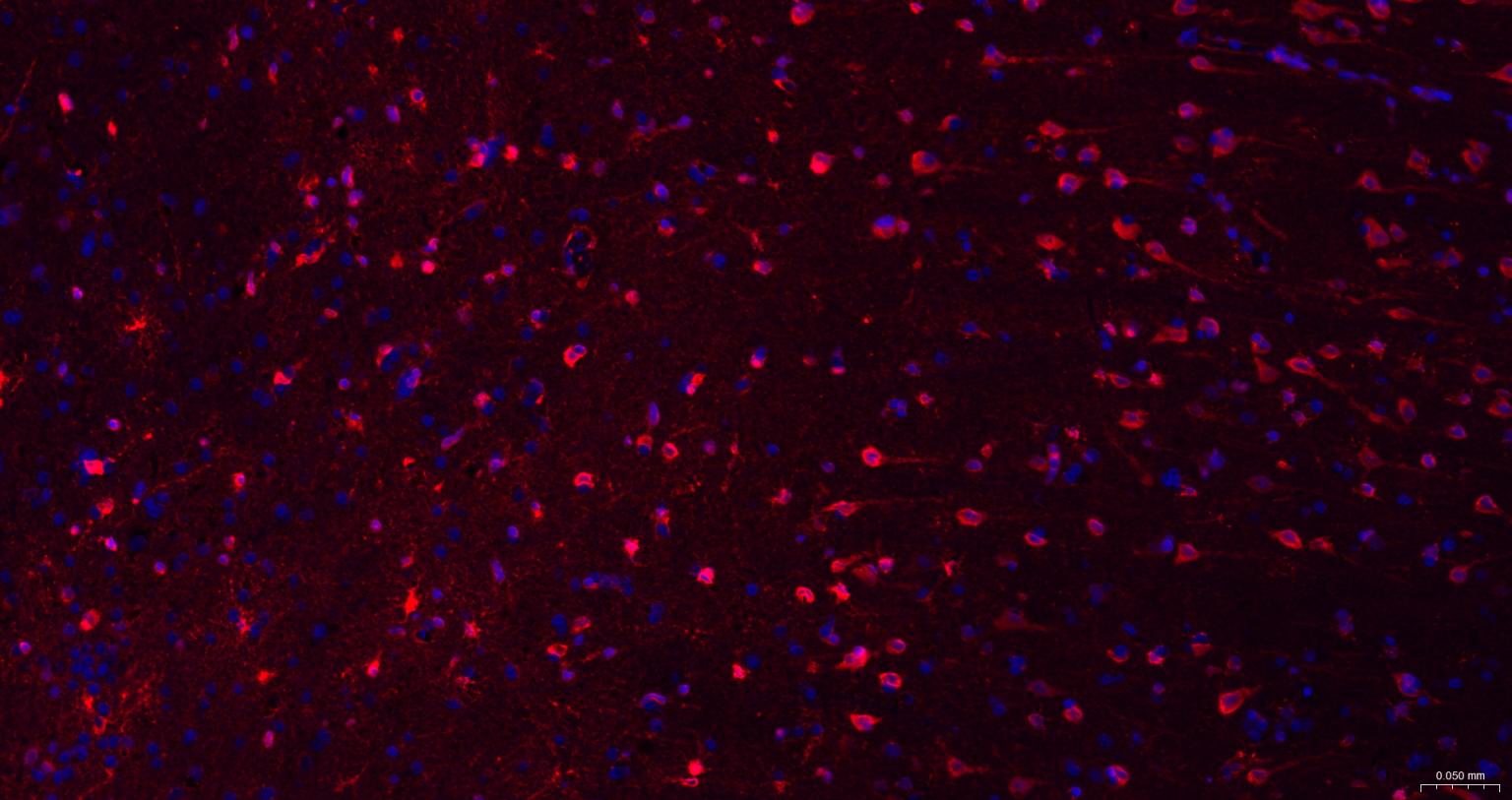
Figure 4. Paraformaldehyde-fixed, paraffin embedded human glioma; Antigen retrieval by boiling in sodium citrate buffer (pH6.0) for 15 min; The section was incubated with Cystatin C Monoclonal Antibody, Unconjugated (SLA0423M) at 1:200 overnight at 4°C. Followed by conjugated Goat Anti-Mouse IgG antibody (red, SL0296G-BF594), DAPI (blue, C02-04002) was used to stain the cell nuclei.
FOR RESEARCH USE ONLY. Not for use in diagnostic or therapeutic procedures.
Partial purchase records(bought amounts latest0)
User Comment(Total0User Comment Num)
- No comment


 +86 571 56623320

 SUNLONG BIOTECH


