Details
Product Name Rabbit Anti-MYH1 Polyclonal Antibody Cat No. SLA0182 Chinese Name 心肌肌球蛋白重链抗体 Alias HEL71; MYHSA1; MYHa; MyHC-2X/D; MyHC-2x; A530084A17Rik; IId; IId/x; MHC-2X/D; MHC2X/D; MYHC-IIX; MdMs; MyHC-IId/x; MyHC-IIx/d; Myhs-f; Myhs-f2; Myhsf2; MYHC; MYH1_BOVIN; MYH1; Myosin heavy chain 1; Myosin heavy chain 2x (MyHC-2x); Myosin heavy chain, skeletal muscle, adult 1; MYH1_HUMAN; Myosin heavy chain IIx/d (MyHC-IIx/d); MYH1_MOUSE; MYH1_PIG; myosin, heavy polypeptide 1, skeletal muscle, adult; myosin heavy chain IIx/d Host Species Rabbit Isotype IgG Clonality Polyclonal React Species Human,Mouse,Rat Applications WB (1:500-2000)
IHC-P (1:100-500)
IHC-F (1:100-500)
IF (1:100-500)
Not yet tested in other applications.
Optimal working dilutions must be determined by the end user.Immunogen Recombinant human MYH1 protein Purification affinity purified by Protein A Concentration 1mg/ml Predicted MW. 223 kDa Observed MW. 44 kDa Subcellular Location Cytoplasm Gene ID 4619 Swiss-Prot P12882 Storage 0.01M TBS (pH7.4) with 1% BSA, 0.02% Proclin300 and 50% Glycerol.
Shipped at 4℃. Store at -20℃ for one year. Avoid repeated freeze/thaw cycles.Background Myosin is a major contractile protein which converts chemical energy into mechanical energy through the hydrolysis of ATP. Myosin is a hexameric protein composed of a pair of myosin heavy chains (MYH) and two pairs of nonidentical light chains. Myosin heavy chains are encoded by a multigene family. In mammals at least 10 different myosin heavy chain (MYH) isoforms have been described from striated, smooth, and nonmuscle cells. These isoforms show expression that is spatially and temporally regulated during development. [provided by RefSeq, Jul 2008]. Attention This product as supplied is intended for research use only, not for use in human, therapeutic or diagnostic applications.
Validation Images
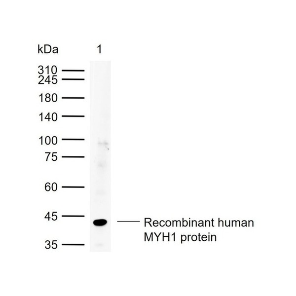
Figure 1. Sample: Lane 1: Recombinant human MYH1 protein, N-His(SL42269P) Primary: Anti-MYH1 (SLA0182) at 1/1000 dilution Secondary: IRDye800CW Goat Anti-Rabbit IgG at 1/20000 dilution Predicted band size: kDa Observed band size: 44 kDa
Figure 2. Paraformaldehyde-fixed, paraffin embedded (mouse tongue); Antigen retrieval by boiling in sodium citrate buffer (pH6.0) for 15min; Block endogenous peroxidase by 3% hydrogen peroxide for 20 minutes; Blocking buffer (normal goat serum) at 37°C for 30min; Incubation with (MYH1) Polyclonal Antibody, Unconjugated (SLA0182) at 1:200 overnight at 4°C, followed by operating according to SP Kit(Rabbit) (sp-0023) instructionsand DAB staining.
Figure 3. Paraformaldehyde-fixed, paraffin embedded (rat tongue); Antigen retrieval by boiling in sodium citrate buffer (pH6.0) for 15min; Block endogenous peroxidase by 3% hydrogen peroxide for 20 minutes; Blocking buffer (normal goat serum) at 37°C for 30min; Incubation with (MYH1) Polyclonal Antibody, Unconjugated (SLA0182) at 1:200 overnight at 4°C, followed by operating according to SP Kit(Rabbit) (sp-0023) instructionsand DAB staining.
Figure 4. Paraformaldehyde-fixed, paraffin embedded (human skeletal muscle); Antigen retrieval by boiling in sodium citrate buffer (pH6.0) for 15min; Block endogenous peroxidase by 3% hydrogen peroxide for 20 minutes; Blocking buffer (normal goat serum) at 37°C for 30min; Incubation with (MYH1) Polyclonal Antibody, Unconjugated (SLA0182) at 1:200 overnight at 4°C, followed by operating according to SP Kit(Rabbit) (sp-0023) instructionsand DAB staining.
Figure 5. Paraformaldehyde-fixed, paraffin embedded (Mouse skeletal muscle); Antigen retrieval by boiling in sodium citrate buffer (pH6.0) for 15min; Block endogenous peroxidase by 3% hydrogen peroxide for 20 minutes; Blocking buffer (normal goat serum) at 37°C for 30min; Incubation with (MYH1) Polyclonal Antibody, Unconjugated (SLA0182) at 1:400 overnight at 4°C, followed by operating according to SP Kit(Rabbit) (sp-0023) instructionsand DAB staining.
Figure 6. Negative sample: Paraformaldehyde-fixed, paraffin embedded (Mouse liver); Antigen retrieval by boiling in sodium citrate buffer (pH6.0) for 15min; Paraformaldehyde-fixed, paraffin embedded (Mouse liver); Antigen retrieval by boiling in sodium citrate buffer (pH6.0) for 15min; Block endogenous peroxidase by 3% hydrogen peroxide for 20 minutes; Blocking buffer (normal goat serum) at 37°C for 30min; Incubation with (MYH1) Polyclonal Antibody, Unconjugated (SLA0182) at 1:400 overnight at 4°C, followed by operating according to SP Kit(Rabbit) (sp-0023) instructionsand DAB staining.
Figure 7. Negative sample: Paraformaldehyde-fixed, paraffin embedded (rat liver);Antigen retrieval by boiling in sodium citrate buffer (pH6.0) for 15min; Paraformaldehyde-fixed, paraffin embedded (rat liver); Antigen retrieval by boiling in sodium citrate buffer (pH6.0) for 15min; Block endogenous peroxidase by 3% hydrogen peroxide for 20 minutes; Blocking buffer (normal goat serum) at 37°C for 30min; Incubation with (MYH1) Polyclonal Antibody, Unconjugated (SLA0182) at 1:400 overnight at 4°C, followed by operating according to SP Kit(Rabbit) (sp-0023) instructionsand DAB staining.
Figure 8. Negative sample: Paraformaldehyde-fixed, paraffin embedded (human brain );Antigen retrieval by boiling in sodium citrate buffer (pH6.0) for 15min; Paraformaldehyde-fixed, paraffin embedded (human brain ); Antigen retrieval by boiling in sodium citrate buffer (pH6.0) for 15min; Block endogenous peroxidase by 3% hydrogen peroxide for 20 minutes; Blocking buffer (normal goat serum) at 37°C for 30min; Incubation with (MYH1) Polyclonal Antibody, Unconjugated (SLA0182) at 1:400 overnight at 4°C, followed by operating according to SP Kit(Rabbit) (sp-0023) instructionsand DAB staining.
Figure 9. Paraformaldehyde-fixed, paraffin embedded Rat Tongue; Antigen retrieval by boiling in sodium citrate buffer (pH6.0) for 15 min; Antibody incubation with MYH1 Polyclonal Antibody, Unconjugated (SLA0182) at 1:200 overnight at 4°C. Followed by conjugated Goat Anti-Rabbit IgG antibody (green, SL0295G-BF488), DAPI (blue, C02-04002) was used to stain the cell nuclei.
Figure 10. Paraformaldehyde-fixed, paraffin embedded Mouse Tongue; Antigen retrieval by boiling in sodium citrate buffer (pH6.0) for 15 min; Antibody incubation with MYH1 Polyclonal Antibody, Unconjugated (SLA0182) at 1:200 overnight at 4°C. Followed by conjugated Goat Anti-Rabbit IgG antibody (green, SL0295G-BF488), DAPI (blue, C02-04002) was used to stain the cell nuclei.
Figure 11. Paraformaldehyde-fixed, paraffin embedded Human Skeletal muscle; Antigen retrieval by boiling in sodium citrate buffer (pH6.0) for 15 min; Antibody incubation with MYH1 Polyclonal Antibody, Unconjugated (SLA0182) at 1:200 overnight at 4°C. Followed by conjugated Goat Anti-Rabbit IgG antibody (green, SL0295G-BF488), DAPI (blue, C02-04002) was used to stain the cell nuclei.
FOR RESEARCH USE ONLY. Not for use in diagnostic or therapeutic procedures.


Partial purchase records(bought amounts latest0)
User Comment(Total0User Comment Num)
- No comment


 +86 571 56623320

 SUNLONG BIOTECH


