Rabbit Anti-GPR125 antibody
FLJ38547; G protein coupled receptor 125; PGR21; Probable G protein coupled receptor 125 precursor; TEM5 like; TEM5L; GP125_HUMAN; GPCR125.
View History [Clear]
Details
Product Name GPR125 Chinese Name G protein-coupled receptor125抗体 Alias FLJ38547; G protein coupled receptor 125; PGR21; Probable G protein coupled receptor 125 precursor; TEM5 like; TEM5L; GP125_HUMAN; GPCR125. Research Area Neurobiology Signal transduction The cell membrane受体 G protein-coupled receptor G protein signal Immunogen Species Rabbit Clonality Polyclonal React Species Mouse, (predicted: Human, Rat, Dog, Cow, Horse, Sheep, ) Applications WB=1:500-2000,IHC-P=1:100-500,IHC-F=1:100-500,IF=1:100-500,Flow-Cyt=1μg/Test (Paraffin sections need antigen repair)
not yet tested in other applications.
optimal dilutions/concentrations should be determined by the end user.Theoretical molecular weight 143kDa Cellular localization The cell membrane Form Liquid Concentration 1mg/ml immunogen KLH conjugated synthetic peptide derived from human GPR125: 422-530/1321 <Extracellular> Lsotype IgG Purification affinity purified by Protein A Buffer Solution 1M TBS(pH7.4) with 1% BSA, 3% Proclin300 and 50% Glycerol. Storage Shipped at 4℃. Store at -20 °C for one year. Avoid repeated freeze/thaw cycles. Attention This product as supplied is intended for research use only, not for use in human, therapeutic or diagnostic applications. PubMed PubMed Product Detail G protein-coupled receptors (GPRs), also known as seven transmembrane receptors, heptahelical receptors or 7TM receptors, comprise a superfamily of proteins that play a role in many different stimulus-response pathways. G protein coupled receptors translate extracellular signals into intracellular signals (G protein activation) and they respond to a variety of signaling molecules, such as hormones and neurotransmitters. GPR125 (G protein-coupled receptor 125), also known as PGR21 or TEM5L, is a 1,321 amino acid multi-pass membrane protein belonging to the G-protein coupled receptor 2 family and the LN-TM7 subfamily. Considered a novel orphan adhesion-type G-protein-coupled receptor, GPR125 has five leucine rich repeats (LRR), an immunoglobulin (Ig) domain and a GPS domain. GPR125 may play a functional role in choroidal and hippocampal response to brain injury. It is also suggested that GPR125 may be a marker for spermatogonial stem cells. Four isoforms of GPR125 exists due to alternative splicing events.
Function:
GPR125 is an orphan receptor which has a leucine rich repeat (LRR),an immunoglobulin (Ig) domain, and a hormone-binding domain (HBD). The Ig domain shows similarities to motilin andtitin, while the LRR domain shows similarities to LRIG1 and SLIT1-2. ESTs have been isolated primarily from amnion,connective tissue, ear, embryo, eye,ganglion, heart, lung,placenta, and skin libraries.
Subunit:
Interacts with DLG1.
Subcellular Location:
Cell membrane; Multi pass membrane protein.
Similarity:
Belongs to the G-protein coupled receptor 2 family. LN-TM7 subfamily.
Contains 1 GPS domain.
Contains 1 Ig-like (immunoglobulin-like) domain.
Contains 5 LRR (leucine-rich) repeats.
Contains 1 LRRCT domain.
SWISS:
Q8IWK6
Gene ID:
166647
Database links:Entrez Gene: 166647 Human
Entrez Gene: 70693 Mouse
Omim: 612303 Human
SwissProt: Q8IWK6 Human
SwissProt: Q7TT36 Mouse
Product Picture 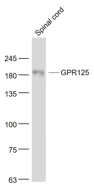 Sample:
Spinal cord (Mouse) Lysate at 40 ug
Primary: Anti- GPR125 (SL12021R) at 1/1000 dilution
Secondary: IRDye800CW Goat Anti-Rabbit IgG at 1/20000 dilution
Predicted band size: 143 kD
Observed band size: 183 kD
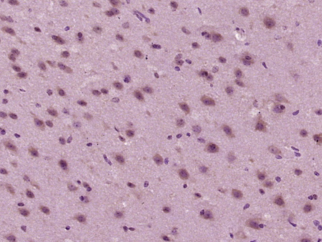 Paraformaldehyde-fixed, paraffin embedded (mouse brain tissue); Antigen retrieval by boiling in sodium citrate buffer (pH6.0) for 15min; Block endogenous peroxidase by 3% hydrogen peroxide for 20 minutes; Blocking buffer (normal goat serum) at 37°C for 30min; Antibody incubation with (GPR125) Polyclonal Antibody, Unconjugated (SL12021R) at 1:400 overnight at 4°C, followed by operating according to SP Kit(Rabbit) (sp-0023) instructionsand DAB staining.
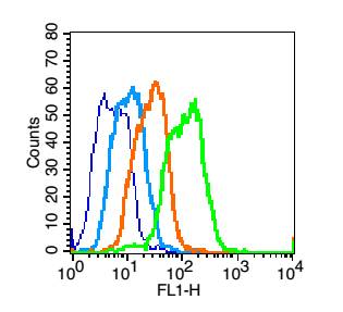 Blank control (blue line): Mouse colon (blue).
Primary Antibody (green line): Rabbit Anti-GPR125 antibody (SL12021R)
Dilution: 1μg /10^6 cells;
Isotype Control Antibody (orange line): Rabbit IgG .
Secondary Antibody (white blue line): F(ab’)2 fragment goat anti-rabbit IgG-FITC
Dilution: 1μg /test.
Protocol
The cells were fixed with 2% paraformaldehyde for 10 min at room temperatureCells stained with Primary Antibody for 30 min at room temperature. The cells were then incubated in 1 X PBS/2%BSA/10% goat serum to block non-specific protein-protein interactions followed by the antibody for 15 min at room temperature. The secondary antibody used for 40 min at room temperature. Acquisition of 20,000 events was performed.
Cartpieces
Totalgoods,subtotals:¥Checkout
Partial purchase records(bought amounts latest0)
No one bought this product
User Comment(Total0User Comment Num)
- No comment


 +86 571 56623320
 SUNLONG BIOTECH

