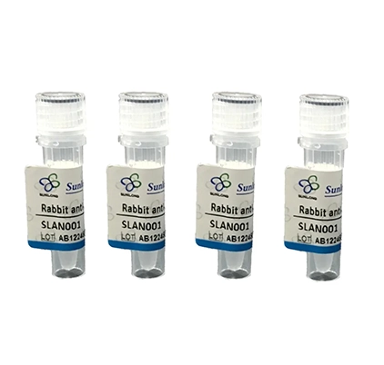
Rabbit Anti-EAR1 antibody
ECP1_MOUSE; Cytotoxic ribonuclease; Eosinophil cationic protein 1; Eosinophil secondary granule ribonuclease 1; EAR-1; Ribonuclease 3-1; RNase 3-1; eosinophil cationic protein 1 precursor; Ear1; ECP 1.
View History [Clear]
Details
Product Name EAR1 Chinese Name 嗜酸性粒细胞阳离子蛋白抗体 Alias ECP1_MOUSE; Cytotoxic ribonuclease; Eosinophil cationic protein 1; Eosinophil secondary granule ribonuclease 1; EAR-1; Ribonuclease 3-1; RNase 3-1; eosinophil cationic protein 1 precursor; Ear1; ECP 1. literatures Research Area Cell biology immunology Channel protein Immunogen Species Rabbit Clonality Polyclonal React Species Human, Mouse, Rat, Applications WB=1:500-2000,IHC-P=1:100-500,IHC-F=1:100-500,IF=1:100-500,Flow-Cyt=1ug/Test (Paraffin sections need antigen repair)
not yet tested in other applications.
optimal dilutions/concentrations should be determined by the end user.Theoretical molecular weight 14kDa Cellular localization The nucleus cytoplasmic Form Liquid Concentration 1mg/ml immunogen KLH conjugated synthetic peptide derived from mouse Eosinophil cationic protein 1: 65-155/155 Lsotype IgG Purification affinity purified by Protein A Buffer Solution 1M TBS(pH7.4) with 1% BSA, 3% Proclin300 and 50% Glycerol. Storage Shipped at 4℃. Store at -20 °C for one year. Avoid repeated freeze/thaw cycles. Attention This product as supplied is intended for research use only, not for use in human, therapeutic or diagnostic applications. PubMed PubMed Product Detail Eosinophil derived neurotoxin (EDN) is a protein belonging to the ribonuclease (RNase) A superfamily. It has recently been found to have antiviral activity against respiratory syncytial virus and human immunodeficiency virus in vitro.
Function:
Cytotoxin and helminthotoxin with ribonuclease activity. Possesses a wide variety of biological activities.
Subunit:
Interacts with bacterial lipopolysaccharide (LPS) and lipoteichoic acid (LTA). In vitro interacts with and insert into lipid bilayers composed of dioleoyl phosphatidylcholine and dioleoyl phosphatidylglycerol. In vitro, tends to form amyloid-like aggregates at pH 3, but not at pH 5, nor 7.
Subcellular Location:
Secreted. Note=Located in the matrix of eosinophil large specific granule, which are released following activation by an immune stimulus.
Similarity:
Belongs to the pancreatic ribonuclease family.
SWISS:
P97426
Gene ID:
13586
Database links:
Entrez Gene: 13586 Mouse
Product Picture 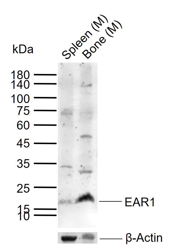 Sample:
Lane 1: Mouse Spleen tissue lysates
Lane 2: Mouse Bone tissue lysates
Primary: Anti-EAR1 (SL1754R) at 1/200 dilution
Secondary: IRDye800CW Goat Anti-Rabbit IgG at 1/20000 dilution
Predicted band size: 14 kDa
Observed band size: 17 kDa
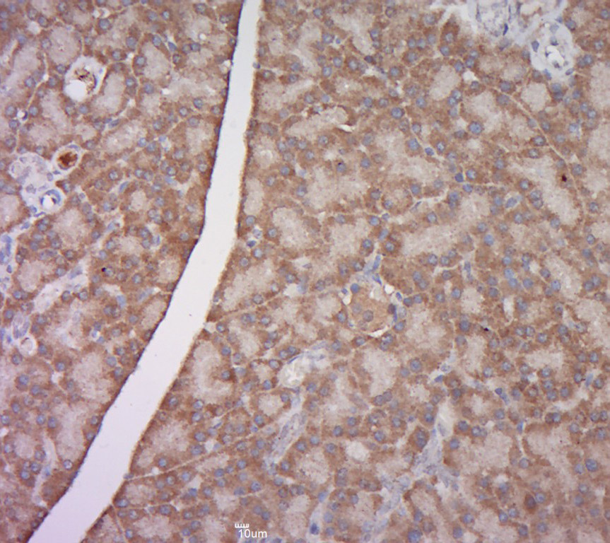 Paraformaldehyde-fixed, paraffin embedded (rat pancreas tissue); Antigen retrieval by boiling in sodium citrate buffer (pH6.0) for 15min; Block endogenous peroxidase by 3% hydrogen peroxide for 20 minutes; Blocking buffer (normal goat serum) at 37°C for 30min; Antibody incubation with (EAR1) Polyclonal Antibody, Unconjugated (SL1754R) at 1:400 overnight at 4°C, followed by a conjugated secondary (sp-0023) for 20 minutes and DAB staining.
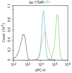 Blank control (Black line): Molt4 (Black).
Primary Antibody (green line): Rabbit Anti-EAR1 antibody (SL1754R)
Dilution: 1μg /10^6 cells;
Isotype Control Antibody (orange line): Rabbit IgG .
Secondary Antibody (white blue line): Goat anti-rabbit IgG-AF647
Dilution: 1μg /test.
Protocol
The cells were fixed with 4% PFA (10min at room temperature)and then permeabilized with 90% ice-cold methanol for 20 min at room temperature. The cells were then incubated in 5%BSA to block non-specific protein-protein interactions for 30 min at room temperature .Cells stained with Primary Antibody for 30 min at room temperature. The secondary antibody used for 40 min at room temperature. Acquisition of 20,000 events was performed.
Cartpieces
Totalgoods,subtotals:¥Checkout
Partial purchase records(bought amounts latest0)
No one bought this product
User Comment(Total0User Comment Num)
- No comment


 +86 571 56623320
 SUNLONG BIOTECH

