Details
Product Name Recombinant Rabbit Anti-MAP2 Monoclonal Antibody Cat No. SLA0988M Chinese Name 微管相关蛋白2重组兔单抗 Alias MAP-2; MAP2A; MAP2B; MAP2C; G1-397-34; Mtap-2; Mtap2; repro4; MAP2R; MTAP2_HUMAN; MAP2; MTAP2_MOUSE; MTAP2_RAT; microtubule associated protein 2 Host Species Rabbit Clonality Monoclonal Clone No. 4A4 React Species Human,Mouse,Rat Applications IHC-P (1:100-500)
IHC-F (1:100-500)
IF (1:100-500)
Not yet tested in other applications.
Optimal working dilutions must be determined by the end user.Immunogen Recombinant human MAP2 protein Purification affinity purified by Protein A Concentration 1mg/ml Predicted MW. 201 kDa Subcellular Location Nucleus Gene ID 4133 Swiss-Prot P11137 Storage 0.01M TBS (pH7.4) with 1% BSA, 0.02% Proclin300 and 50% Glycerol.
Shipped at 4℃. Store at -20℃ for one year. Avoid repeated freeze/thaw cycles.Background MAP2 is the major microtubule associated protein of brain tissue. There are three forms of MAP2; two are similarily sized with apparent molecular weights of 280 kDa (MAP2a and MAP2b) and the third with a lower molecular weight of 70 kDa (MAP2c). In the newborn rat brain, MAP2b and MAP2c are present, while MAP2a is absent. Between postnatal days 10 and 20, MAP2a appears. At the same time, the level of MAP2c drops by 10-fold. This change happens during the period when dendrite growth is completed and when neurons have reached their mature morphology. MAP2 is degraded by a Cathepsin D-like protease in the brain of aged rats. There is some indication that MAP2 is expressed at higher levels in some types of neurons than in other types. MAP2 is known to promote microtubule assembly and to form side-arms on microtubules. It also interacts with neurofilaments, actin, and other elements of the cytoskeleton. Attention This product as supplied is intended for research use only, not for use in human, therapeutic or diagnostic applications.
Validation Images
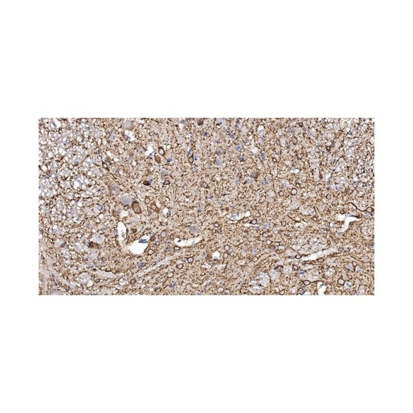
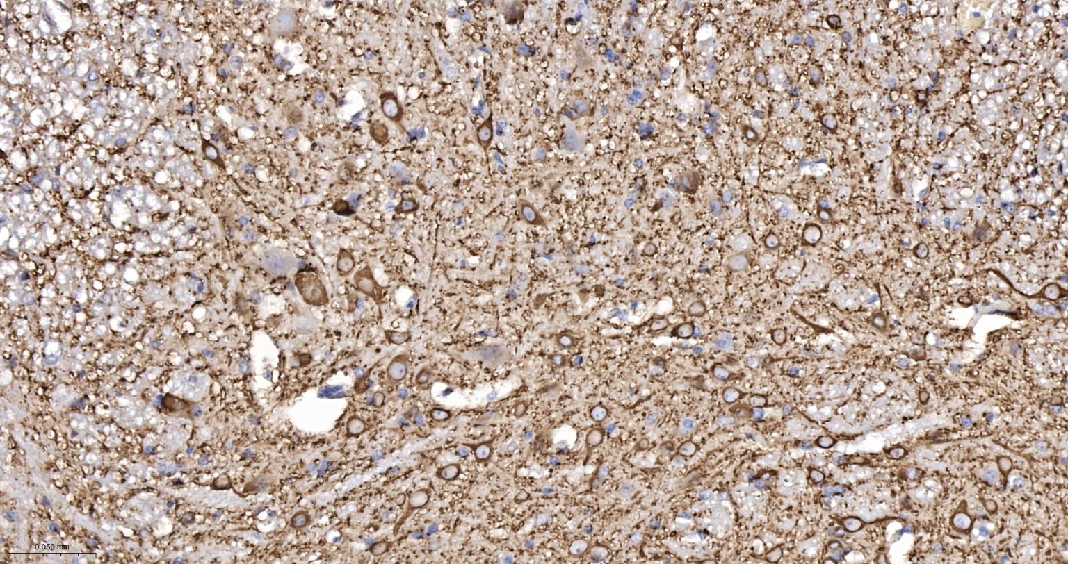
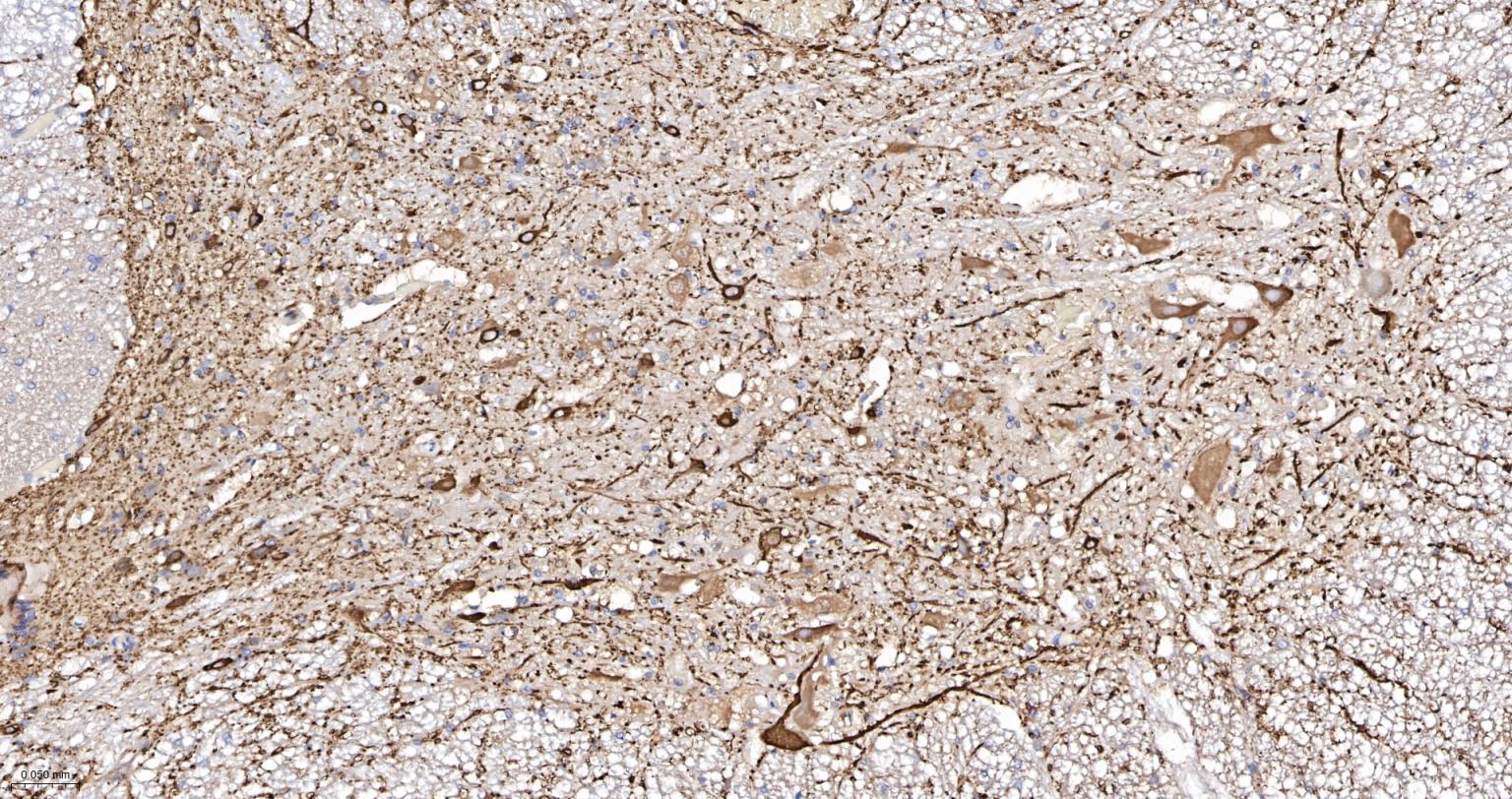
Figure 1. Paraformaldehyde-fixed, paraffin embedded Mouse Spinal Cord; Antigen retrieval by boiling in sodium citrate buffer (pH6.0) for 15 min; The section was incubated with MAP2 Monoclonal Antibody, Unconjugated (SLA0988M) at 1:200 overnight at 4°C, followed by conjugation to the SL0295G-HRP and DAB (C-0010) staining.
Figure 2. Paraformaldehyde-fixed, paraffin embedded Rat Spinal Cord; Antigen retrieval by boiling in sodium citrate buffer (pH6.0) for 15 min; The section was incubated with MAP2 Monoclonal Antibody, Unconjugated (SLA0988M) at 1:200 overnight at 4°C, followed by conjugation to the SL0295G-HRP and DAB (C-0010) staining.
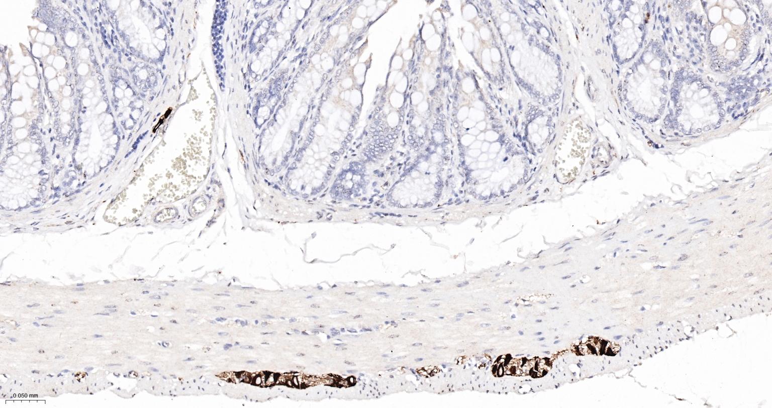
Figure 3. Paraformaldehyde-fixed, paraffin embedded Mouse Colon; Antigen retrieval by boiling in sodium citrate buffer (pH6.0) for 15 min; The section was incubated with MAP2 Monoclonal Antibody, Unconjugated (SLA0988M) at 1:200 overnight at 4°C, followed by conjugation to the SL0295G-HRP and DAB (C-0010) staining.
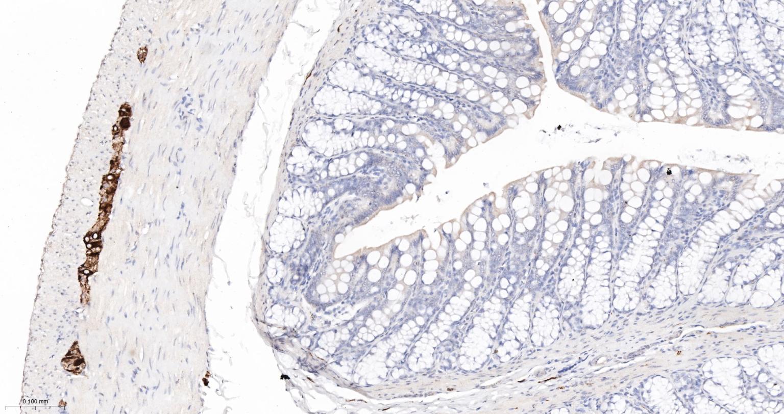
Figure 4. Paraformaldehyde-fixed, paraffin embedded Rat Colon; Antigen retrieval by boiling in sodium citrate buffer (pH6.0) for 15 min; The section was incubated with MAP2 Monoclonal Antibody, Unconjugated (SLA0988M) at 1:200 overnight at 4°C, followed by conjugation to the SL0295G-HRP and DAB (C-0010) staining.
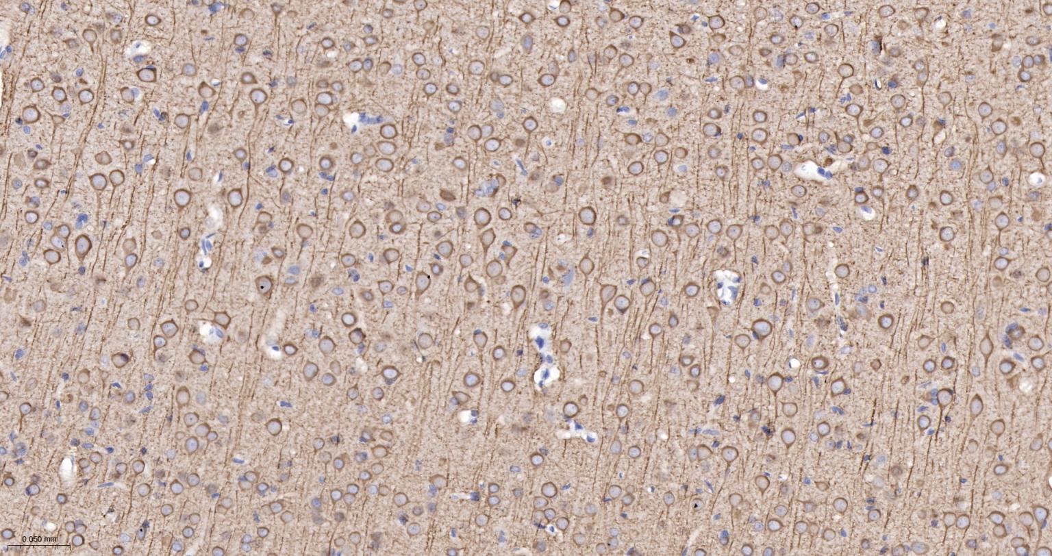
Figure 5. Paraformaldehyde-fixed, paraffin embedded Mouse Cerebrum; Antigen retrieval by boiling in sodium citrate buffer (pH6.0) for 15 min; The section was incubated with MAP2 Monoclonal Antibody, Unconjugated (SLA0988M) at 1:200 overnight at 4°C, followed by conjugation to the SL0295G-HRP and DAB (C-0010) staining.
Figure 6. Paraformaldehyde-fixed, paraffin embedded Rat Cerebrum; Antigen retrieval by boiling in sodium citrate buffer (pH6.0) for 15 min; The section was incubated with MAP2 Monoclonal Antibody, Unconjugated (SLA0988M) at 1:200 overnight at 4°C, followed by conjugation to the SL0295G-HRP and DAB (C-0010) staining.
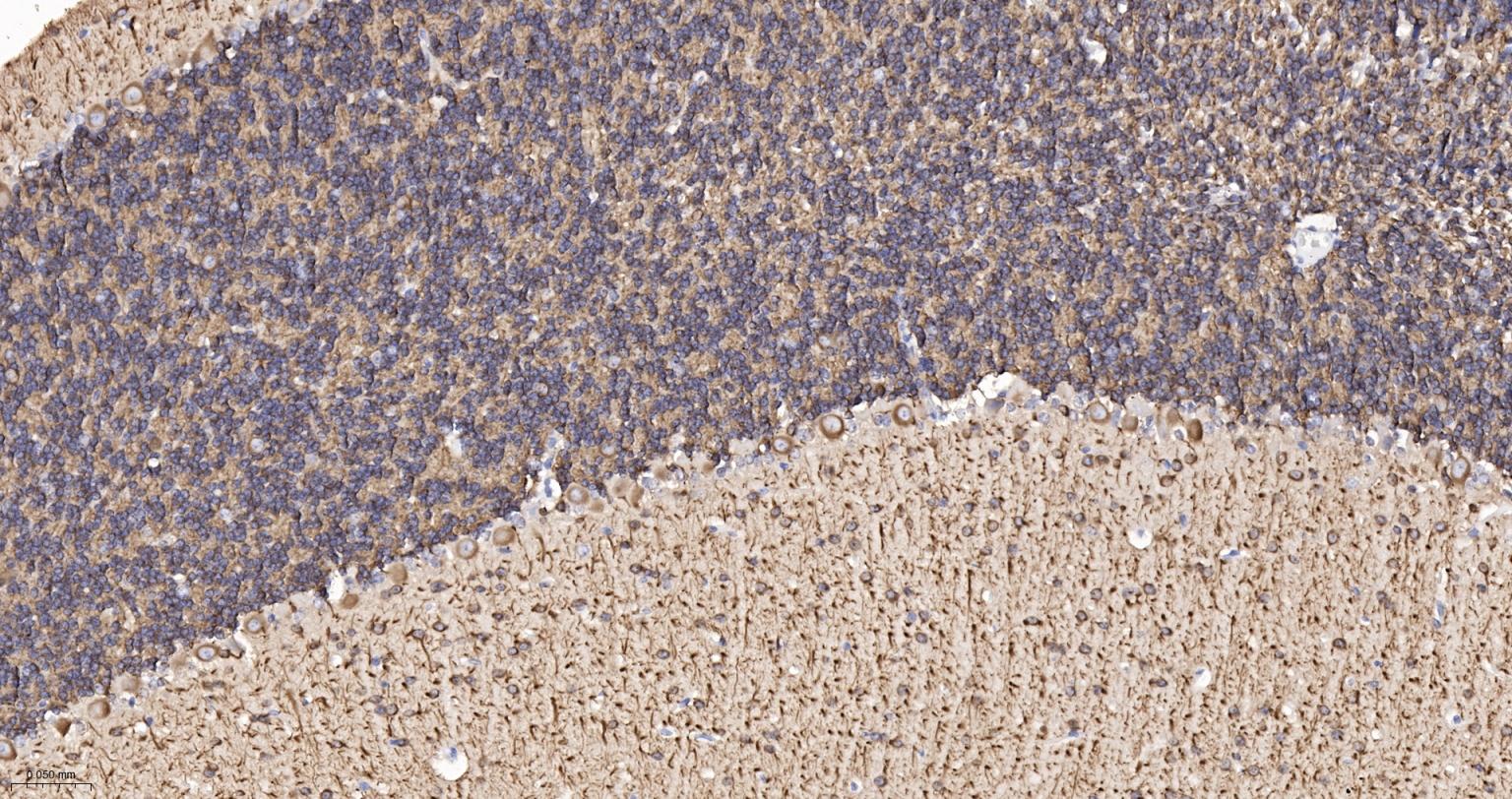
Figure 7. Paraformaldehyde-fixed, paraffin embedded Mouse Cerebellum; Antigen retrieval by boiling in sodium citrate buffer (pH6.0) for 15 min; The section was incubated with MAP2 Monoclonal Antibody, Unconjugated (SLA0988M) at 1:200 overnight at 4°C, followed by conjugation to the SL0295G-HRP and DAB (C-0010) staining.
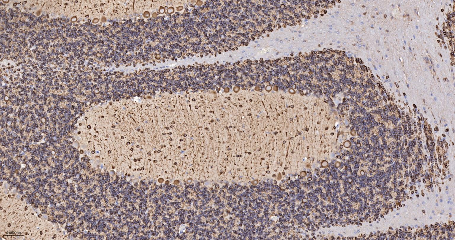
Figure 8. Paraformaldehyde-fixed, paraffin embedded Rat Cerebellum; Antigen retrieval by boiling in sodium citrate buffer (pH6.0) for 15 min; The section was incubated with MAP2 Monoclonal Antibody, Unconjugated (SLA0988M) at 1:200 overnight at 4°C, followed by conjugation to the SL0295G-HRP and DAB (C-0010) staining.
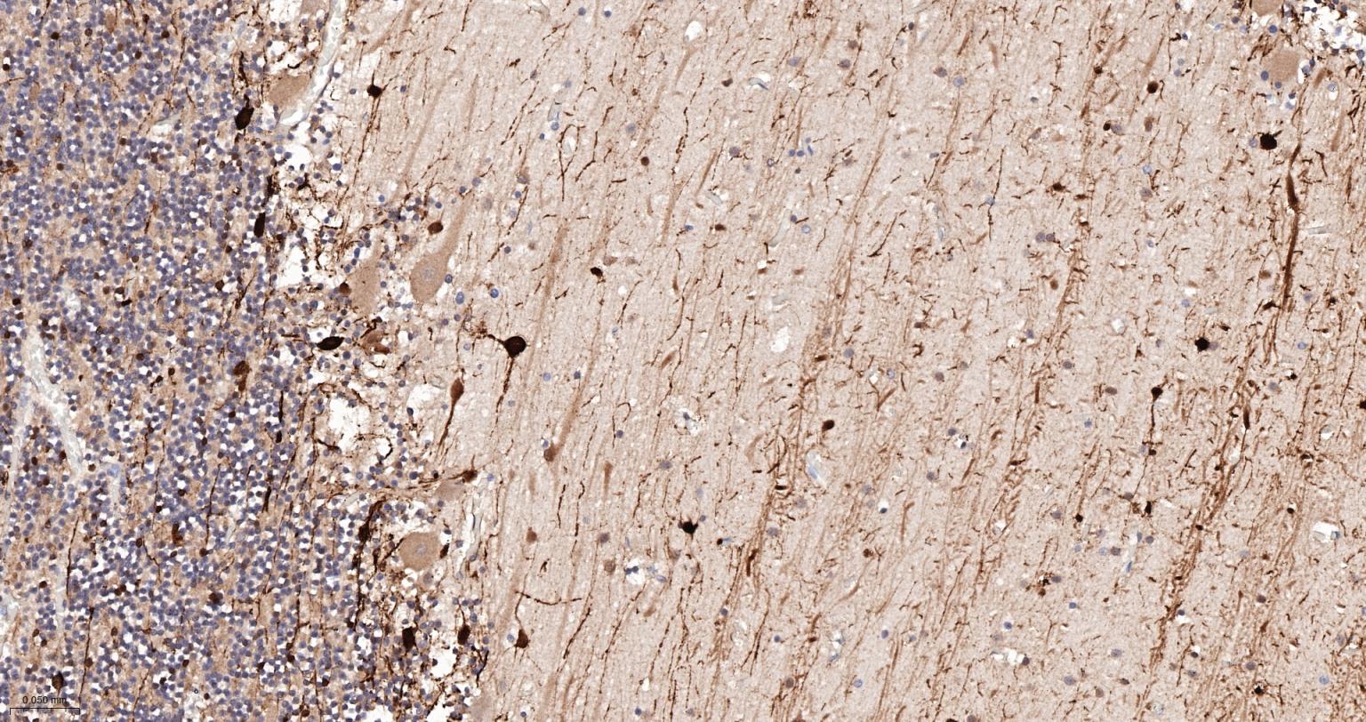
Figure 9. Paraformaldehyde-fixed, paraffin embedded Human Cerebellum; Antigen retrieval by boiling in sodium citrate buffer (pH6.0) for 15 min; The section was incubated with MAP2 Monoclonal Antibody, Unconjugated (SLA0988M) at 1:200 overnight at 4°C, followed by conjugation to the SL0295G-HRP and DAB (C-0010) staining.
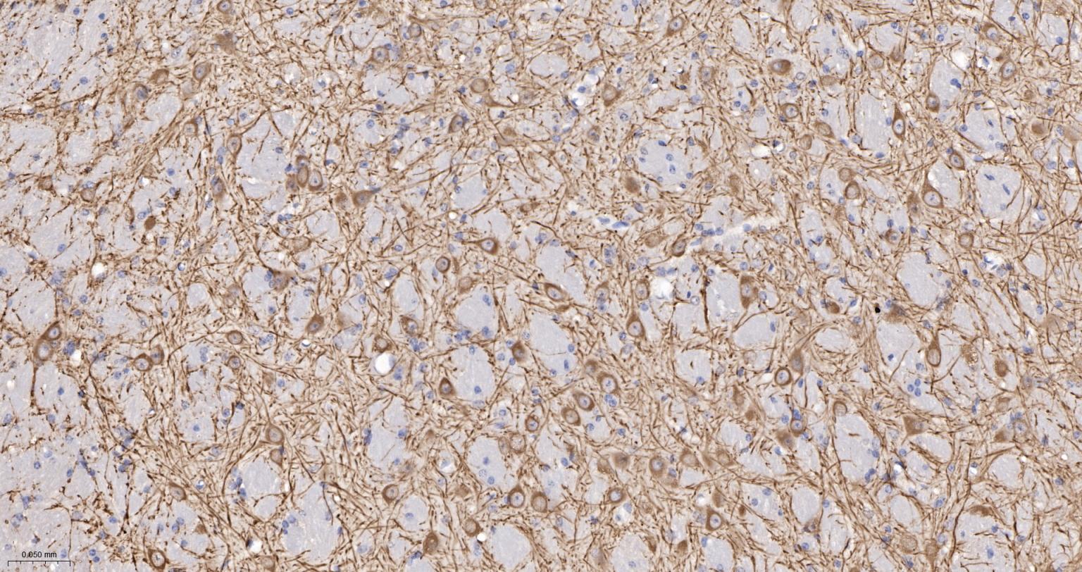
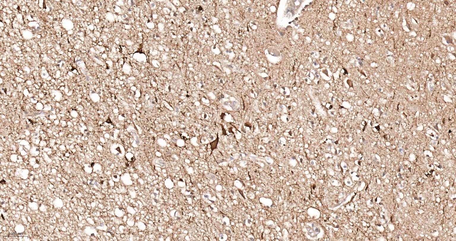
Figure 10. Paraformaldehyde-fixed, paraffin embedded Human Cerebrum; Antigen retrieval by boiling in sodium citrate buffer (pH6.0) for 15 min; The section was incubated with MAP2 Monoclonal Antibody, Unconjugated (SLA0988M) at 1:200 overnight at 4°C, followed by conjugation to the SL0295G-HRP and DAB (C-0010) staining.
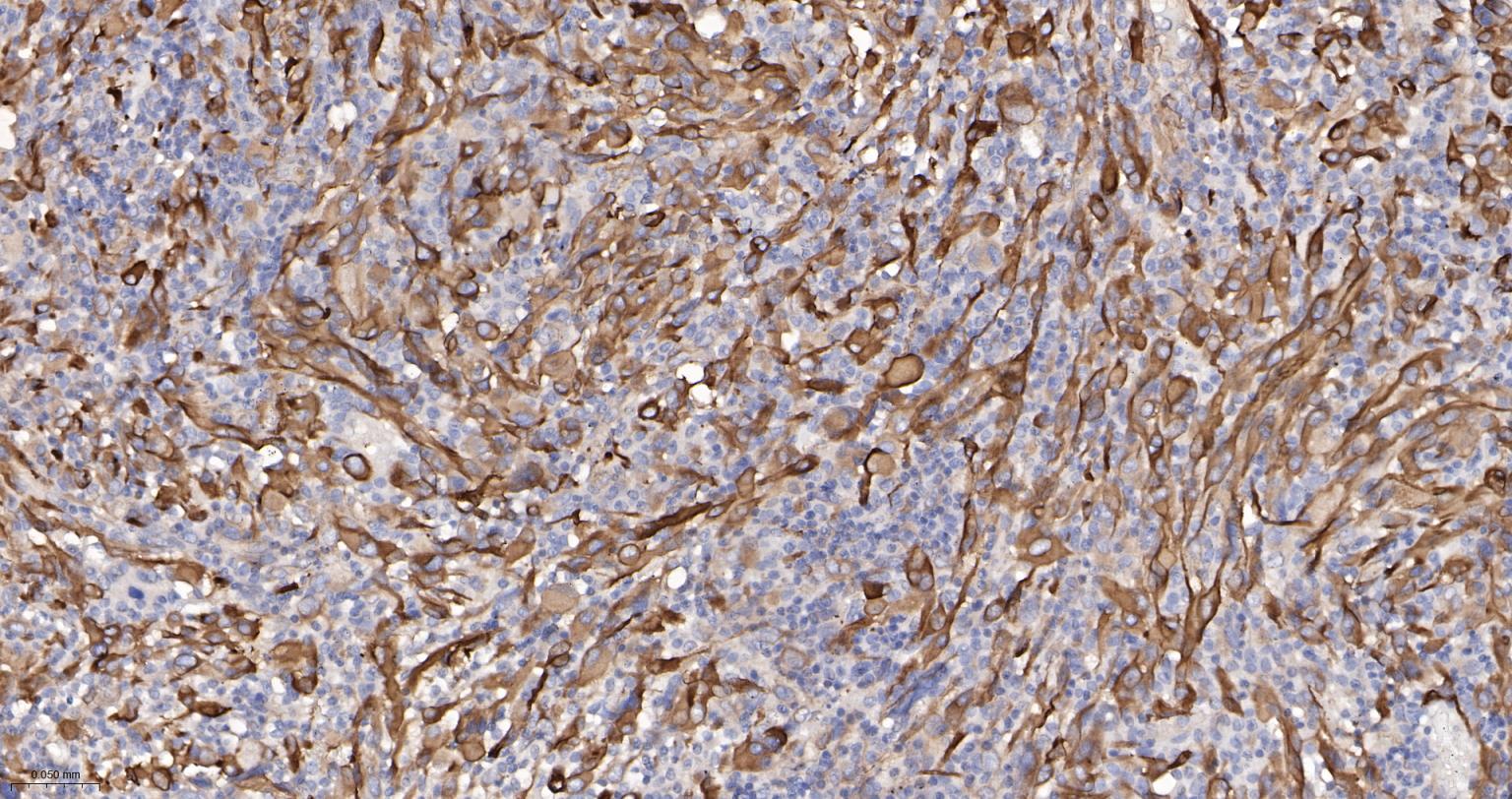
Figure 11. Paraformaldehyde-fixed, paraffin embedded Human Glioma; Antigen retrieval by boiling in sodium citrate buffer (pH6.0) for 15 min; The section was incubated with MAP2 Monoclonal Antibody, Unconjugated (SLA0988M) at 1:200 overnight at 4°C, followed by conjugation to the SL0295G-HRP and DAB (C-0010) staining.
FOR RESEARCH USE ONLY. Not for use in diagnostic or therapeutic procedures.
Partial purchase records(bought amounts latest0)
User Comment(Total0User Comment Num)
- No comment


 +86 571 56623320

 SUNLONG BIOTECH


