Details
Product Name Recombinant Rabbit Anti-MAPT Monoclonal Antibody Cat No. SLA0832M Chinese Name 微管相关蛋白重组兔单抗 Alias tau; DDPAC; FTD1; FTDP-17; MAPTL; MSTD; MTBT1; MTBT2; PPND; PPP1R103; Tau-PHF6; tau-40; Mtapt; PHF-tau; MAPT_0N4R; MAPT_1N4R; MAPT_2N4R; RNPTAU; pTau; TAU_BOVIN; MAPT; Neurofibrillary tangle protein; Paired helical filament-tau (PHF-tau); TAU_HUMAN; TAU_MOUSE; TAU_RAT; microtubule associated protein tau; G protein beta1/gamma2 subunit-interacting factor 1; microtubule-associated protein tau, isoform 4; protein phosphatase 1, regulatory subunit 103; Tau-derived paired helical filament hexapeptide Host Species Rabbit Isotype IgG Clonality Monoclonal Clone No. 1G4 React Species Human,Mouse,Rat Applications WB (1:500-2000)
IHC-P (1:50-200)
IHC-F (1:50-200)
IF (1:50-200)
Not yet tested in other applications.
Optimal working dilutions must be determined by the end user.Immunogen A synthesized peptide derived from human Tau Purification affinity purified by Protein A Concentration 1mg/ml Predicted MW. 79 kDa Observed MW. 50-65 kDa Subcellular Location Cytoplasm Gene ID 4137 Swiss-Prot P10636 Storage 0.01M TBS (pH7.4) with 1% BSA, 0.02% Proclin300 and 50% Glycerol.
Shipped at 4℃. Store at -20℃ for one year. Avoid repeated freeze/thaw cycles.Background Tau proteins are important Promotes microtubule assembly and stability, and might be involved in the establishment and maintenance of neuronal polarity. The C-terminus binds axonal microtubules while the N-terminus binds neural plasma membrane components, suggesting that tau functions as a linker protein between both. Axonal polarity is predetermined by tau localization (in the neuronal cell) in the domain of the cell body defined by the centrosome. The short isoforms allow plasticity of the cytoskeleton whereas the longer isoforms may preferentially play a role in its stabilization. Tau proteins subcellular located in the axons of neurons, in the cytoso l and in association with plasma membrane components. It expressed in neurons. PNS-tau is expressed in the peripheral nervous system while the others are expressed in the central nervous system.
Attention This product as supplied is intended for research use only, not for use in human, therapeutic or diagnostic applications.
Validation Images
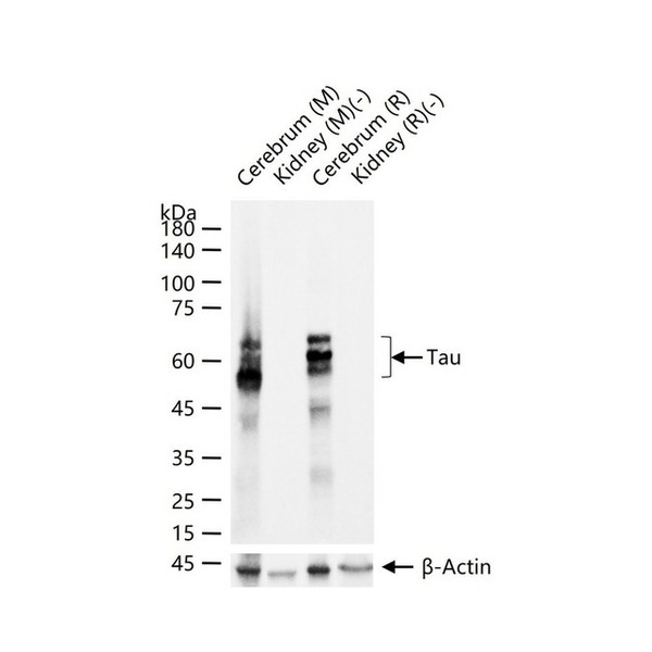
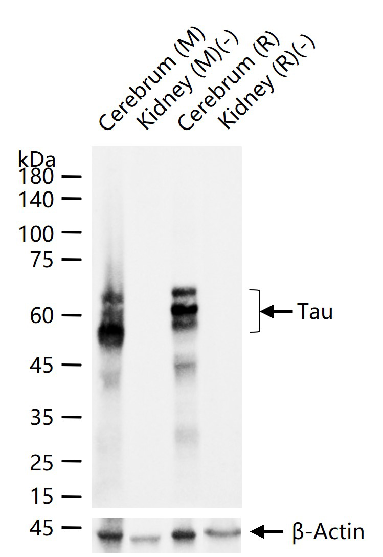
Figure 1. 25 ug total protein per lane of various lysates (see on figure) probed with Tau monoclonal antibody, unconjugated (SLA0832M) at 1:1000 dilution and 4°C overnight incubation. Followed by conjugated secondary antibody incubation at r.t. for 60 min.
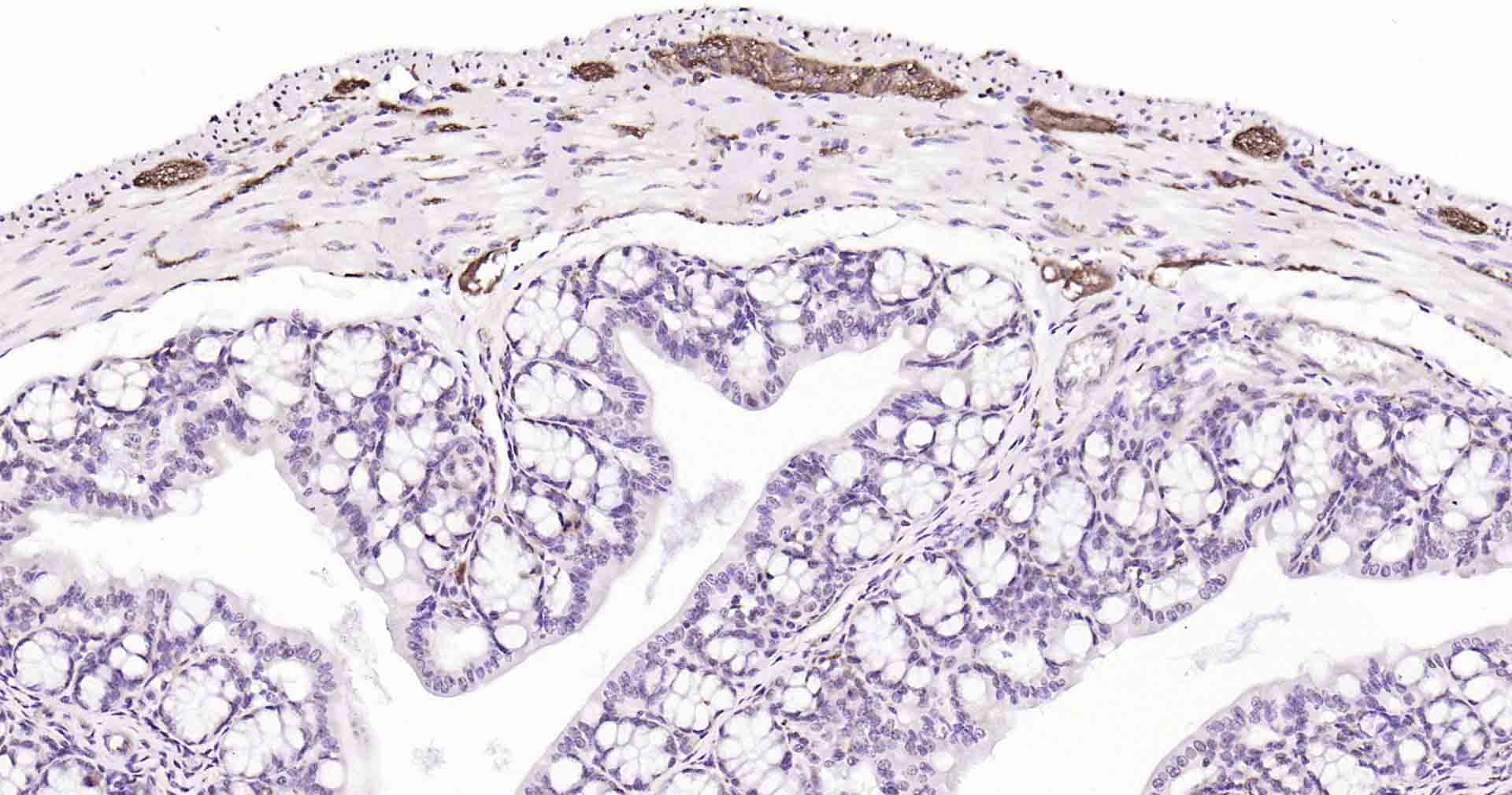
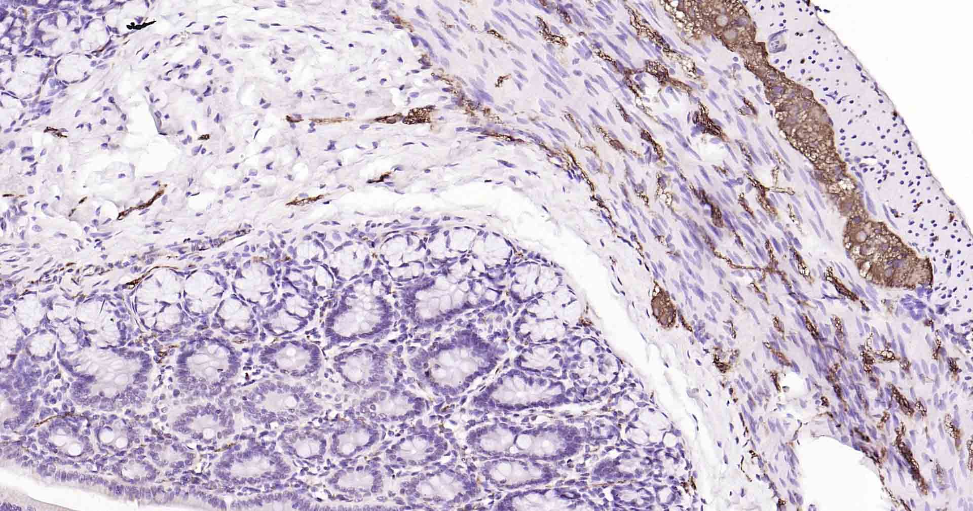
Figure 2. Paraformaldehyde-fixed, paraffin embedded Mouse Colon; Antigen retrieval by boiling in sodium citrate buffer (pH6.0) for 15 min; Antibody incubation with Tau Monoclonal Antibody, Unconjugated(SLA0832M) at 1:200overnight at 4°C, followed by conjugation to the SP Kit (Rabbit, SP-0023) and DAB (C-0010) staining.
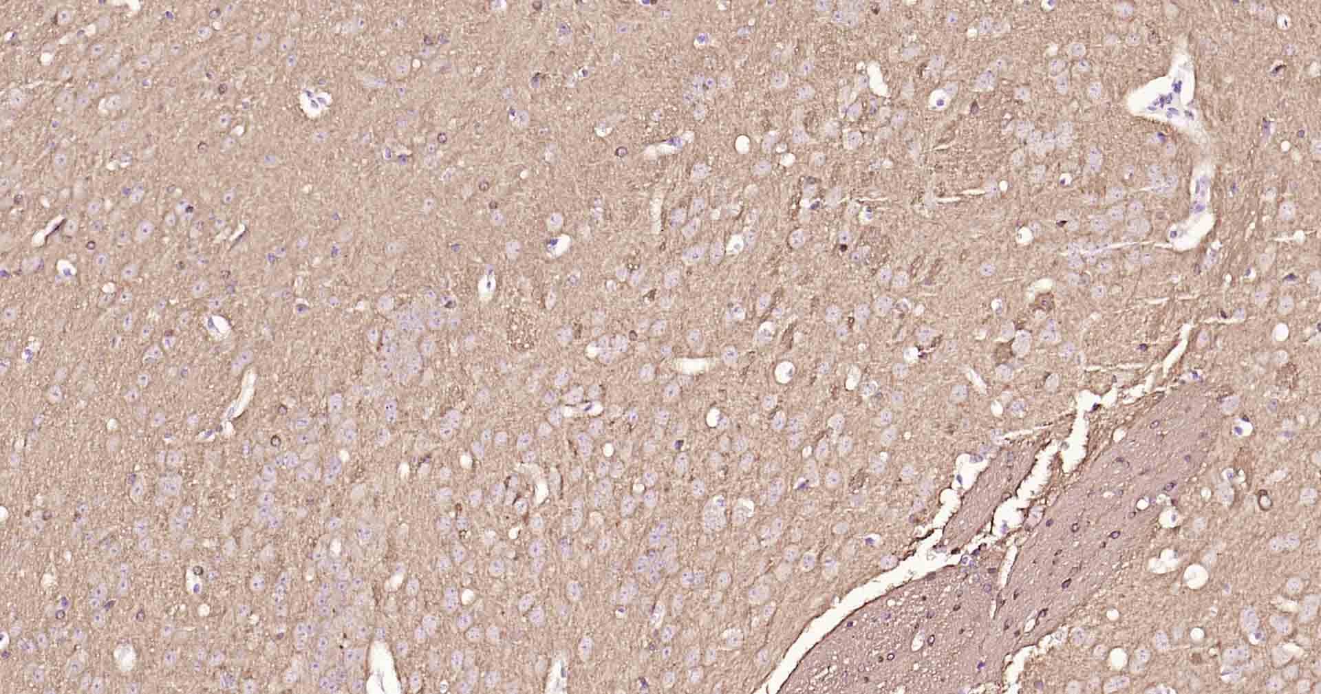
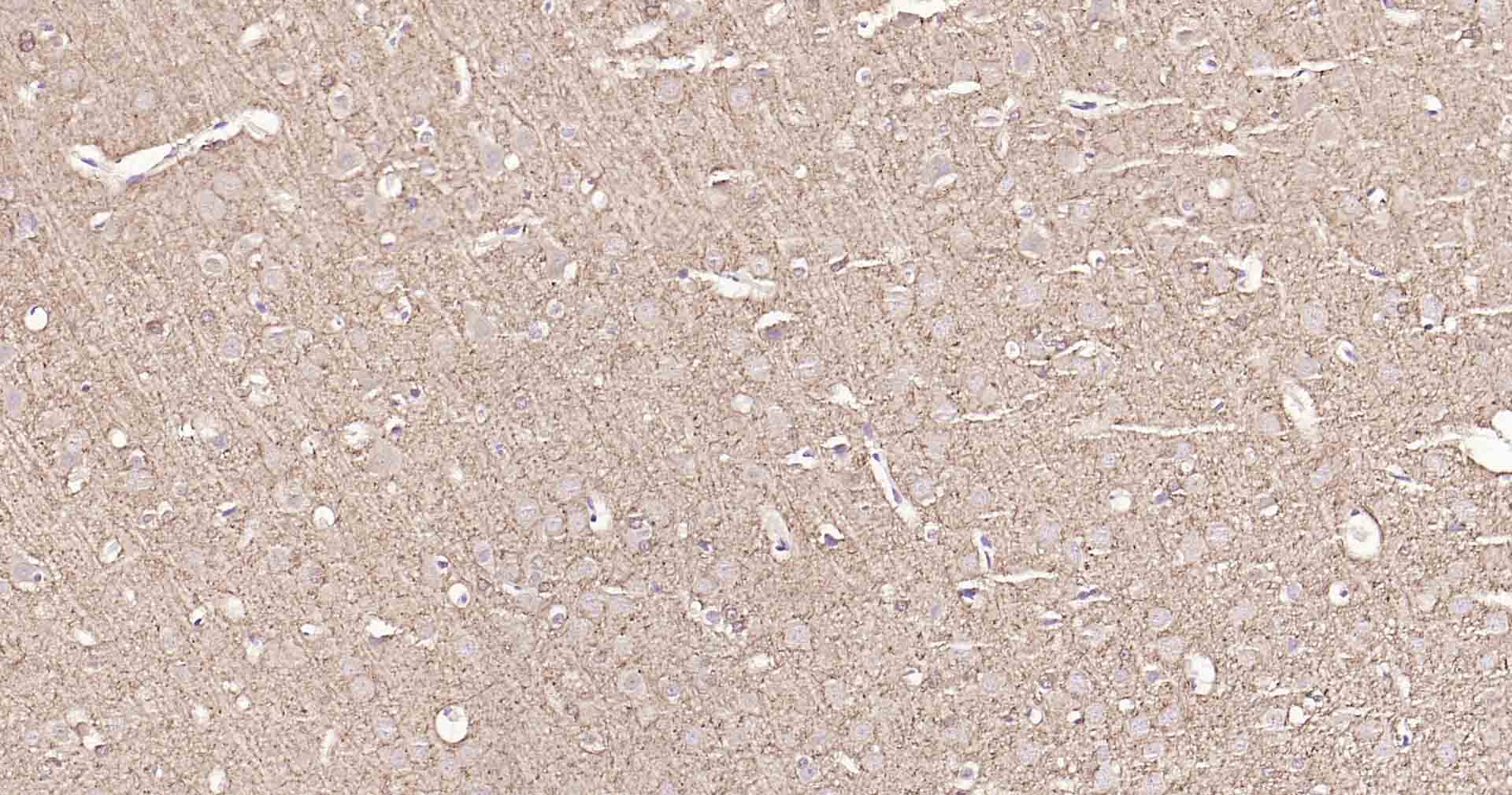
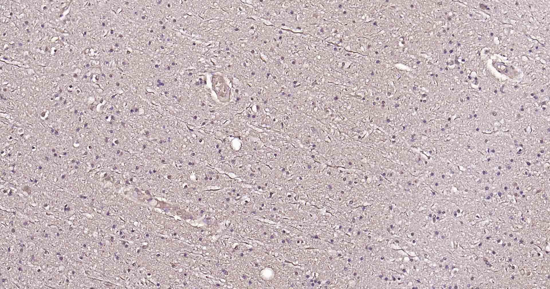
Figure 3. Paraformaldehyde-fixed, paraffin embedded Mouse Cerebrum; Antigen retrieval by boiling in sodium citrate buffer (pH6.0) for 15 min; Antibody incubation with Tau Monoclonal Antibody, Unconjugated(SLA0832M) at 1:200overnight at 4°C, followed by conjugation to the SP Kit (Rabbit, SP-0023) and DAB (C-0010) staining.
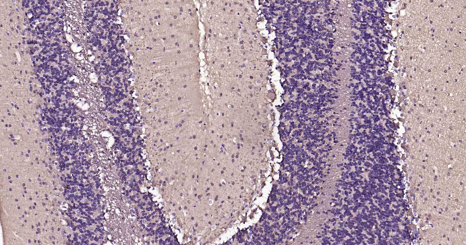
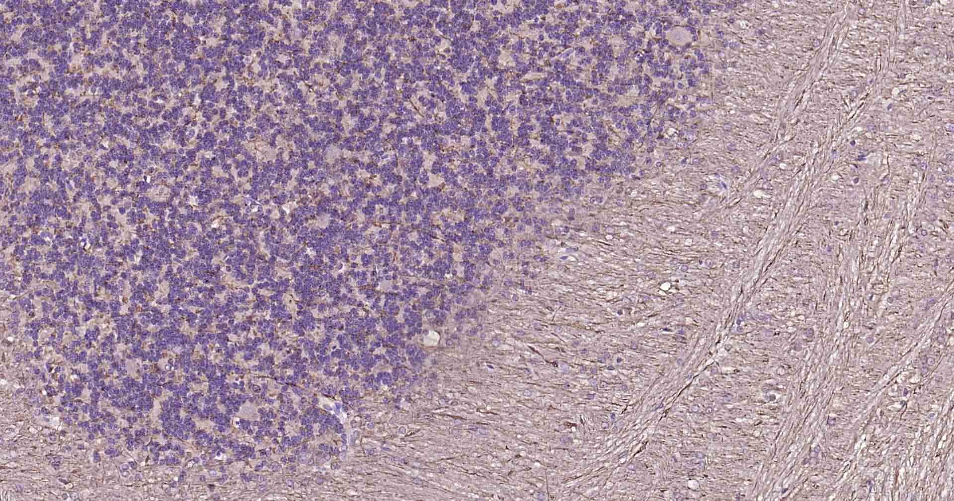
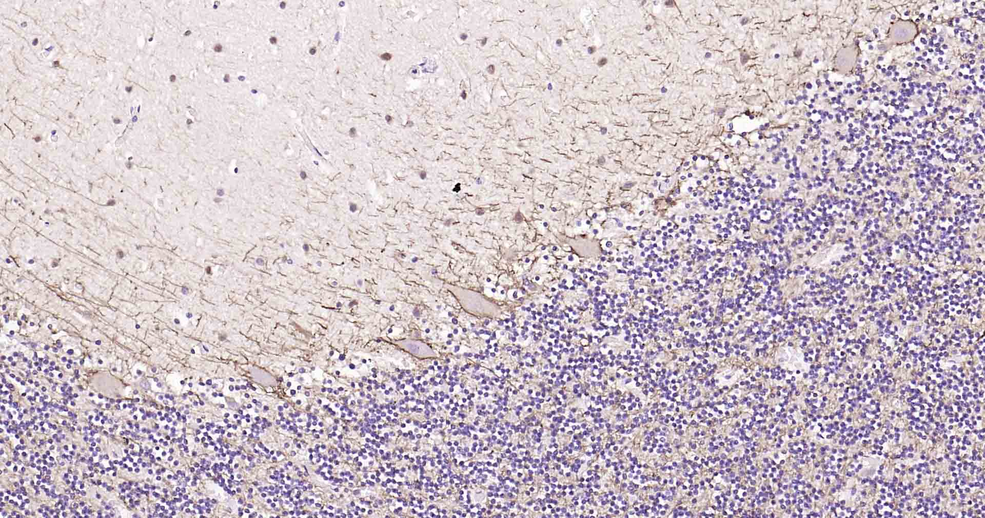
Figure 4. Paraformaldehyde-fixed, paraffin embedded Mouse Cerebellum; Antigen retrieval by boiling in sodium citrate buffer (pH6.0) for 15 min; Antibody incubation with Tau Monoclonal Antibody, Unconjugated(SLA0832M) at 1:200overnight at 4°C, followed by conjugation to the SP Kit (Rabbit, SP-0023) and DAB (C-0010) staining.
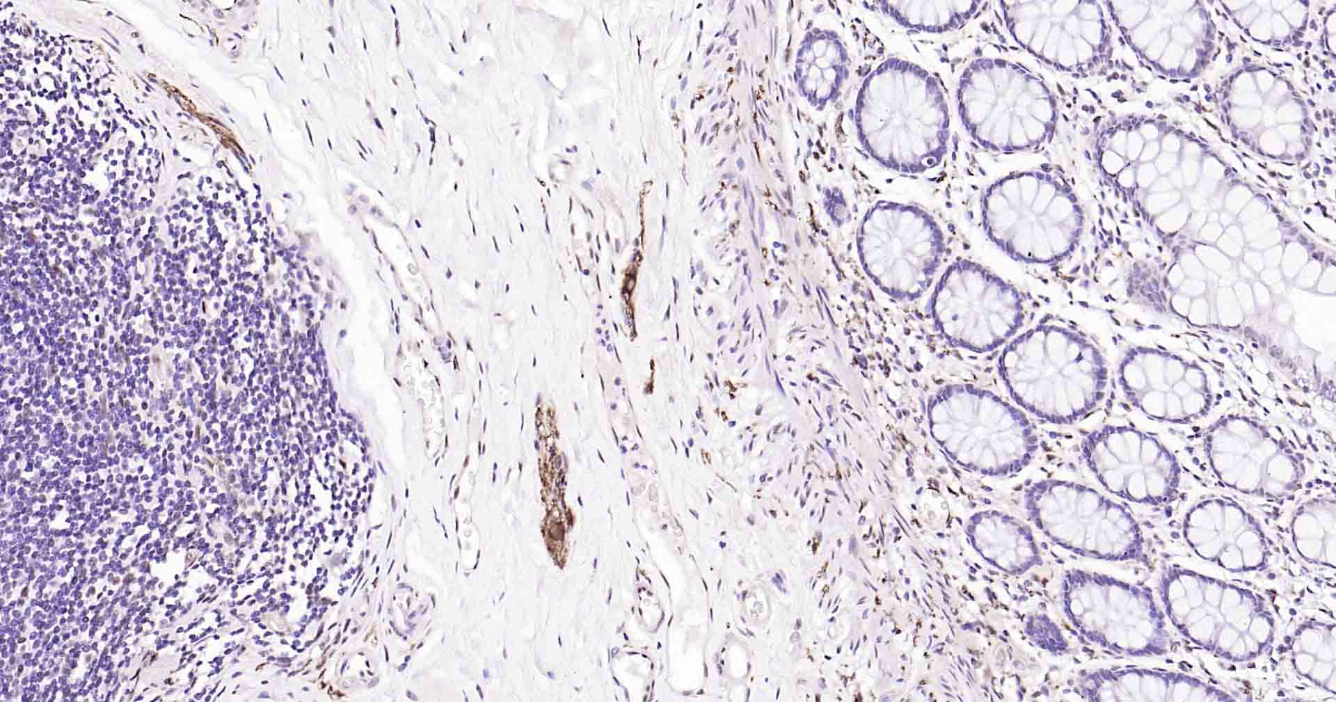
Figure 5. Paraformaldehyde-fixed, paraffin embedded Rat Colon; Antigen retrieval by boiling in sodium citrate buffer (pH6.0) for 15 min; Antibody incubation with Tau Monoclonal Antibody, Unconjugated(SLA0832M) at 1:200overnight at 4°C, followed by conjugation to the SP Kit (Rabbit, SP-0023) and DAB (C-0010) staining.
Figure 6. Paraformaldehyde-fixed, paraffin embedded Mouse Cerebrum; Antigen retrieval by boiling in sodium citrate buffer (pH6.0) for 15 min; Antibody incubation with Tau Monoclonal Antibody, Unconjugated(SLA0832M) at 1:200overnight at 4°C, followed by conjugation to the SP Kit (Rabbit, SP-0023) and DAB (C-0010) staining.
Figure 7. Paraformaldehyde-fixed, paraffin embedded Rat Cerebellum; Antigen retrieval by boiling in sodium citrate buffer (pH6.0) for 15 min; Antibody incubation with Tau Monoclonal Antibody, Unconjugated(SLA0832M) at 1:200overnight at 4°C, followed by conjugation to the SP Kit (Rabbit, SP-0023) and DAB (C-0010) staining.
Figure 8. Paraformaldehyde-fixed, paraffin embedded Human Colon; Antigen retrieval by boiling in sodium citrate buffer (pH6.0) for 15 min; Antibody incubation with Tau Monoclonal Antibody, Unconjugated(SLA0832M) at 1:200overnight at 4°C, followed by conjugation to the SP Kit (Rabbit, SP-0023) and DAB (C-0010) staining.
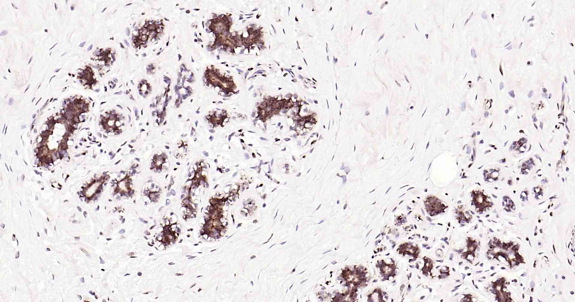
Figure 9. Paraformaldehyde-fixed, paraffin embedded Human Breast; Antigen retrieval by boiling in sodium citrate buffer (pH6.0) for 15 min; Antibody incubation with Tau Monoclonal Antibody, Unconjugated(SLA0832M) at 1:200overnight at 4°C, followed by conjugation to the SP Kit (Rabbit, SP-0023) and DAB (C-0010) staining.
Figure 10. Paraformaldehyde-fixed, paraffin embedded Human Cerebellum; Antigen retrieval by boiling in sodium citrate buffer (pH6.0) for 15 min; Antibody incubation with Tau Monoclonal Antibody, Unconjugated(SLA0832M) at 1:200overnight at 4°C, followed by conjugation to the SP Kit (Rabbit, SP-0023) and DAB (C-0010) staining.
Figure 11. Paraformaldehyde-fixed, paraffin embedded Human Left Parietal Lobe; Antigen retrieval by boiling in sodium citrate buffer (pH6.0) for 15 min; Antibody incubation with Tau Monoclonal Antibody, Unconjugated(SLA0832M) at 1:200overnight at 4°C, followed by conjugation to the SP Kit (Rabbit, SP-0023) and DAB (C-0010) staining.
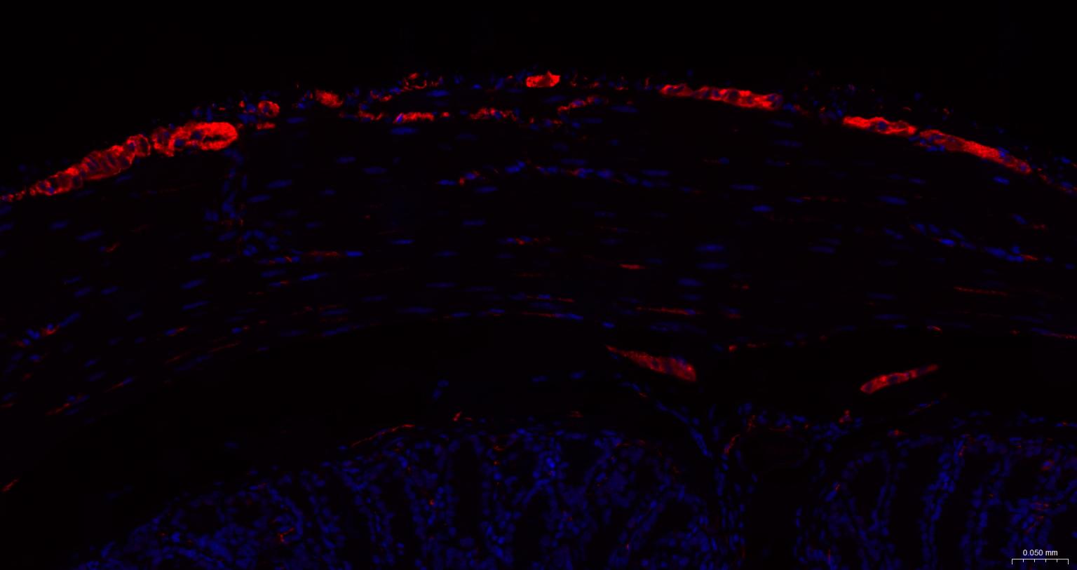
Figure 12. Paraformaldehyde-fixed, paraffin embedded Mouse Colon; Antigen retrieval by boiling in sodium citrate buffer (pH6.0) for 15 min; The section was incubated with MAPT/Tau Monoclonal Antibody, Unconjugated (SLA0832M) at 1:200 overnight at 4°C. Followed by conjugated Goat Anti-Rabbit IgG antibody (Red, SL0295G-BF594), DAPI (blue, C02-04002) was used to stain the cell nuclei.
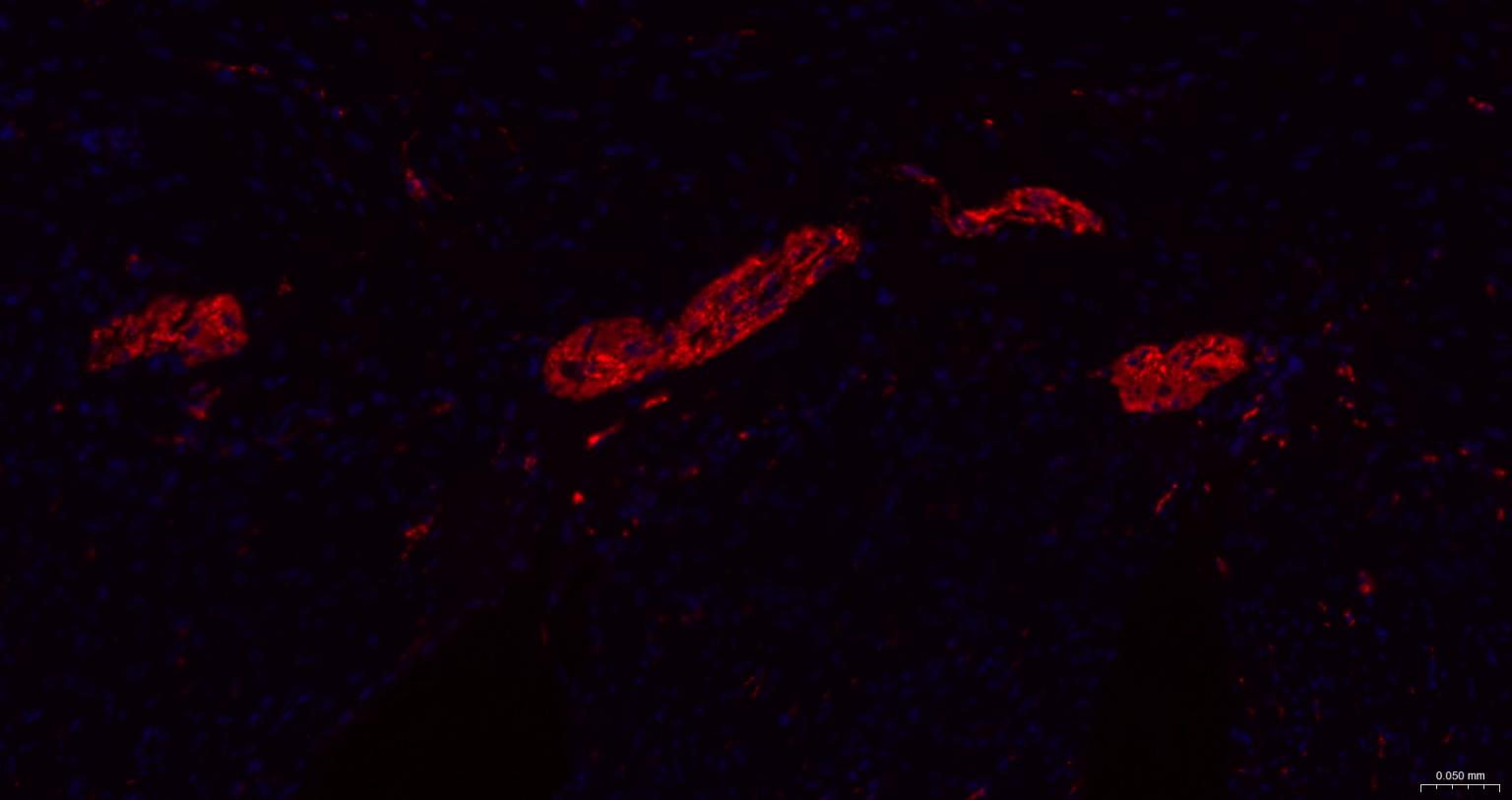
Figure 13. Paraformaldehyde-fixed, paraffin embedded Human Colon; Antigen retrieval by boiling in sodium citrate buffer (pH6.0) for 15 min; The section was incubated with MAPT/Tau Monoclonal Antibody, Unconjugated (SLA0832M) at 1:200 overnight at 4°C. Followed by conjugated Goat Anti-Rabbit IgG antibody (Red, SL0295G-BF594), DAPI (blue, C02-04002) was used to stain the cell nuclei.
FOR RESEARCH USE ONLY. Not for use in diagnostic or therapeutic procedures.
Partial purchase records(bought amounts latest0)
User Comment(Total0User Comment Num)
- No comment


 +86 571 56623320

 SUNLONG BIOTECH


