Details
Product Name Recombinant Rabbit Anti-CDH1 Monoclonal Antibody Cat No. SLA1156M Chinese Name 上皮钙粘附分子重组兔单抗 Alias Arc-1; BCDS1; CD324; CDHE; ECAD; LCAM; UVO; E-cad; L-CAM; Um; Cadherin-1; Uvomorulin; CADH1_BOVIN; CDH1; Epithelial cadherin (E-cadherin); CADH1_HUMAN; CAM 120/80; CADH1_MOUSE; CADH1_RAT; cadherin 1; cadherin 1, type 1, E-cadherin (epithelial); E-Cadherin Host Species Rabbit Isotype IgG Clonality Monoclonal Clone No. 6E1 React Species Human Applications Flow-Cyt (1:100-200)
ICC/IF (1:100-200)
Not yet tested in other applications.
Optimal working dilutions must be determined by the end user.Immunogen KLH conjugated synthetic peptide derived from human E-cadherin Purification affinity purified by Protein A Concentration 1mg/ml Predicted MW. Observed MW. Subcellular Location Membrane Gene ID 999 Swiss-Prot P12830 Storage 0.01M TBS (pH7.4) with 1% BSA, 0.02% Proclin300 and 50% Glycerol.
Shipped at 4℃. Store at -20℃ for one year. Avoid repeated freeze/thaw cycles.Background This gene encodes a classical cadherin of the cadherin superfamily. Alternative splicing results in multiple transcript variants, at least one of which encodes a preproprotein that is proteolytically processed to generate the mature glycoprotein. This calcium-dependent cell-cell adhesion protein is comprised of five extracellular cadherin repeats, a transmembrane region and a highly conserved cytoplasmic tail. Mutations in this gene are correlated with gastric, breast, colorectal, thyroid and ovarian cancer. Loss of function of this gene is thought to contribute to cancer progression by increasing proliferation, invasion, and/or metastasis. The ectodomain of this protein mediates bacterial adhesion to mammalian cells and the cytoplasmic domain is required for internalization. This gene is present in a gene cluster with other members of the cadherin family on chromosome 16. [provided by RefSeq, Nov 2015] Attention This product as supplied is intended for research use only, not for use in human, therapeutic or diagnostic applications.
Validation Images
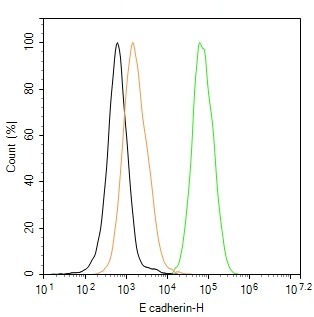
Figure 1. The T-47D (H) cells were incubated in 5%BSA to block non-specific protein-protein interactions (30 min at r.t.), followed by secondary antibody incubation for 40 min at room temperature. Primary Antibody (green): Rabbit Anti-E cadherin antibody (SLA1156M,1:100); Isotype Control (orange): Rabbit IgG (SL0295P). Blank control (black): PBS. Acquisition of 20,000 events was performed
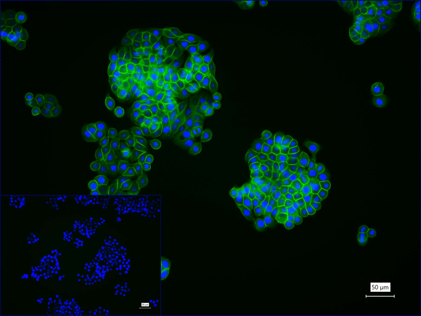
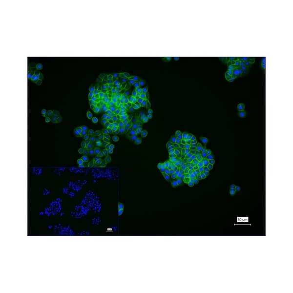
Figure 2. 4% Paraformaldehyde-fixed HT-29 (H) cell; Triton X-100 at r.t. for 20 min; Antibody incubation with (E cadherin) monoclonal Antibody, unconjugated (SLA1156M) 1:100, 90 min at 37°C; followed by BF488 conjugated Goat Anti-Rabbit IgG antibody (green, SL60295G-BF488) at 37°C for 90 min, DAPI (blue, C02-04002) was used to stain the cell nuclei. PBS instead of the primary antibody was used as the blank control
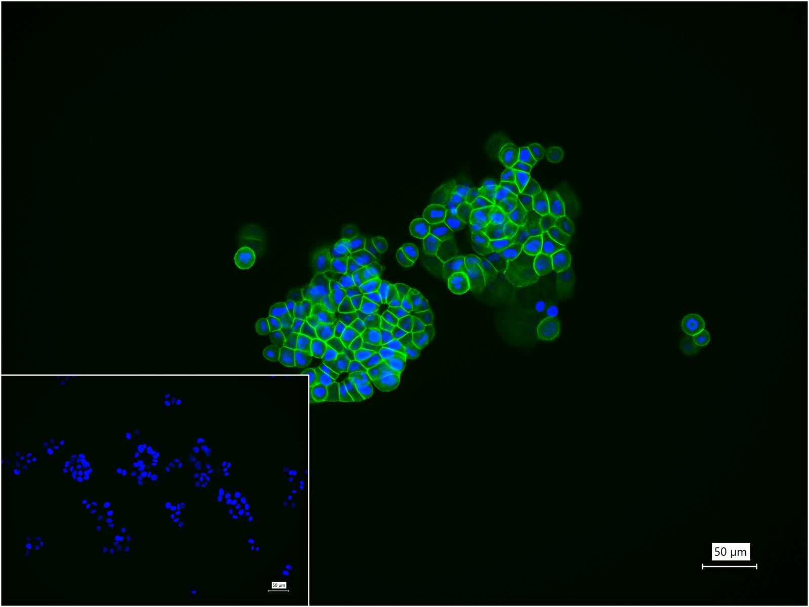
Figure 3. 4% Paraformaldehyde-fixed T-47D (H) cell; Triton X-100 at r.t. for 20 min; Antibody incubation with (E cadherin) monoclonal Antibody, unconjugated (SLA1156M) 1:100, 90 min at 37°C; followed by BF488 conjugated Goat Anti-Rabbit IgG antibody (green, SL60295G-BF488) at 37°C for 90 min, DAPI (blue, C02-04002) was used to stain the cell nuclei. PBS instead of the primary antibody was used as the blank control
FOR RESEARCH USE ONLY. Not for use in diagnostic or therapeutic procedures.
Partial purchase records(bought amounts latest0)
User Comment(Total0User Comment Num)
- No comment


 +86 571 56623320

 SUNLONG BIOTECH


