Details
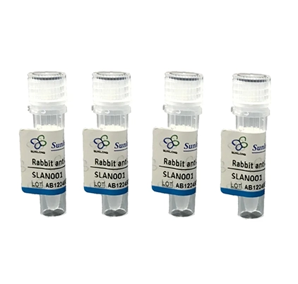
Product Name Recombinant Rabbit Anti-NEFL Monoclonal Antibody Cat No. SLA0531M Chinese Name 低分子量神经丝蛋白重组兔单抗 Alias CMT1F; CMT2E; CMTDIG; NF-L; NF68; NFL; PPP1R110; NFL_HUMAN; NEFL; 68 kDa neurofilament protein; Neurofilament triplet L protein; NFL_MOUSE; NFL_RAT; neurofilament light chain; neurofilament, light polypeptide 68kDa; neurofilament, light polypeptide; neurofilament light; protein phosphatase 1, regulatory subunit 110 Host Species Rabbit Isotype IgG Clonality Monoclonal Clone No. 4A11 React Species Human Applications Flow-Cyt (1μg/Test)
ICC/IF (1:50-200)
Not yet tested in other applications.
Optimal working dilutions must be determined by the end user.Immunogen Recombinant human NF-L protein Purification affinity purified by Protein A Concentration 1mg/ml Predicted MW. 68 kDa Observed MW. Subcellular Location Cytoplasm Gene ID 4747 Swiss-Prot P07196 Storage 0.01M TBS (pH7.4) with 1% BSA, 0.02% Proclin300 and 50% Glycerol.
Shipped at 4℃. Store at -20℃ for one year. Avoid repeated freeze/thaw cycles.Background Neurofilament light polypeptide also called NF-L; Neurofilament triplet L protein; 68 kDa neurofilament protein. Neurofilaments usually contain three intermediate filament proteins: L, M, and H which are involved in the maintenance of neuronal caliber. The extra mass and high charge density that distinguish the neurofilament proteins from all other intermediate filament proteins are due to the tailpiece extensions. This region may form a charged scaffolding structure suitable for interaction with other neuronal components or ions. NF-L is the most abundant of the three neurofilament proteins and, as the other nonepithelial intermediate filament proteins, it can form homopolymeric 10-nm filaments. Belongs to the intermediate filament family. Involved in the maintenance of neuronal caliber, neurofilaments are the intermediate filament proteins found specifically in neurons, and are composed predominantly of three major proteins called NF-L, NF-M and NF-H. Like most other intermediate filament proteins (IFPs), the expression of the different neuronal IFPs is both tissue-specific and developmentally regulated. NF-L is the light or low molecular weight microfilament subunit and runs on SDS-PAGE gels at approximately 70 kDa. Neurofilament are the 10nm or intermediate filament proteins found specifically in neurons, and are composed predominantly of three major proteins called NF-L, NF-M and NF-H. NF-H is the heavy or high molecular weight microfilament subunit and runs on SDS-PAGE gels in the range 180-220 kDa, with some variation in different species. Attention This product as supplied is intended for research use only, not for use in human, therapeutic or diagnostic applications.
Validation Images

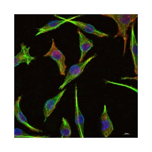
Figure 1. 4% Paraformaldehyde-fixed SH-SY5Y (H) cell; Triton X-100 at r.t. for 20 min; Antibody incubation with (NF-L) monoclonal Antibody, unconjugated (SLA0531M) 1:100, 90 min at 37°C; followed by BF488 conjugated Goat Anti-Rabbit IgG antibody (green, SL60295G-BF488) at 37°C for 90 min, then stained with Phalloidin/BF555 for 30 min at room temperature; DAPI (blue, C02-04002) was used to stain the cell nuclei

Figure 2. The SH-SY5Y (H) cells were fixed with 4% PFA (10 min at r.t.) and then permeabilized with 90% ice-cold methanol for 20 min at -20℃,the cells then were incubated in 5%BSA to block non-specific protein-protein interactions (30 min at r.t.).Primary Antibody (green):Rabbit Anti-NF-L antibody (SLA0531M): 1 μg/10^6 cells; Secondary Antibody (white blue): Goat anti-Rabbit IgG-FITC (SL40295G-FITC): 1 μg/test. Blank control (black): PBS. Acquisition of 20,000 events was performed

Figure 3. 4% Paraformaldehyde-fixed SH-SY5Y (H) cell; Triton X-100 at r.t. for 20 min; Antibody incubation with (NF-L) monoclonal Antibody, unconjugated (SLA0531M) 1:100, 90 min at 37°C; followed by conjugated Goat Anti-Rabbit IgG antibody (green, SL40295G-FITC) at 37°C for 90 min, DAPI (blue, C02-04002) was used to stain the cell nuclei
FOR RESEARCH USE ONLY. Not for use in diagnostic or therapeutic procedures.
Partial purchase records(bought amounts latest0)
User Comment(Total0User Comment Num)
- No comment


 +86 571 56623320

 SUNLONG BIOTECH


