Details
Product Name FBLN5 | FIBL-5 Rabbit pAb Cat No. SL42060R Chinese Name 衰老关键蛋白抗体 Alias ADCL2; ARCL1A; ARMD3; CMT1H; DANCE; EVEC; FIBL-5; HNARMD; UP50; A55; FBLN5_HUMAN; FBLN5; Developmental arteries and neural crest EGF-like protein (Dance); Urine p50 protein (UP50); FBLN5_MOUSE; fibulin 5; embryonic vascular EGF-like repeat-containing protein; developmental arteries and neural crest EGF-like Host Species Rabbit Isotype IgG Clonality Polyclonal React Species Human Applications WB (1:500-2000)
IHC-P (1:100-500)
IHC-F (1:100-500)
IF (1:100-500)
ICC/IF (1:50-200)
Not yet tested in other applications.
Optimal working dilutions must be determined by the end user.Immunogen Recombinant human Fibulin 5 protein Purification affinity purified by Protein A Concentration 1mg/ml Predicted MW. 48 kDa Observed MW. 30 kDa Subcellular Location Secreted Gene ID 10516 Swiss-Prot Q9UBX5 Storage 0.01M TBS (pH7.4) with 1% BSA, 0.02% Proclin300 and 50% Glycerol.
Shipped at 4℃. Store at -20℃ for one year. Avoid repeated freeze/thaw cycles.Background Fibulin 5: A protein that belongs to a family of extracellular proteins expressed in the basement membranes of blood vessels. Fibulin 5 may be essential for the polymerization of elastin. Missense mutations in FBLN5, the gene that encodes fibulin 5, appear responsible for 1-2% of cases of age-related macular degeneration (AMD). FBLN5 is located on chromosome 14 in band 14q32.1. See also: Fibulin 3. May play a role in vascular growth and maturation during development and in lesions of injured vessels. Attention This product as supplied is intended for research use only, not for use in human, therapeutic or diagnostic applications.
Validation Images
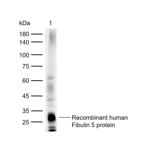
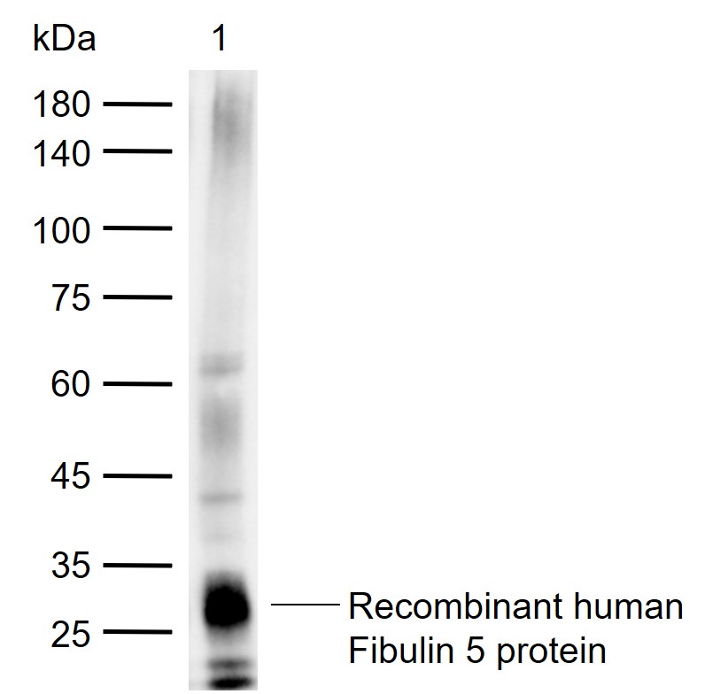
Figure 1. Sample: Lane 1: Recombinant human Fibulin 5 protein, C-His(SL42060P) Primary: Anti-Fibulin 5 (SL42060R) at 1/1000 dilution Secondary: IRDye800CW Goat Anti-Rabbit IgG at 1/20000 dilution Predicted band size: 48 kDa Observed band size: 30 kDa
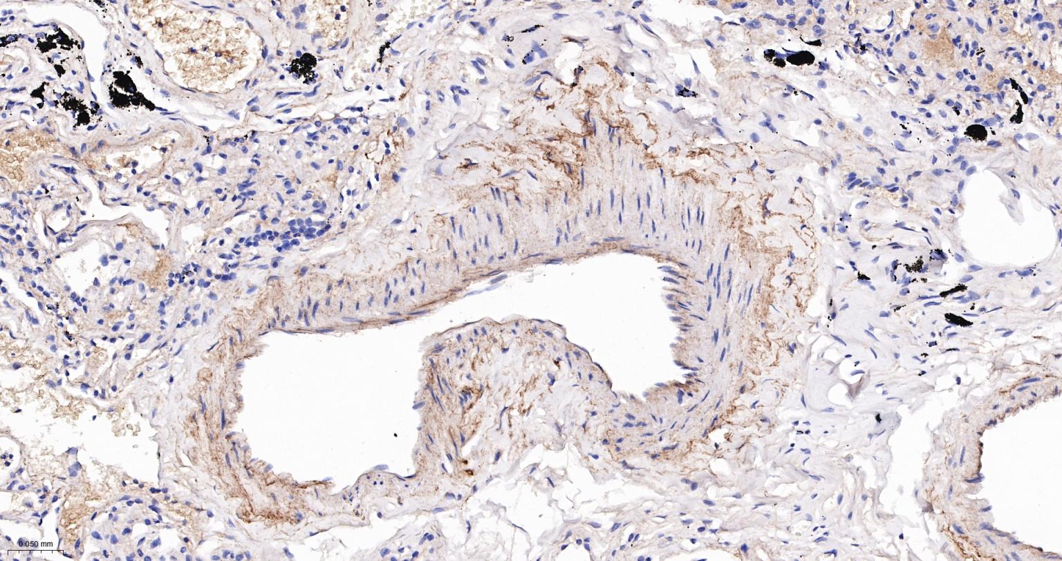
Figure 2. Paraformaldehyde-fixed, paraffin embedded Human Lung; Antigen retrieval by boiling in sodium citrate buffer (pH6.0) for 15 min; The section was incubated with Fibulin 5 Polyclonal Antibody, Unconjugated (SL42060R) at 1:200 overnight at 4°C, followed by conjugation to the SL0295G-HRP and DAB (C-0010) staining.
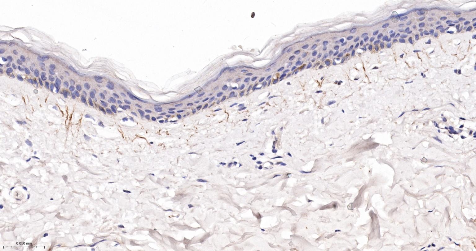
Figure 3. Paraformaldehyde-fixed, paraffin embedded Human Skin; Antigen retrieval by boiling in sodium citrate buffer (pH6.0) for 15 min; The section was incubated with Fibulin 5 Polyclonal Antibody, Unconjugated (SL42060R) at 1:200 overnight at 4°C, followed by conjugation to the SL0295G-HRP and DAB (C-0010) staining.
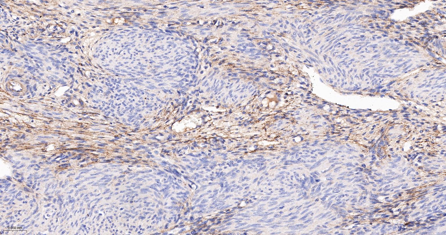
Figure 4. Paraformaldehyde-fixed, paraffin embedded Human Uterus; Antigen retrieval by boiling in sodium citrate buffer (pH6.0) for 15 min; The section was incubated with Fibulin 5 Polyclonal Antibody, Unconjugated (SL42060R) at 1:200 overnight at 4°C, followed by conjugation to the SL0295G-HRP and DAB (C-0010) staining.
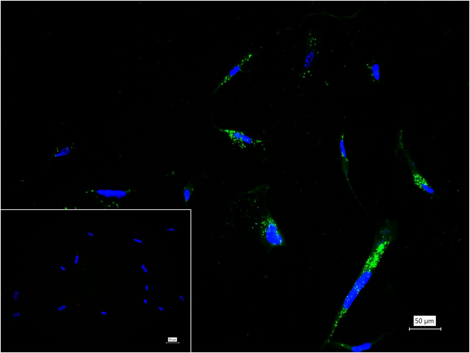
Figure 5. 4% Paraformaldehyde-fixed WI-38 (H) cell; Triton X-100 at r.t. for 20 min; Antibody incubation with (Fibulin 5) polyclonal Antibody, unconjugated (SL42060R) 1:100, 90 min at 37°C; followed by BF488 conjugated Goat Anti-Rabbit IgG antibody (green, SL60295G-BF488) at 37°C for 90 min, DAPI (blue, C02-04002) was used to stain the cell nuclei. PBS instead of the primary antibody was used as the blank control.
FOR RESEARCH USE ONLY. Not for use in diagnostic or therapeutic procedures.
Partial purchase records(bought amounts latest0)
User Comment(Total0User Comment Num)
- No comment


 +86 571 56623320

 SUNLONG BIOTECH


