Details
Product Name Rabbit Anti-GLUL Polyclonal Antibody Cat No. SLA0192 Chinese Name 谷氨酰胺合成酶/谷氨酸氨连接酶抗体 Alias DEE116; GLNS; GS; PIG43; PIG59; GLNA_BOVIN; GLUL; Glutamate--ammonia ligase; Palmitoyltransferase GLUL; 6.3.1.2; GLNA_CANLF; GLNA_HUMAN; GLNA_MOUSE; GLNA_RAT; glutamate-ammonia ligase; glutamate-ammonia ligase (glutamine synthase); glutamine synthetase Host Species Rabbit Isotype IgG Clonality Polyclonal React Species Human,Mouse,Rat Applications WB (1:500-2000)
IHC-P (1:100-500)
IHC-F (1:100-500)
IF (1:100-500)
Not yet tested in other applications.
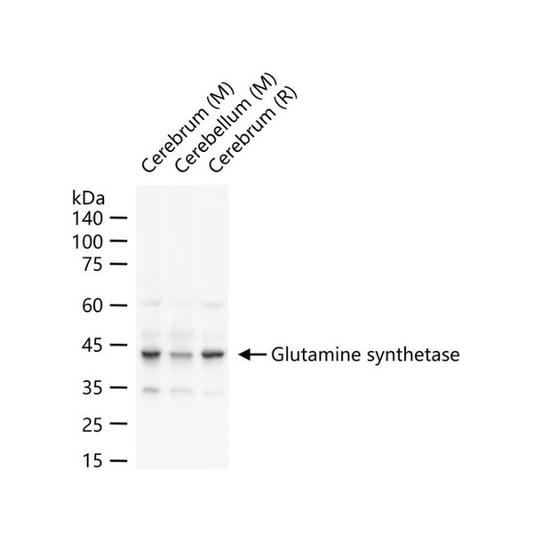
Optimal working dilutions must be determined by the end user.Immunogen Recombinant human Glutamine synthetase protein Purification affinity purified by Protein A Concentration 1mg/ml Predicted MW. 42 kDa Observed MW. 42 kDa Subcellular Location Cytoplasm Gene ID 2752 Swiss-Prot P15104 Storage 0.01M TBS (pH7.4) with 1% BSA, 0.02% Proclin300 and 50% Glycerol.
Shipped at 4℃. Store at -20℃ for one year. Avoid repeated freeze/thaw cycles.Background Glutamine Synthetase catalyzes the conversion of ammonia and glutamate to glutamine. It is found in astrocytes as an octamer of identical 42 kDa subunits. The function of Glutamine Synthetase is the detoxification of brain ammonia. It also has an important role in the metabolic regulation of neurotransmitter glutamate. Because of the multiple functions and importance of Glutamine Synthetase in cellular metabolism, both catalytic activities and synthesis are highly regulated. The activity of Glutamine Synthetase is controlled by adenylylation. Its activity is decreased in the cerebral cortex of brains affected by Alzheimer's disease, particularly in the vicinity of senile plaques. It is also decreased under conditions of glucose deprivation. The level of expression of Glutamine Synthetase is increased during ischemia in vivo or hypoxia in culture.
Attention This product as supplied is intended for research use only, not for use in human, therapeutic or diagnostic applications.
Validation Images
Figure 1. 25 ug total protein per lane of various lysates (see on figure) probed with Glutamine synthetase polyclonal antibody, unconjugated (SLA0192) at 1:1000 dilution and 4°C overnight incubation. Followed by conjugated secondary antibody incubation at r.t. for 60 min.
Figure 2. Paraformaldehyde-fixed, paraffin embedded Rat Liver; Antigen retrieval by boiling in sodium citrate buffer (pH6.0) for 15 min; Antibody incubation with Glutamine synthetase Polyclonal Antibody, Unconjugated (SLA0192) at 1:200 overnight at 4°C, followed by conjugation to the SP Kit (Rabbit, SP-0023) and DAB (C-0010) staining.
Figure 3. Paraformaldehyde-fixed, paraffin embedded Mouse Liver; Antigen retrieval by boiling in sodium citrate buffer (pH6.0) for 15 min; Antibody incubation with Glutamine synthetase Polyclonal Antibody, Unconjugated (SLA0192) at 1:200 overnight at 4°C, followed by conjugation to the SP Kit (Rabbit, SP-0023) and DAB (C-0010) staining.
Figure 4. Paraformaldehyde-fixed, paraffin embedded Human Liver; Antigen retrieval by boiling in sodium citrate buffer (pH6.0) for 15 min; Antibody incubation with Glutamine synthetase Polyclonal Antibody, Unconjugated (SLA0192) at 1:200 overnight at 4°C, followed by conjugation to the SP Kit (Rabbit, SP-0023) and DAB (C-0010) staining.
Figure 5. Paraformaldehyde-fixed, paraffin embedded Rat Liver; Antigen retrieval by boiling in sodium citrate buffer (pH6.0) for 15 min; The section was incubated with Glutamine synthetase Polyclonal Antibody, Unconjugated (SLA0192) at 1:200 overnight at 4°C. Followed by conjugated Goat Anti-Rabbit IgG antibody (Red, SL0295G-BF594), DAPI (blue, C02-04002) was used to stain the cell nuclei.
Figure 6. Paraformaldehyde-fixed, paraffin embedded Mouse Liver; Antigen retrieval by boiling in sodium citrate buffer (pH6.0) for 15 min; The section was incubated with Glutamine synthetase Polyclonal Antibody, Unconjugated (SLA0192) at 1:200 overnight at 4°C. Followed by conjugated Goat Anti-Rabbit IgG antibody (Red, SL0295G-BF594), DAPI (blue, C02-04002) was used to stain the cell nuclei.
Figure 7. Paraformaldehyde-fixed, paraffin embedded Human Liver; Antigen retrieval by boiling in sodium citrate buffer (pH6.0) for 15 min; The section was incubated with Glutamine synthetase Polyclonal Antibody, Unconjugated (SLA0192) at 1:200 overnight at 4°C. Followed by conjugated Goat Anti-Rabbit IgG antibody (Red, SL0295G-BF594), DAPI (blue, C02-04002) was used to stain the cell nuclei.
FOR RESEARCH USE ONLY. Not for use in diagnostic or therapeutic procedures.
Ordering & shipping
- Minimum order: 1 unit
- Lead time: varies with order quantity and specifications. Please consult your local distributor or our sales team for confirmation
- Payment: T/T 100% in advance; PayPal; Alibaba Trade Assurance
- Shipping: FedEx / DHL / UPS express; cold-chain packing available on request
- Price: USD, per unit (50ul / 100ul / 200ul)


Partial purchase records(bought amounts latest0)
User Comment(Total0User Comment Num)
- No comment


 +86 571 56623320

 SUNLONG BIOTECH


