Details
Product Name Recombinant Rabbit Anti-CD8A Monoclonal Antibody Cat No. SLA1209M Chinese Name CD8重组兔单抗 Alias CD8; CD8alpha; IMD116; Leu2; p32; Ly-2; Ly-35; Ly-B; Lyt-2; CD8A_HUMAN; CD8A; T-lymphocyte differentiation antigen T8/Leu-2; MAL; CD8A_MOUSE; T-cell surface glycoprotein Lyt-2; Lyt2; G1SX34_RABIT; CD8 subunit alpha; CD8 antigen, alpha polypeptide (p32); T-cell surface glycoprotein CD8 alpha chain; CD8a molecule Host Species Rabbit Isotype IgG Clonality Monoclonal Clone No. 9A6 React Species Human Applications IHC-P (1:200-1000)
IHC-F (1:200-1000)
IF (1:200-1000)
Not yet tested in other applications.
Optimal working dilutions must be determined by the end user.Immunogen KLH conjugated synthetic peptide derived from human CD8 Purification affinity purified by Protein A Concentration 1mg/ml Predicted MW. 26 kDa Subcellular Location Membrane Gene ID 925 Swiss-Prot P01732 Storage 0.01M TBS (pH7.4) with 1% BSA, 0.02% Proclin300 and 50% Glycerol.
Shipped at 4℃. Store at -20℃ for one year. Avoid repeated freeze/thaw cycles.Background The CD8 antigen is a cell surface glycoprotein found on most cytotoxic T lymphocytes that mediates efficient cell-cell interactions within the immune system. The CD8 antigen acts as a coreceptor with the T-cell receptor on the T lymphocyte to recognize antigens displayed by an antigen presenting cell in the context of class I MHC molecules. The coreceptor functions as either a homodimer composed of two alpha chains or as a heterodimer composed of one alpha and one beta chain. Both alpha and beta chains share significant homology to immunoglobulin variable light chains. This gene encodes the CD8 alpha chain. Multiple transcript variants encoding different isoforms have been found for this gene. The major protein isoforms of this gene differ by the presence or absence of a transmembrane domain and thus differ in being a membrane-anchored or secreted protein. [provided by RefSeq, May 2020] Attention This product as supplied is intended for research use only, not for use in human, therapeutic or diagnostic applications.
Validation Images
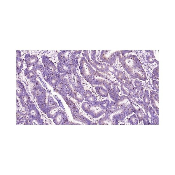
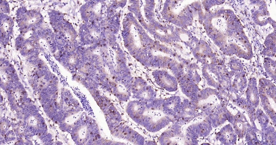
Figure 1. Paraformaldehyde-fixed, paraffin embedded Human Colon Cancer; Antigen retrieval by boiling in sodium citrate buffer (pH6.0) for 15 min; Antibody incubation with CD8 Monoclonal Antibody, Unconjugated(SLA1209M) at 1:300 overnight at 4°C, followed by conjugation to the SP Kit (Rabbit, SP-0023)and DAB (C-0010) staining.
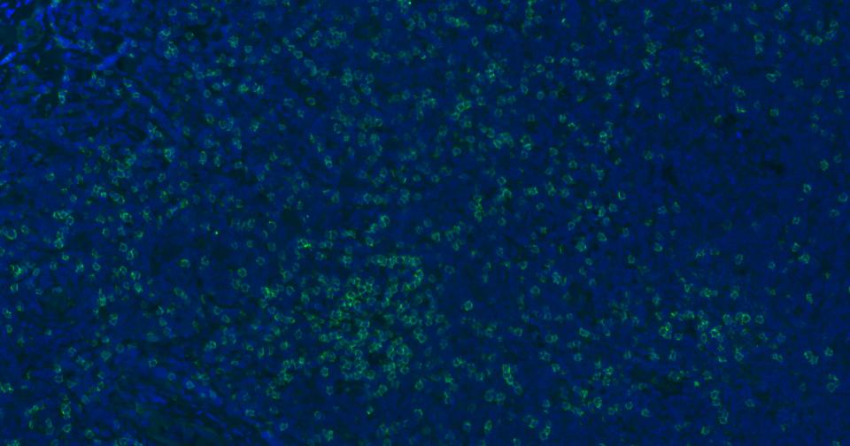
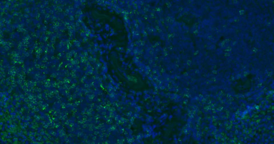
Figure 2. Paraformaldehyde-fixed, paraffin embedded Human Tonsil; Antigen retrieval by boiling in sodium citrate buffer (pH6.0) for 15 min; Antibody incubation with CD8 Polyclonal Antibody, Unconjugated (SLA1209M) at 1:200 overnight at 4°C. Followed by conjugated Goat Anti-Rabbit IgG antibody (green, SL0295G-BF488), DAPI (blue, C02-04002) was used to stain the cell nuclei.
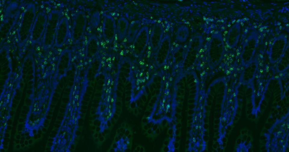
Figure 3. Paraformaldehyde-fixed, paraffin embedded Human Small Intestine; Antigen retrieval by boiling in sodium citrate buffer (pH6.0) for 15 min; Antibody incubation with CD8 Polyclonal Antibody, Unconjugated (SLA1209M) at 1:200 overnight at 4°C. Followed by conjugated Goat Anti-Rabbit IgG antibody (green, SL0295G-BF488), DAPI (blue, C02-04002) was used to stain the cell nuclei.
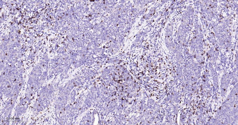
Figure 4. Paraformaldehyde-fixed, paraffin embedded Human Lung squamous cell Cancer; Antigen retrieval by boiling in sodium citrate buffer (pH6.0) for 15 min; The section was incubated with CD8 Monoclonal Antibody, Unconjugated (SLA1209M) at 1:800 overnight at 4°C, followed by conjugation to the SL0295G-HRP and DAB (C-0010) staining.
Figure 5. Paraformaldehyde-fixed, paraffin embedded Human Gastric Cancer; Antigen retrieval by boiling in sodium citrate buffer (pH6.0) for 15 min; The section was incubated with CD8 Monoclonal Antibody, Unconjugated (SLA1209M) at 1:800 overnight at 4°C, followed by conjugation to the SL0295G-HRP and DAB (C-0010) staining.
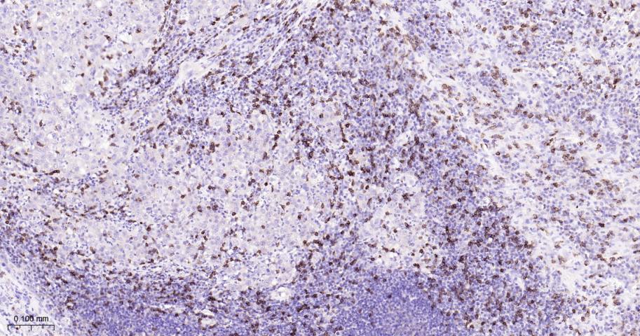
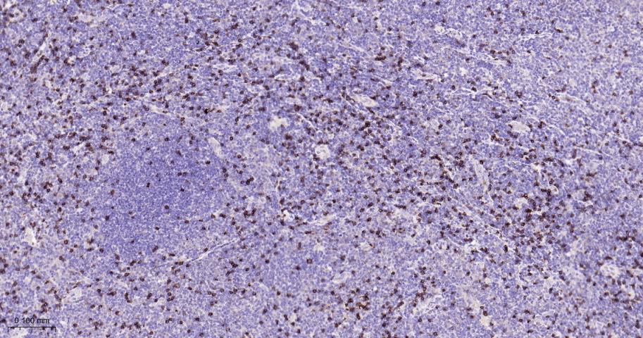
Figure 6. Paraformaldehyde-fixed, paraffin embedded Human Tonsil; Antigen retrieval by boiling in sodium citrate buffer (pH6.0) for 15 min; The section was incubated with CD8 Monoclonal Antibody, Unconjugated (SLA1209M) at 1:800 overnight at 4°C, followed by conjugation to the SL0295G-HRP and DAB (C-0010) staining.
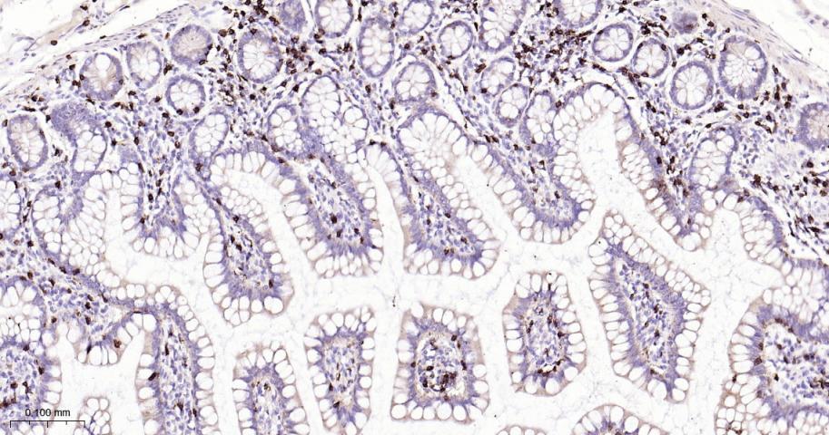
Figure 7. Paraformaldehyde-fixed, paraffin embedded Human Small Intestine; Antigen retrieval by boiling in sodium citrate buffer (pH6.0) for 15 min; The section was incubated with CD8 Monoclonal Antibody, Unconjugated (SLA1209M) at 1:800 overnight at 4°C, followed by conjugation to the SL0295G-HRP and DAB (C-0010) staining.
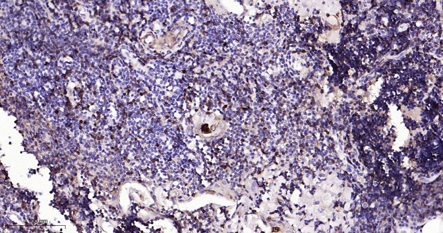
Figure 8. Paraformaldehyde-fixed, paraffin embedded Human Thymus; Antigen retrieval by boiling in sodium citrate buffer (pH6.0) for 15 min; The section was incubated with CD8 Monoclonal Antibody, Unconjugated (SLA1209M) at 1:800 overnight at 4°C, followed by conjugation to the SL0295G-HRP and DAB (C-0010) staining.
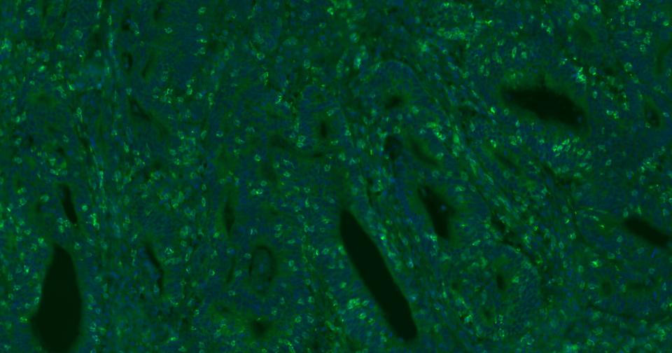
Figure 9. Paraformaldehyde-fixed, paraffin embedded Human Colon Cancer; Antigen retrieval by boiling in sodium citrate buffer (pH6.0) for 15 min; Antibody incubation with CD8 Polyclonal Antibody, Unconjugated (SLA1209M) at 1:200 overnight at 4°C. Followed by conjugated Goat Anti-Rabbit IgG antibody (green, SL0295G-BF488), DAPI (blue, C02-04002) was used to stain the cell nuclei.
Figure 10. Paraformaldehyde-fixed, paraffin embedded Human Spleen; Antigen retrieval by boiling in sodium citrate buffer (pH6.0) for 15 min; Antibody incubation with CD8 Polyclonal Antibody, Unconjugated (SLA1209M) at 1:200 overnight at 4°C. Followed by conjugated Goat Anti-Rabbit IgG antibody (green, SL0295G-BF488), DAPI (blue, C02-04002) was used to stain the cell nuclei.
FOR RESEARCH USE ONLY. Not for use in diagnostic or therapeutic procedures.
Partial purchase records(bought amounts latest0)
User Comment(Total0User Comment Num)
- No comment


 +86 571 56623320

 SUNLONG BIOTECH


