Molecular Weight: Predicted, 14 kDa, observed, 14 kDa
Clonality: Rabbit monoclonal antibody
Clone ID: 23GB1960
Species Reactivity: Human, Mouse, Rat
Applications Tested: Western Blotting (WB), Immunocytochemistry (IC)
Immunogen
A synthesized peptide derived from human MAP1LC3A
Isotype
Rabbit IgG
Storage Buffer
Supplied in PBS (pH 7.4) containing 50% glycerol, and 0.02% sodium azide.
Storage
Store at -20 °C for one year.
Recommended Dilutions
Western Blotting (WB): 1:1,000-1:20,000
Immunocytochemistry (IC): 1:1,000
Protocols
For general and specific antibody protocols please visit our website. Read all instructions before using this product.
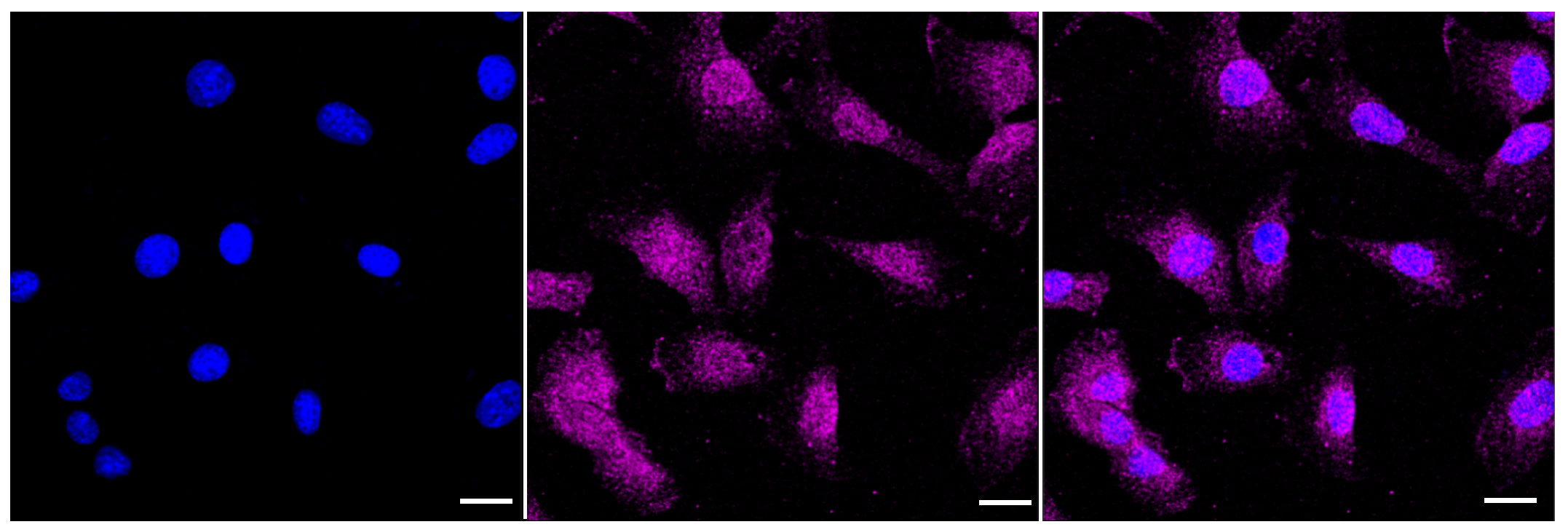
Immunocytochemical staining of C2C12 cells with MAP1LC3A antibody 1:1,000. Nuclei were stained blue with DAPI; MAP1LC3A was stained magenta with Alexa Fluor® 647. Images were taken using Leica stellaris 5. Protein abundance based on laser Intensity and smart gain: Medium. Scale bar: 20 μm.
Western blotting analysis using anti-MAP1LC3A antibody 1:20,000 and HRP-conjugated goat anti-rabbit secondary antibody 1:20,000 respectively.
Western blotting analysis using anti-MAP1LC3A antibody 1:20,000 and HRP-conjugated goat anti-rabbit secondary antibody 1:20,000 respectively.
![[KD-Validated] Anti-MAP1LC3A Rabbit Monoclonal Antibody](images/default_images/52.jpg)
 Immunocytochemical staining of C2C12 cells with MAP1LC3A antibody 1:1,000. Nuclei were stained blue with DAPI; MAP1LC3A was stained magenta with Alexa Fluor® 647. Images were taken using Leica stellaris 5. Protein abundance based on laser Intensity and smart gain: Medium. Scale bar: 20 μm.
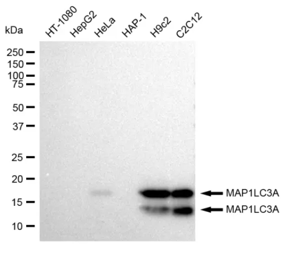 Western blotting analysis using anti-MAP1LC3A antibody 1:20,000 and HRP-conjugated goat anti-rabbit secondary antibody 1:20,000 respectively.
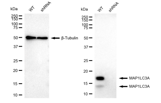 Western blotting analysis using anti-MAP1LC3A antibody 1:20,000 and HRP-conjugated goat anti-rabbit secondary antibody 1:20,000 respectively.

 +86 571 56623320
 [email protected]
 SUNLONG BIOTECH

