For general and specific antibody protocols please visit our website. Read all instructions before using this product.
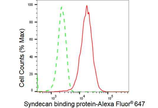
Flow cytometric analysis of Syndecan binding protein expression in HepG2 cells using Syndecan binding protein antibody 1:2,000. Green, isotype control; red, Syndecan binding protein.
Immunocytochemical staining of HepG2 cells with Syndecan binding protein antibody 1:1,000. Nuclei were stained blue with DAPI; Syndecan binding protein was stained magenta with Alexa Fluor® 647. Images were taken using Leica stellaris 5. Protein abundance based on laser Intensity and smart gain: Medium. Scale bar: 20 μm.
Western blotting analysis using anti-Syndecan binding protein antibody 1:5,000 and HRP-conjugated goat anti-rabbit secondary antibody 1:20,000 respectively.
Western blotting analysis using anti-Syndecan binding protein antibody 1:5,000 and HRP-conjugated goat anti-rabbit secondary antibody 1:20,000 respectively.
![[KD-Validated] Anti-Syndecan binding protein Rabbit Monoclonal Antibody](images/default_images/52.jpg)
 Flow cytometric analysis of Syndecan binding protein expression in HepG2 cells using Syndecan binding protein antibody 1:2,000. Green, isotype control; red, Syndecan binding protein.
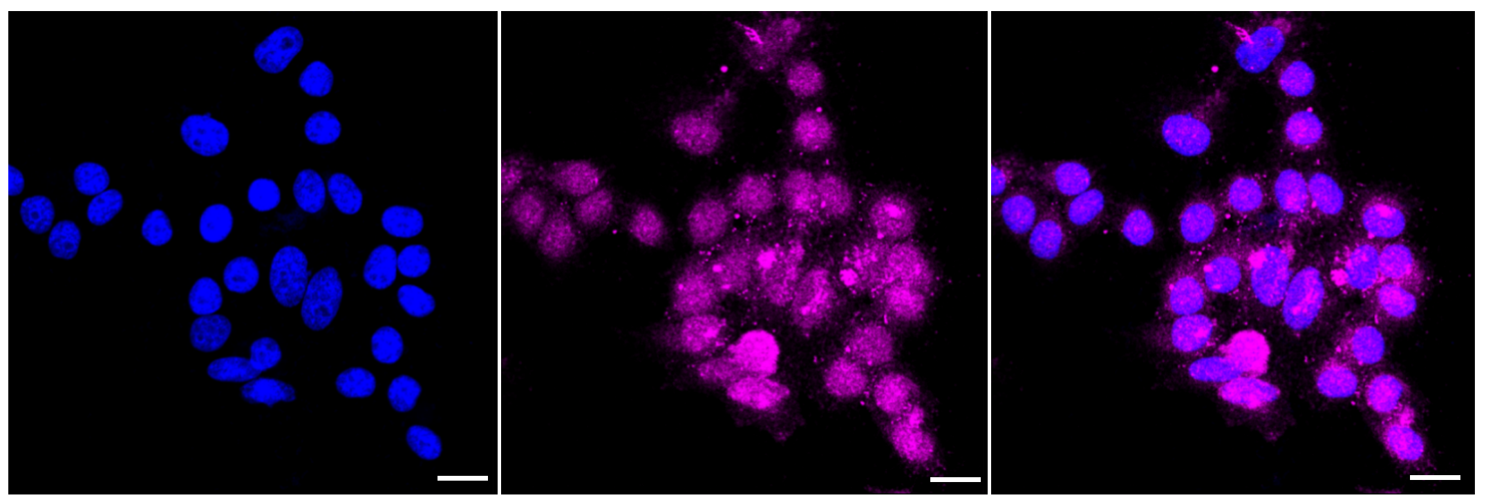 Immunocytochemical staining of HepG2 cells with Syndecan binding protein antibody 1:1,000. Nuclei were stained blue with DAPI; Syndecan binding protein was stained magenta with Alexa Fluor® 647. Images were taken using Leica stellaris 5. Protein abundance based on laser Intensity and smart gain: Medium. Scale bar: 20 μm.
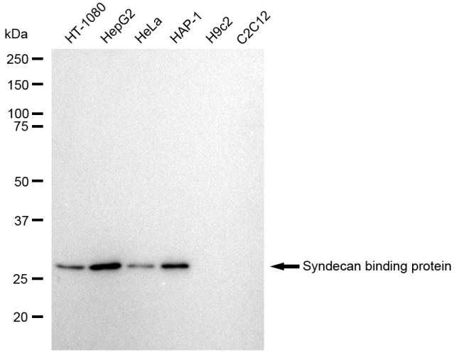 Western blotting analysis using anti-Syndecan binding protein antibody 1:5,000 and HRP-conjugated goat anti-rabbit secondary antibody 1:20,000 respectively.
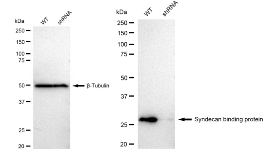 Western blotting analysis using anti-Syndecan binding protein antibody 1:5,000 and HRP-conjugated goat anti-rabbit secondary antibody 1:20,000 respectively.

 +86 571 56623320
 [email protected]
 SUNLONG BIOTECH

