Rabbit Anti-CD44 antibody
LHR; BA-1; CD 44; CD44 antigen; CD44 molecule; CD44_HUMAN; CDw44; Cell surface glycoprotein CD44; chondroitin sulfate proteoglycan 8; ECMR-III; Epican; Extracellular matrix receptor III; GP90 lymphocyte homing/adhesion receptor; hematopoietic cell E- and
View History [Clear]
Details
Product Name CD44 Chinese Name CD44抗体 Alias LHR; BA-1; CD 44; CD44 antigen; CD44 molecule; CD44_HUMAN; CDw44; Cell surface glycoprotein CD44; chondroitin sulfate proteoglycan 8; ECMR-III; Epican; Extracellular matrix receptor III; GP90 lymphocyte homing/adhesion receptor; hematopoietic cell E- and L-selectin ligand; Heparan sulfate proteoglycan; Hermes antigen; homing function and Indian blood group system; HSA; HUTCH-I; HUTCH1; Hyaluronate receptor; MDU2; MDU3; MIC4; MUTCH1; PGP-1; PGP1; Phagocytic glycoprotein 1; Phagocytic glycoprotein I; CD44 antigen isoform 1 precursor. literatures Research Area Tumour immunology Stem cells Cell Surface Molecule glycoprotein Cell type markers t-lymphocyte Immunogen Species Rabbit Clonality Polyclonal React Species Human(predicted:Mouse,Rat,Dog,Pig,Cow,Horse,Rabbit) Applications WB=1:500-2000 (Paraffin sections need antigen repair)
not yet tested in other applications.
optimal dilutions/concentrations should be determined by the end user.Theoretical molecular weight 85kDa Detection molecular weight 80-95kDa Cellular localization The cell membrane Form Liquid Concentration 1mg/ml immunogen KLH conjugated synthetic peptide derived from human CD44: 701-742/742 <Cytoplasmic> Lsotype IgG Purification affinity purified by Protein A Buffer Solution Human(predicted:Mouse,Rat,Dog,Pig,Cow,Horse,Rabbit)1M TBS(pH7.4) with 1% BSA, Human(predicted:Mouse,Rat,Dog,Pig,Cow,Horse,Rabbit)3% Proclin300 and 50% Glycerol. Storage Shipped at 4℃. Store at -20 °C for one year. Avoid repeated freeze/thaw cycles. Attention This product as supplied is intended for research use only, not for use in human, therapeutic or diagnostic applications. PubMed PubMed Product Detail The protein encoded by this gene is a cell-surface glycoprotein involved in cell-cell interactions, cell adhesion and migration. It is a receptor for hyaluronic acid (HA) and can also interact with other ligands, such as osteopontin, collagens, and matrix metalloproteinases (MMPs). This protein participates in a wide variety of cellular functions including lymphocyte activation, recirculation and homing, hematopoiesis, and tumor metastasis. Transcripts for this gene undergo complex alternative splicing that results in many functionally distinct isoforms, however, the full length nature of some of these variants has not been determined. Alternative splicing is the basis for the structural and functional diversity of this protein, and may be related to tumor metastasis. [provided by RefSeq, Jul 2008].
Function:
Receptor for hyaluronic acid (HA). Mediates cell-cell and cell-matrix interactions through its affinity for HA, and possibly also through its affinity for other ligands such as osteopontin, collagens, and matrix metalloproteinases (MMPs). Adhesion with HA plays an important role in cell migration, tumor growth and progression. Also involved in lymphocyte activation, recirculation and homing, and in hematopoiesis. Altered expression or dysfunction causes numerous pathogenic phenotypes. Great protein heterogeneity due to numerous alternative splicing and post-translational modification events.
Subunit:
Interacts with PKN2 (By similarity). Interacts with HA, as well as other glycosaminoglycans, collagen, laminin, and fibronectin via its N-terminal segment. Interacts with ANK, the ERM proteins (VIL2, RDX and MSN), and NF2 via its C-terminal segment.
Subcellular Location:
Membrane; Single-pass type I membrane protein. Note=Colocalizes with actin in membrane protrusions at wounding edges.
Tissue Specificity:
Isoform 10 (epithelial isoform) is expressed by cells of epithelium and highly expressed by carcinomas. Expression is repressed in neuroblastoma cells.
Post-translational modifications:
Proteolytically cleaved in the extracellular matrix by specific proteinases (possibly MMPs) in several cell lines and tumors.
N- and O-glycosylated. O-glycosylation contains more-or-less-sulfated chondroitin sulfate glycans, whose number may affect the accessibility of specific proteinases to their cleavage site(s). It is uncertain if O-glycosylation occurs on Thr-637 or Thr-638.
Phosphorylated; activation of PKC results in the dephosphorylation of Ser-706 (constitutive phosphorylation site), and the phosphorylation of Ser-672.
Similarity:
Contains 1 Link domain.
SWISS:
P16070
Gene ID:
960
Database links:
Entrez Gene: 960 Human
Entrez Gene: 12505 Mouse
Entrez Gene: 100301546 Rabbit
Omim: 107269 Human
SwissProt: P16070 Human
SwissProt: P15379 Mouse
Unigene: 502328 Human
Unigene: 423621 Mouse
Unigene: 1120 Rat
CD44是一个重要的细胞表面粘附分子。它在许多类型的人类细胞上都表达,在很多生理和病理过程中有复杂的作用,例如细胞迁移和在Tumour细胞的生长调控中。CD44传递着诸多的细胞通路,有研究者认为CD44是一个新的信号通路蛋白。CD44介导lymphocyte与vascular endothelial cell包括粘膜组织中高内皮小静脉的粘连,这可能是通过激活其它粘附分子而间接造成的。CD44也是Tumour细胞浸润的重要因素。Product Picture 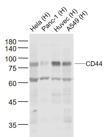 Sample:
Lane 1: Hela (Human) Cell Lysate at 30 ug
Lane 2: Panc-1 (Human) Cell Lysate at 30 ug
Lane 3: Huvec (Human) Cell Lysate at 30 ug
Lane 4: A549 (Human) Cell Lysate at 30 ug
Primary: Anti-CD44 (SL0521R) at 1/1000 dilution
Secondary: IRDye800CW Goat Anti-Rabbit IgG at 1/20000 dilution
Predicted band size: 83 kD
Observed band size: 83 kD
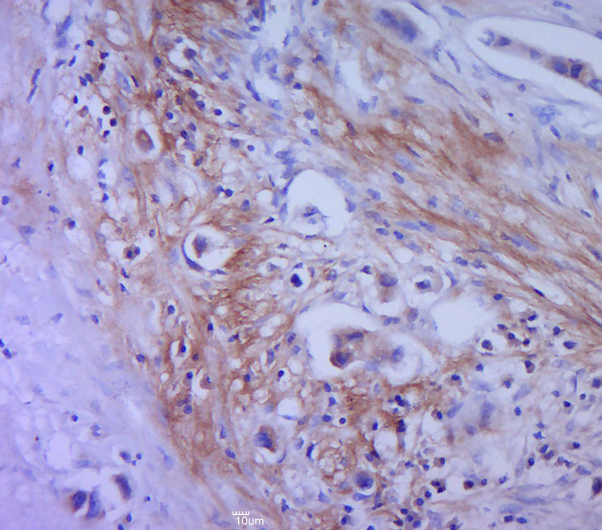 Paraformaldehyde-fixed, paraffin embedded (human cervical cancer); Antigen retrieval by boiling in sodium citrate buffer (pH6.0) for 15min; Block endogenous peroxidase by 3% hydrogen peroxide for 20 minutes; Blocking buffer (normal goat serum) at 37°C for 30min; Antibody incubation with (CD44) Polyclonal Antibody, Unconjugated (SL0521R) at 1:400 overnight at 4°C, followed by a conjugated secondary (sp-0023) for 20 minutes and DAB staining.
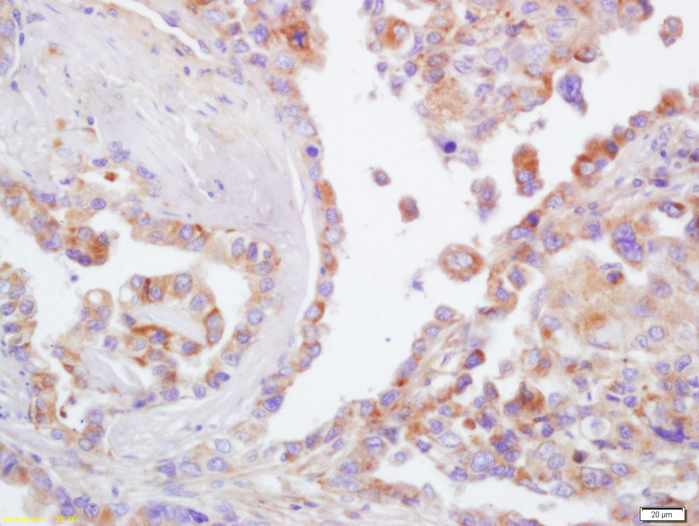 Tissue/cell: human lung carcinoma; 4% Paraformaldehyde-fixed and paraffin-embedded;
Antigen retrieval: citrate buffer ( Human(predicted:Mouse,Rat,Dog,Pig,Cow,Horse,Rabbit)1M, pH 6.0 ), Boiling bathing for 15min; Block endogenous peroxidase by 3% Hydrogen peroxide for 30min; Blocking buffer (normal goat serum,C-0005) at 37℃ for 20 min;
Incubation: Anti-CD44 Polyclonal Antibody, Unconjugated(SL0521R) 1:200, overnight at 4°C, followed by conjugation to the secondary antibody(SP-0023) and DAB(C-0010) staining
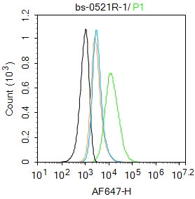 Blank control: HELA.
Primary Antibody (green line): Rabbit Anti-CD44 antibody (SL0521R)
Dilution: 1μg /10^6 cells;
Isotype Control Antibody (orange line): Rabbit IgG .
Secondary Antibody : Goat anti-rabbit IgG-AF647
Dilution: 1μg /test.
Protocol
The cells were incubated in 5%BSA to block non-specific protein-protein interactions for 30 min at room temperature .Cells stained with Primary Antibody for 30 min at room temperature. The secondary antibody used for 40 min at room temperature. Acquisition of 20,000 events was performed.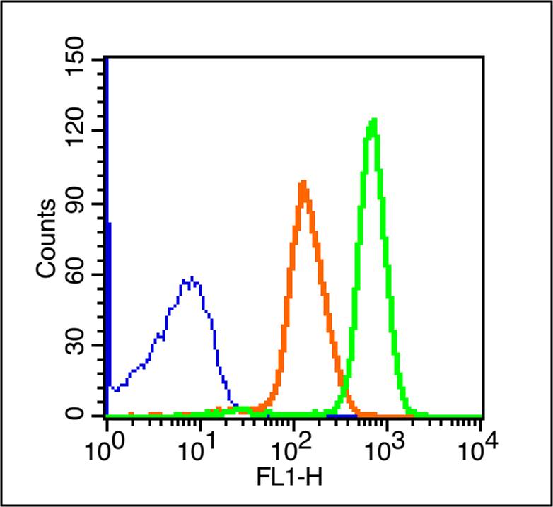 Blank control (blue line): Rabbit spleen cells(blue).
Primary Antibody (green line): Rabbit Anti-CD44/FITC Conjugated antibody (SL0521R-FITC)
Dilution: 1μg /10^6 cells;
Isotype Control Antibody (orange line): Rabbit IgG-FITC.
Protocol
The cells were fixed with 70% ice-cold methanol overnight at 4℃ . The cells were then incubated in 1 X PBS/2%BSA/10% goat serum to block non-specific protein-protein interactions followed by the antibody for 15 min at room temperature. Cells stained with Primary Antibody for 30 min at room temperature.Acquisition of 20,000 events was performed.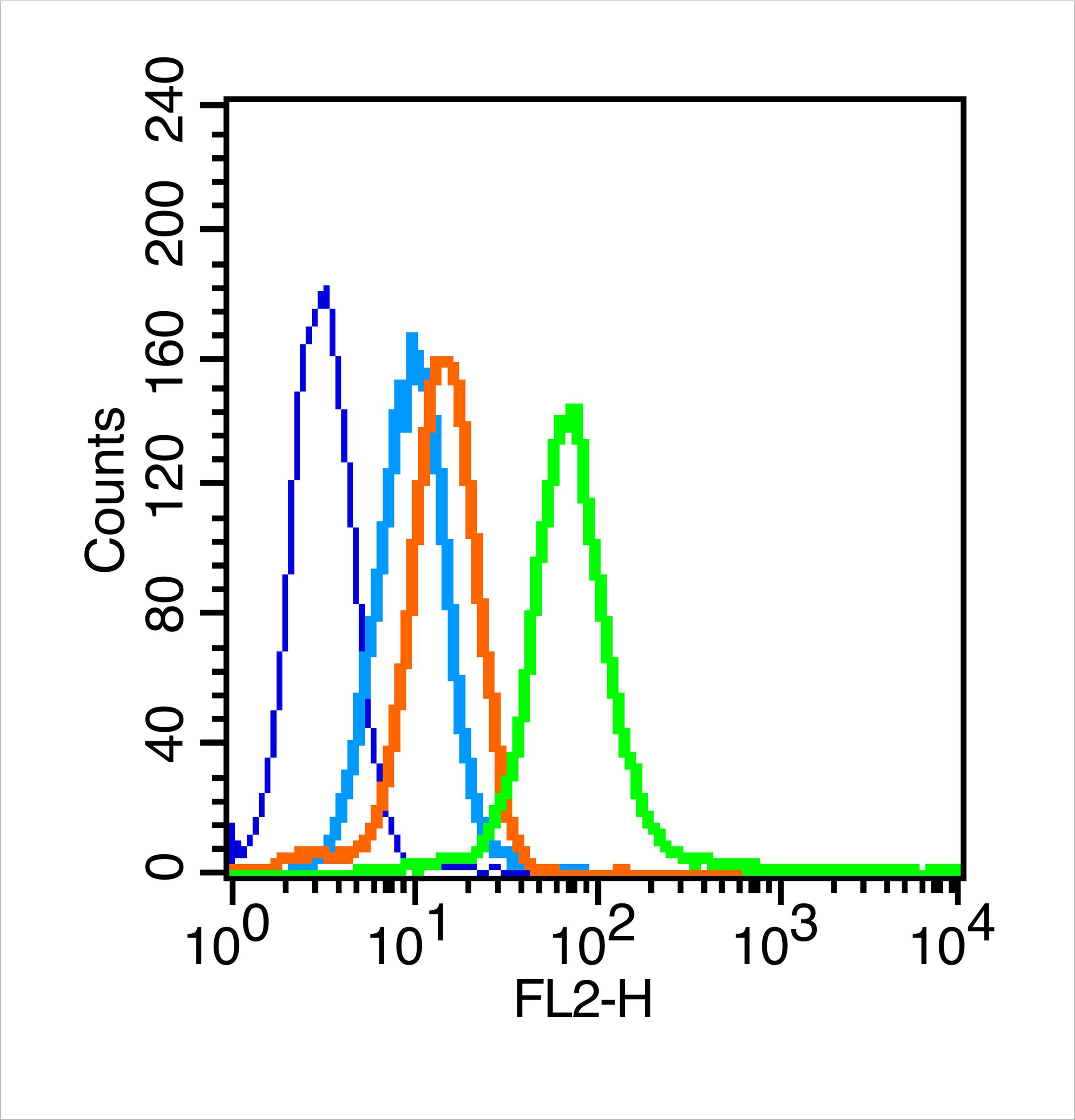 Blank control: U251 (blue).
Primary Antibody:Rabbit Anti-CD44 antibody (SL0521R,Green); Dilution: 3μg in 100 μL 1X PBS containing 0.5% BSA;
Isotype Control Antibody: Rabbit IgG(orange), used under the same conditions;
Secondary Antibody: Goat anti-rabbit IgG-FITC(white blue), Dilution: 1:200 in 1 X PBS containing 0.5% BSA.
Protocol
The cells were fixed with Ice-cold 70% ethanol overnight at 4 °C. Primary were incubated for 30 min at room temperature, followed by 1 X PBS containing 0.5% BSA + 1 0% goat serum (15 min) to block non-specific protein-protein interactions. Then the Goat Anti-rabbit IgG/FITC antibody was added into the blocking buffer mentioned above to react with the primary antibody at 1/200 dilution for 40 min on ice. Acquisition of 20,000 events was performed.
Cartpieces
Totalgoods,subtotals:¥Checkout
Partial purchase records(bought amounts latest0)
No one bought this product
User Comment(Total0User Comment Num)
- No comment


 +86 571 56623320
 SUNLONG BIOTECH

