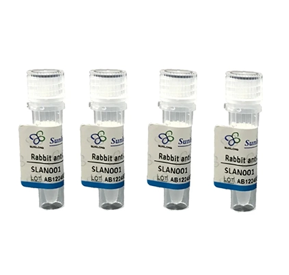
Details
Product Name MSH2 Chinese Name 错配修复蛋白2Recombinant rabbit monoclonal anti Alias MSH-2; BAT26; DNA mismatch repair protein Msh2; FCC1; COCA1; HNPCC; LCFS2; HNPCC1; BAT26; COCA 1; COCA1; DNA mismatch repair protein Msh2; FCC 1; FCC1; hMSH2; HNPCC 1; HNPCC; HNPCC1; LCFS2; MSH 2; MSH2_HUMAN; MutS homolog 2; MutS homolog 2 colon cancer nonpolyposis type 1; MutS protein homolog 2. Research Area Tumour Cell biology Epigenetics Immunogen Species Rabbit Clonality Monoclonal Clone NO. B4F3 React Species Human, Applications WB=1:1000-5000,IHC-P=1:200-1000,IHC-F=1:200-1000,IF=1:200-1000,Flow-Cyt=1ug/Test
not yet tested in other applications.
optimal dilutions/concentrations should be determined by the end user.Theoretical molecular weight 105kDa Cellular localization The nucleus Form Liquid Concentration 1mg/ml immunogen KLH conjugated synthetic peptide derived from human MSH2 Lsotype IgG Purification affinity purified by Protein A Buffer Solution 1M TBS(pH7.4) with 1% BSA, 3% Proclin300 and 50% Glycerol. Storage Shipped at 4℃. Store at -20 °C for one year. Avoid repeated freeze/thaw cycles. Attention This product as supplied is intended for research use only, not for use in human, therapeutic or diagnostic applications. PubMed PubMed Product Detail Component of the post-replicative DNA mismatch repair system (MMR). Forms two different heterodimers: MutS alpha (MSH2-MSH6 heterodimer) and MutS beta (MSH2-MSH3 heterodimer) which binds to DNA mismatches thereby initiating DNA repair. When bound, heterodimers bend the DNA helix and shields approximately 20 base pairs. MutS alpha recognizes single base mismatches and dinucleotide insertion-deletion loops (IDL) in the DNA. MutS beta recognizes larger insertion-deletion loops up to 13 nucleotides long. After mismatch binding, MutS alpha or beta forms a ternary complex with the MutL alpha heterodimer, which is thought to be responsible for directing the downstream MMR events, including strand discrimination, excision, and resynthesis. ATP binding and hydrolysis play a pivotal role in mismatch repair functions. The ATPase activity associated with MutS alpha regulates binding similar to a molecular switch: mismatched DNA provokes ADP-->ATP exchange, resulting in a discernible conformational transition that converts MutS alpha into a sliding clamp capable of hydrolysis-independent diffusion along the DNA backbone. This transition is crucial for mismatch repair. MutS alpha may also play a role in DNA homologous recombination repair. In melanocytes may modulate both UV-B-induced cell cycle regulation and apoptosis.
Function:
Component of the post-replicative DNA mismatch repair system (MMR). Forms two different heterodimers: MutS alpha (MSH2-MSH6 heterodimer) and MutS beta (MSH2-MSH3 heterodimer) which binds to DNA mismatches thereby initiating DNA repair. When bound, heterodimers bend the DNA helix and shields approximately 20 base pairs. MutS alpha recognizes single base mismatches and dinucleotide insertion-deletion loops (IDL) in the DNA. MutS beta recognizes larger insertion-deletion loops up to 13 nucleotides long. After mismatch binding, MutS alpha or beta forms a ternary complex with the MutL alpha heterodimer, which is thought to be responsible for directing the downstream MMR events, including strand discrimination, excision, and resynthesis. ATP binding and hydrolysis play a pivotal role in mismatch repair functions. The ATPase activity associated with MutS alpha regulates binding similar to a molecular switch: mismatched DNA provokes ADP-->ATP exchange, resulting in a discernible conformational transition that converts MutS alpha into a sliding clamp capable of hydrolysis-independent diffusion along the DNA backbone. This transition is crucial for mismatch repair. MutS alpha may also play a role in DNA homologous recombination repair. In melanocytes may modulate both UV-B-induced cell cycle regulation and apoptosis.
Subunit:
Heterodimer consisting of MSH2-MSH6 (MutS alpha) or MSH2-MSH3 (MutS beta). Both heterodimer form a ternary complex with MutL alpha (MLH1-PMS1). Interacts with EXO1. Part of the BRCA1-associated genome surveillance complex (BASC), which contains BRCA1, MSH2, MSH6, MLH1, ATM, BLM, PMS2 and the RAD50-MRE11-NBS1 protein complex. This association could be a dynamic process changing throughout the cell cycle and within subnuclear domains. Interacts with ATR. Interacts with SLX4/BTBD12; this interaction is direct and links MutS beta to SLX4, a subunit of different structure-specific endonucleases. Interacts with SMARCAD1.
Subcellular Location:
Nucleus.
Tissue Specificity:
Ubiquitously expressed.
Post-translational modifications:
Phosphorylated by PRKCZ, which may prevent MutS alpha degradation by the ubiquitin-proteasome pathway. Phosphorylated upon DNA damage, probably by ATM or ATR.
DISEASE:
Hereditary non-polyposis colorectal cancer 1 (HNPCC1) [MIM:120435]: An autosomal dominant disease associated with marked increase in cancer susceptibility. It is characterized by a familial predisposition to early-onset colorectal carcinoma (CRC) and extra-colonic tumors of the gastrointestinal, urological and female reproductive tracts. HNPCC is reported to be the most common form of inherited colorectal cancer in the Western world. Clinically, HNPCC is often divided into two subgroups. Type I is characterized by hereditary predisposition to colorectal cancer, a young age of onset, and carcinoma observed in the proximal colon. Type II is characterized by increased risk for cancers in certain tissues such as the uterus, ovary, breast, stomach, small intestine, skin, and larynx in addition to the colon. Diagnosis of classical HNPCC is based on the Amsterdam criteria: 3 or more relatives affected by colorectal cancer, one a first degree relative of the other two; 2 or more generation affected; 1 or more colorectal cancers presenting before 50 years of age; exclusion of hereditary polyposis syndromes. The term 'suspected HNPCC' or 'incomplete HNPCC' can be used to describe families who do not or only partially fulfill the Amsterdam criteria, but in whom a genetic basis for colon cancer is strongly suspected. Note=The disease is caused by mutations affecting the gene represented in this entry.
Muir-Torre syndrome (MRTES) [MIM:158320]: Rare autosomal dominant disorder characterized by sebaceous neoplasms and visceral malignancy. Note=The disease is caused by mutations affecting the gene represented in this entry.
Endometrial cancer (ENDMC) [MIM:608089]: A malignancy of endometrium, the mucous lining of the uterus. Most endometrial cancers are adenocarcinomas, cancers that begin in cells that make and release mucus and other fluids. Note=Disease susceptibility is associated with variations affecting the gene represented in this entry.
Similarity:
Belongs to the DNA mismatch repair MutS family.
SWISS:
P43246
Gene ID:
4436
Database links:Entrez Gene: 4436 Human
Entrez Gene: 17685 Mouse
Omim: 120435 Human
SwissProt: P43246 Human
SwissProt: P43247 Mouse
Unigene: 597656 Human
Unigene: 4619 Mouse
Unigene: 471165 Mouse
Unigene: 3174 Rat
MSH2 is one of the nucleotide mismatch repair enzymes, and its mutations are associated with the development of various Tumours. It is mainly used in research on congenital colon cancer, sporadic colon cancer, and some digestive system Tumours. The DNA mismatch repair system refers to a group of enzymes that specifically repair DNA base mismatches, ensuring the high fidelity of DNA genetic material. It is composed of the MLH1, MSH2, PMS1, PMS2, MSH6, and MSH3 genes. If mutations or inactivation occur in these genes, it will cause defects in cellular mismatch repair function, generate genetic instability, and lead to Tumour susceptibility. MSH2 gene mutations occur in sporadic colon cancer.
Both hMSH-2 and hMLH1 are associated with the development of Tumours.Product Picture
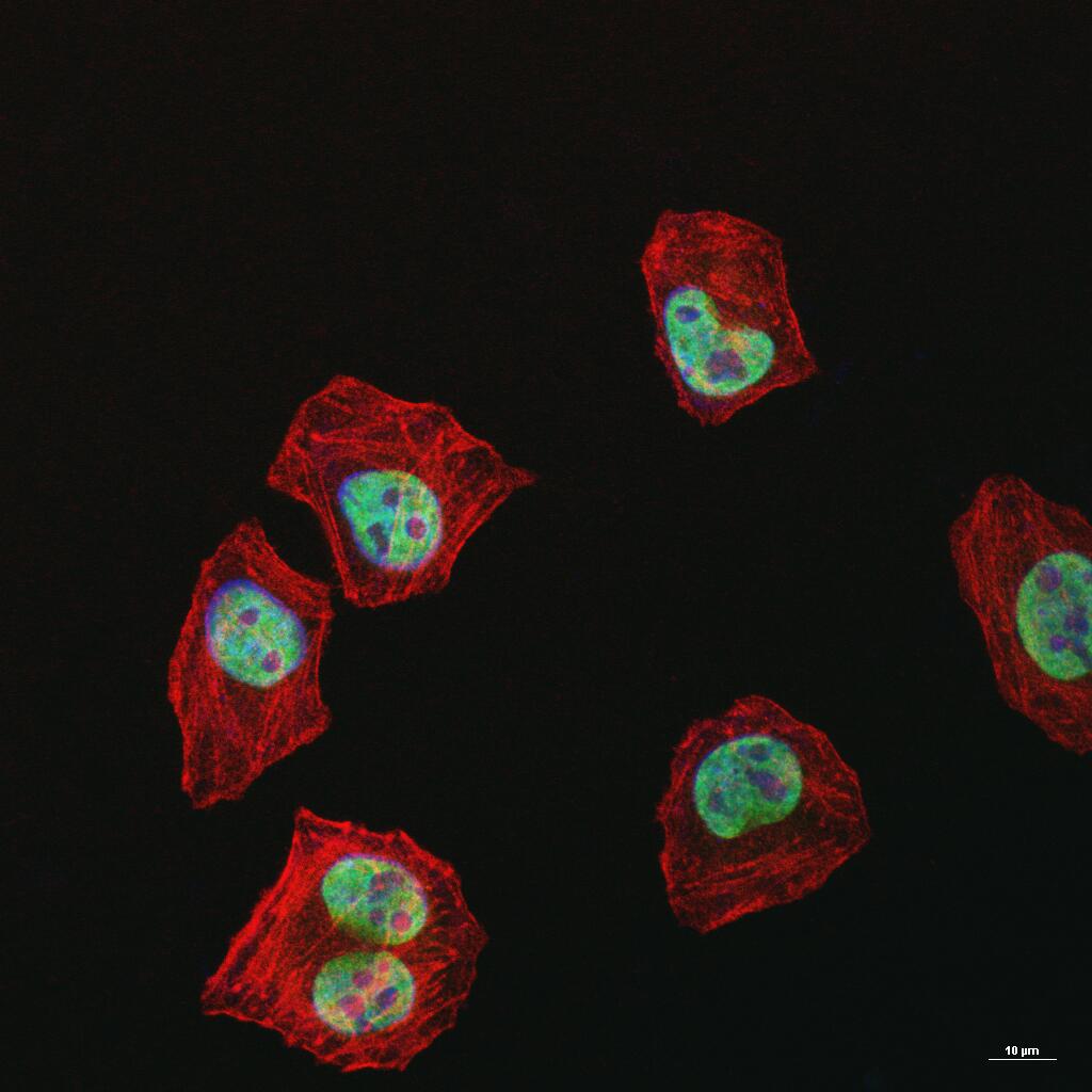
Figure 1. 4% Paraformaldehyde-fixed HeLa (H) cell; Triton X-100 at r.t. for 20 min; Antibody incubation with (MSH2) monoclonal Antibody, unconjugated (SLM-60222R) 1:100, 90 min at 37°C; followed by BF488 conjugated Goat Anti-Rabbit IgG antibody (green, SL60295G-BF488) at 37°C for 90 min, then stained with Phalloidin/BF555 for 30 min at room temperature; DAPI (blue, C02-04002) was used to stain the cell nuclei
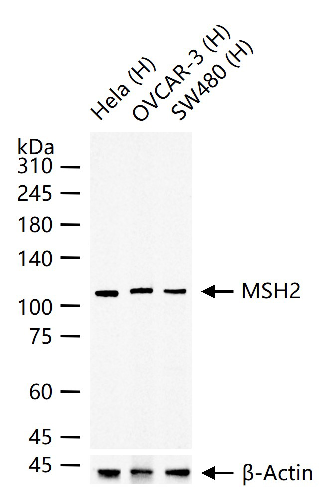
Figure 2. 25 ug total protein per lane of various lysates (see on figure) probed with MSH2 monoclonal antibody, unconjugated (SLM-60222R) at 1:1000 dilution and 4°C overnight incubation. Followed by conjugated secondary antibody incubation at r.t. for 60 min
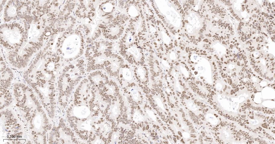
Figure 3. Paraformaldehyde-fixed, paraffin embedded Human Colon Cancer; Antigen retrieval by boiling in sodium citrate buffer (pH6.0) for 15 min; Antibody incubation with MSH2 Monoclonal Antibody, Unconjugated(SLM-60222R) at 1:200 overnight at 4°C, followed by conjugation to the SP Kit (Rabbit, SP-0023) and DAB (C-0010) staining
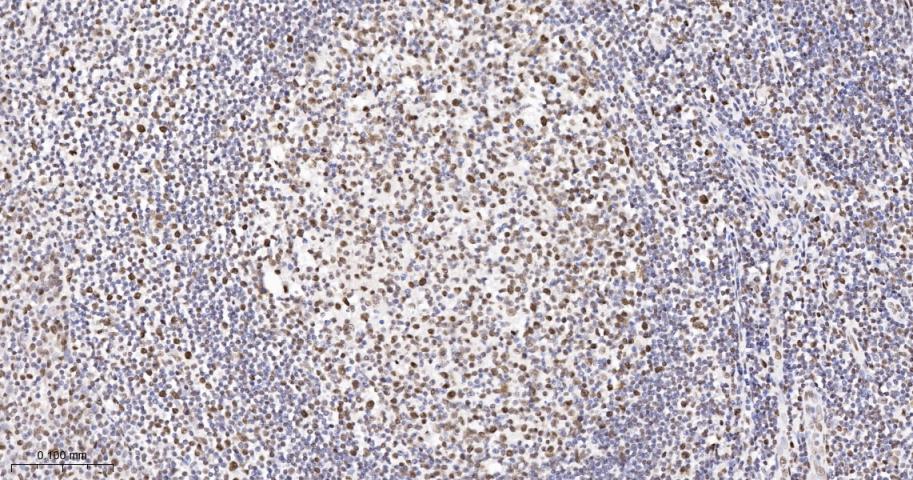
Figure 4. Paraformaldehyde-fixed, paraffin embedded Human Tonsil; Antigen retrieval by boiling in sodium citrate buffer (pH6.0) for 15 min; Antibody incubation with MSH2 Monoclonal Antibody, Unconjugated(SLM-60222R) at 1:200 overnight at 4°C, followed by conjugation to the SP Kit (Rabbit, SP-0023) and DAB (C-0010) staining
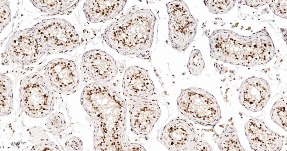
Figure 5. Paraformaldehyde-fixed, paraffin embedded Human Testicles; Antigen retrieval by boiling in sodium citrate buffer (pH6.0) for 15 min; Antibody incubation with MSH2 Monoclonal Antibody, Unconjugated(SLM-60222R) at 1:200 overnight at 4°C, followed by conjugation to the SP Kit (Rabbit, SP-0023) and DAB (C-0010) staining
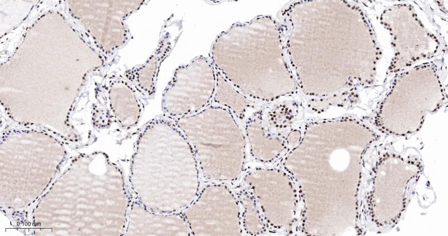
Figure 6. Paraformaldehyde-fixed, paraffin embedded Human Thyroid Gland; Antigen retrieval by boiling in sodium citrate buffer (pH6.0) for 15 min; Antibody incubation with MSH2 Monoclonal Antibody, Unconjugated(SLM-60222R) at 1:200 overnight at 4°C, followed by conjugation to the SP Kit (Rabbit, SP-0023) and DAB (C-0010) staining
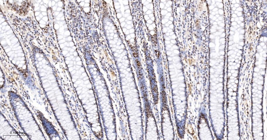
Figure 7. Paraformaldehyde-fixed, paraffin embedded Human Colon; Antigen retrieval by boiling in sodium citrate buffer (pH6.0) for 15 min; Antibody incubation with MSH2 Monoclonal Antibody, Unconjugated(SLM-60222R) at 1:200 overnight at 4°C, followed by conjugation to the SP Kit (Rabbit, SP-0023) and DAB (C-0010) staining
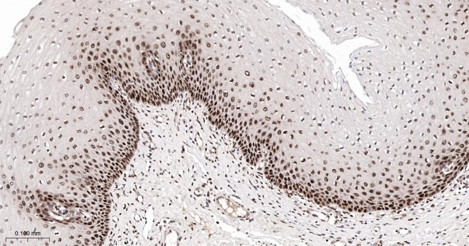
Figure 8. Paraformaldehyde-fixed, paraffin embedded Human Esophagus; Antigen retrieval by boiling in sodium citrate buffer (pH6.0) for 15 min; Antibody incubation with MSH2 Monoclonal Antibody, Unconjugated(SLM-60222R) at 1:200 overnight at 4°C, followed by conjugation to the SP Kit (Rabbit, SP-0023) and DAB (C-0010) staining
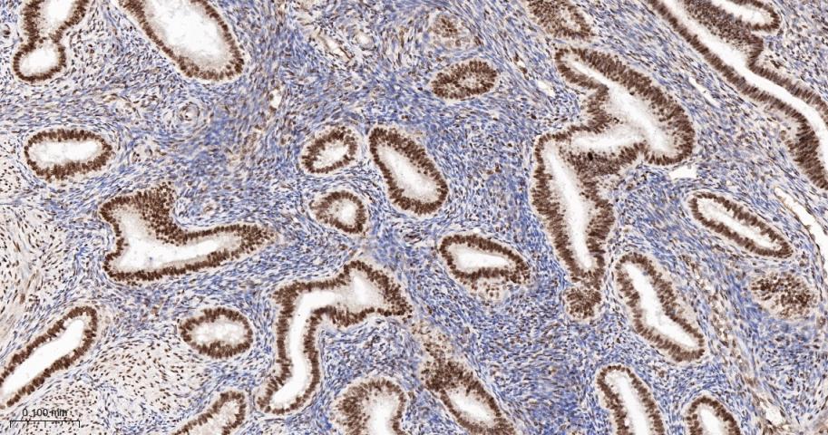
Figure 9. Paraformaldehyde-fixed, paraffin embedded Human Endometrial; Antigen retrieval by boiling in sodium citrate buffer (pH6.0) for 15 min; Antibody incubation with MSH2 Monoclonal Antibody, Unconjugated(SLM-60222R) at 1:200 overnight at 4°C, followed by conjugation to the SP Kit (Rabbit, SP-0023) and DAB (C-0010) staining
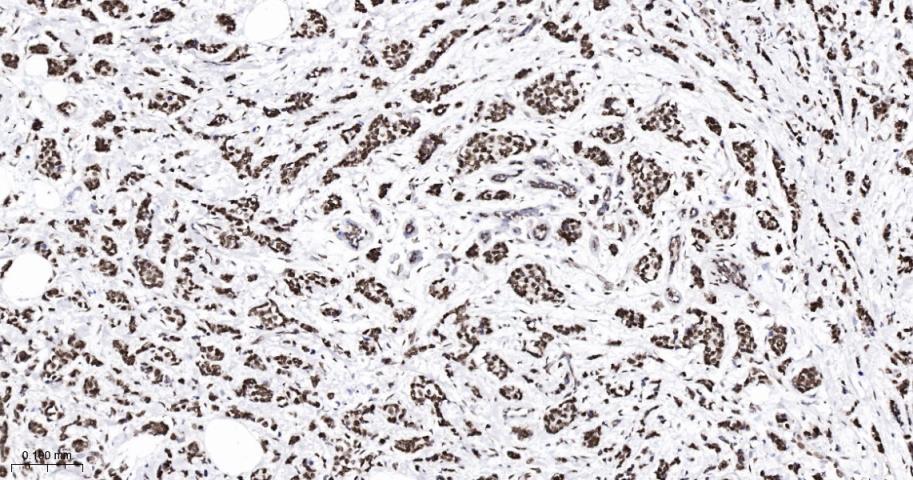
Figure 10. Paraformaldehyde-fixed, paraffin embedded Human Breast Cancer; Antigen retrieval by boiling in sodium citrate buffer (pH6.0) for 15 min; Antibody incubation with MSH2 Monoclonal Antibody, Unconjugated(SLM-60222R) at 1:200 overnight at 4°C, followed by conjugation to the SP Kit (Rabbit, SP-0023) and DAB (C-0010) staining
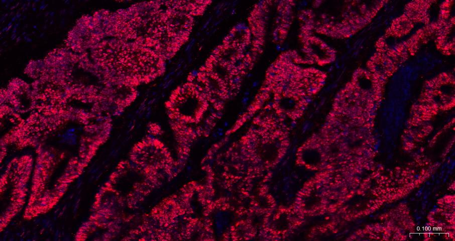
Figure 11. Paraformaldehyde-fixed, paraffin embedded Human Colon Cancer; Antigen retrieval by boiling in sodium citrate buffer (pH6.0) for 15 min; The section was incubated with MSH2 Antibody, Unconjugated (SLM-60222R) at 1:200 overnight at 4°C. Followed by conjugated Goat Anti-Rabbit IgG antibody (Red, SL0295G-BF594), DAPI (blue, C02-04002) was used to stain the cell nuclei
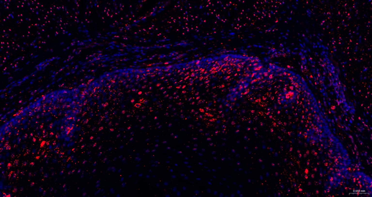
Figure 12. Paraformaldehyde-fixed, paraffin embedded Human Esophagus; Antigen retrieval by boiling in sodium citrate buffer (pH6.0) for 15 min; The section was incubated with MSH2 Antibody, Unconjugated (SLM-60222R) at 1:200 overnight at 4°C. Followed by conjugated Goat Anti-Rabbit IgG antibody (Red, SL0295G-BF594), DAPI (blue, C02-04002) was used to stain the cell nuclei
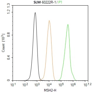
Figure 13. The Hela (H) cells were fixed with 4% PFA (10 min at r.t.) and then permeabilized with 90% ice-cold methanol for 20 min at -20℃,the cells then were incubated in 5%BSA to block non-specific protein-protein interactions (30 min at r.t.), followed by secondary antibody incubation for 40 min at room temperature. Primary Antibody (green):Rabbit Anti-MSH2 antibody (SLM-60222R): 1 μg/10^6 cells; Isotype Control (orange): Rabbit IgG (SL0295P). Blank control (black): PBS. Acquisition of 20,000 events was performed
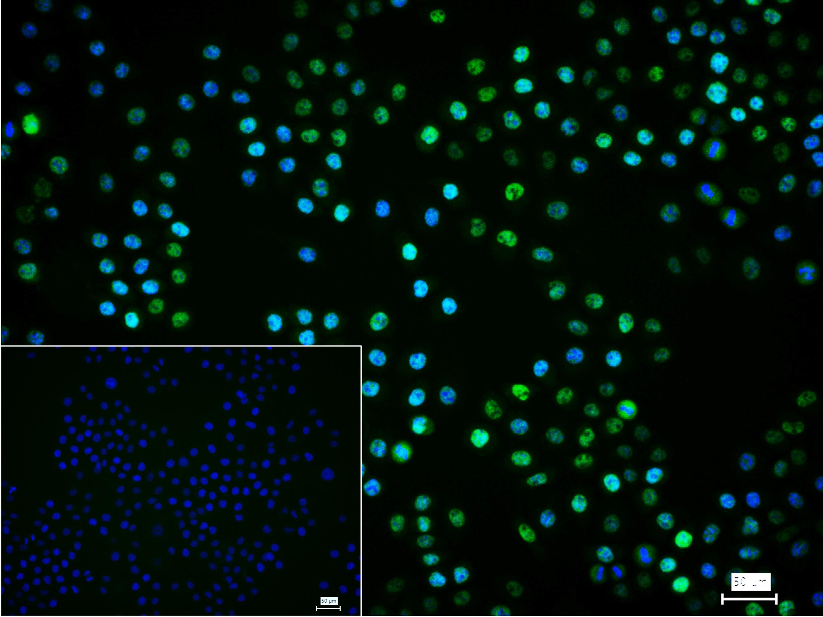
Figure 14. 4% Paraformaldehyde-fixed Hela (H) cell; Triton X-100 at r.t. for 20 min; Antibody incubation with (MSH2) monoclonal Antibody, unconjugated (SLM-60222R) 1:100, 90 min at 37°C; followed by BF488 conjugated Goat Anti-Rabbit IgG antibody (green, SL60295G-BF488) at 37°C for 90 min, DAPI (blue, C02-04002) was used to stain the cell nuclei. PBS instead of the primary antibody was used as the blank control
Ordering & shipping
- Minimum order: 1 unit
- Lead time: varies with order quantity and specifications. Please consult your local distributor or our sales team for confirmation
- Payment: T/T 100% in advance; PayPal; Alibaba Trade Assurance
- Shipping: FedEx / DHL / UPS express; cold-chain packing available on request
- Price: USD, per unit (50ul / 100ul)
Partial purchase records(bought amounts latest4)
| Username | Quantity | bought time |
| Br*** | 1 | 2026-08-25 |
| Ni*** | 1 | 2026-05-24 |
| Ta*** | 1 | 2026-01-26 |
| Ow*** | 1 | 2026-01-07 |
User Comment(Total0User Comment Num)
- No comment


 +86 571 56623320

 SUNLONG BIOTECH


