Details
Product Name CD13 Chinese Name CD13Recombinant rabbit monoclonal anti Alias ANPEN; Aminopeptidase N; Alanyl aminopeptidase; Alanyl membrane aminopeptidase; Aminopeptidase M; Aminopeptidase N; ANPEP; APN; CD 13; CD13 antigen; gp150; hAPN; Lap 1; Lap1; Alanyl (membrane) aminopeptidase; AMPN_HUMAN; AP M; AP N; AP-M; AP-N; CD13; LAP 1; LAP1; PEPN; Microsomal aminopeptidase; Myeloid plasma membrane glycoprotein CD13; p150. Research Area Tumour Cell biology Stem cells Cell Surface Molecule Immunogen Species Rabbit Clonality Monoclonal Clone NO. C4F12 React Species Human,Mouse,Rat Applications WB=1:500-2000,IHC-P=1:50-200,IHC-F=1:50-200,ICC/IF=1:50-200,IF=1:50-200,Flow-Cyt=1:50-100 (Paraffin sections need antigen repair)
not yet tested in other applications.
optimal dilutions/concentrations should be determined by the end user.Theoretical molecular weight 109kDa Cellular localization The cell membrane Form Liquid Concentration 1mg/ml immunogen KLH conjugated synthetic peptide derived from human CD13 Lsotype IgG Purification affinity purified by Protein A Buffer Solution 1M TBS(pH7.4) with 1% BSA, 3% Proclin300 and 50% Glycerol. Storage Shipped at 4℃. Store at -20 °C for one year. Avoid repeated freeze/thaw cycles. Attention This product as supplied is intended for research use only, not for use in human, therapeutic or diagnostic applications. PubMed PubMed Product Detail Aminopeptidase N is located in the small-intestinal and renal microvillar membrane, and also in other plasma membranes. In the small intestine aminopeptidase N plays a role in the final digestion of peptides generated from hydrolysis of proteins by gastric and pancreatic proteases. Its function in proximal tubular epithelial cells and other cell types is less clear. The large extracellular carboxyterminal domain contains a pentapeptide consensus sequence characteristic of members of the zinc-binding metalloproteinase superfamily. Sequence comparisons with known enzymes of this class showed that CD13 and aminopeptidase N are identical. The latter enzyme was thought to be involved in the metabolism of regulatory peptides by diverse cell types, including small intestinal and renal tubular epithelial cells, macrophages, granulocytes, and synaptic membranes from the CNS. This membrane-bound zinc metalloprotease is known to serve as a receptor for the HCoV-229E alphacoronavirus as well as other non-human coronaviruses. This gene has also been shown to promote angiogenesis, tumor growth, and metastasis and defects in this gene are associated with various types of leukemia and lymphoma. [provided by RefSeq, Apr 2020]
SWISS:
P15144
Gene ID:
290
Product Picture
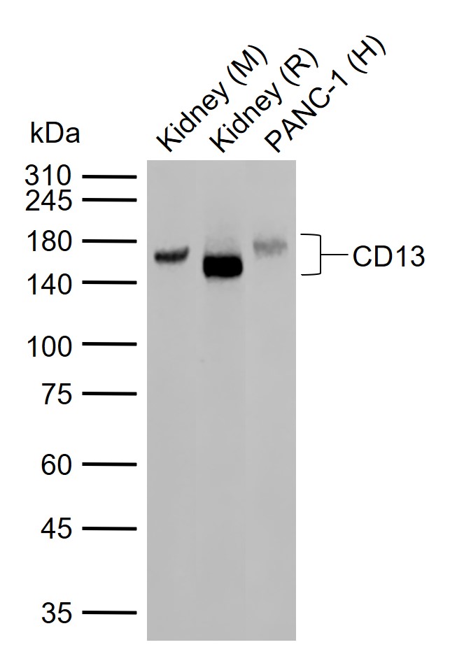
Figure 1. Sample: Lane 1: Mouse Kidney tissue lysates Lane 2: Rat Kidney tissue lysates Lane 3: Human PANC-1 cell lysates Primary: Anti-CD13 (SLM-60371R) at 1/1000 dilution Secondary: IRDye800CW Goat Anti-Rabbit IgG at 1/20000 dilution Predicted band size: 109 kDa Observed band size: 150 kDa
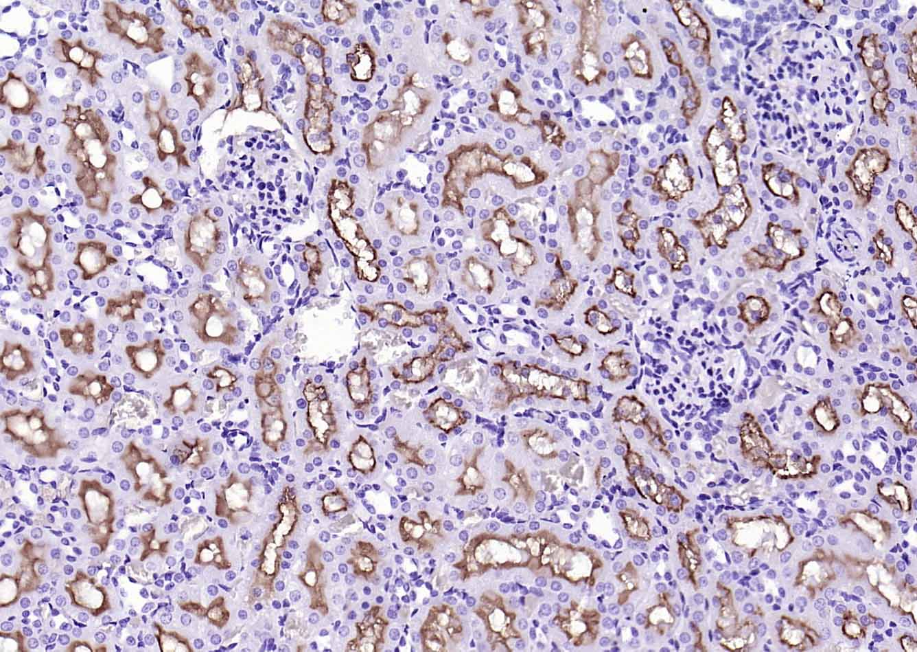
Figure 2. Paraformaldehyde-fixed, paraffin embedded (rat kidney); Antigen retrieval by boiling in sodium citrate buffer (pH6.0) for 15min; Block endogenous peroxidase by 3% hydrogen peroxide for 20 minutes; Blocking buffer (normal goat serum) at 37°C for 30min; Antibody incubation with (CD13) Monoclonal Antibody, Unconjugated (SLM-60371R) at 1:200 overnight at 4°C, followed by operating according to SP Kit(Rabbit) (sp-0023) instructionsand DAB staining
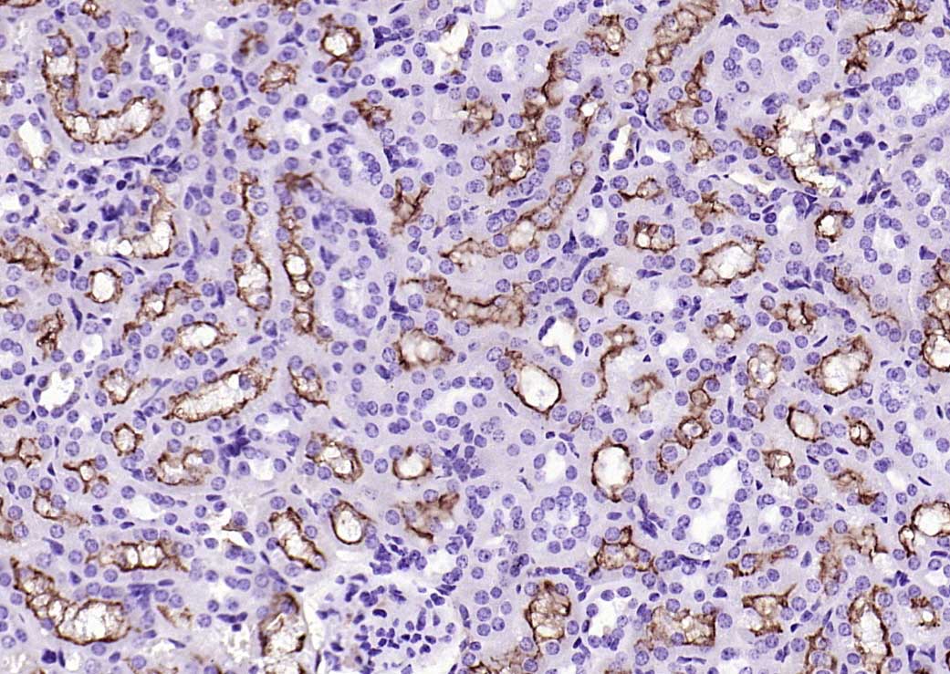
Figure 3. Paraformaldehyde-fixed, paraffin embedded (mouse kidney); Antigen retrieval by boiling in sodium citrate buffer (pH6.0) for 15min; Block endogenous peroxidase by 3% hydrogen peroxide for 20 minutes; Blocking buffer (normal goat serum) at 37°C for 30min; Antibody incubation with (CD13) Monoclonal Antibody, Unconjugated (SLM-60371R) at 1:200 overnight at 4°C, followed by operating according to SP Kit(Rabbit) (sp-0023) instructionsand DAB staining
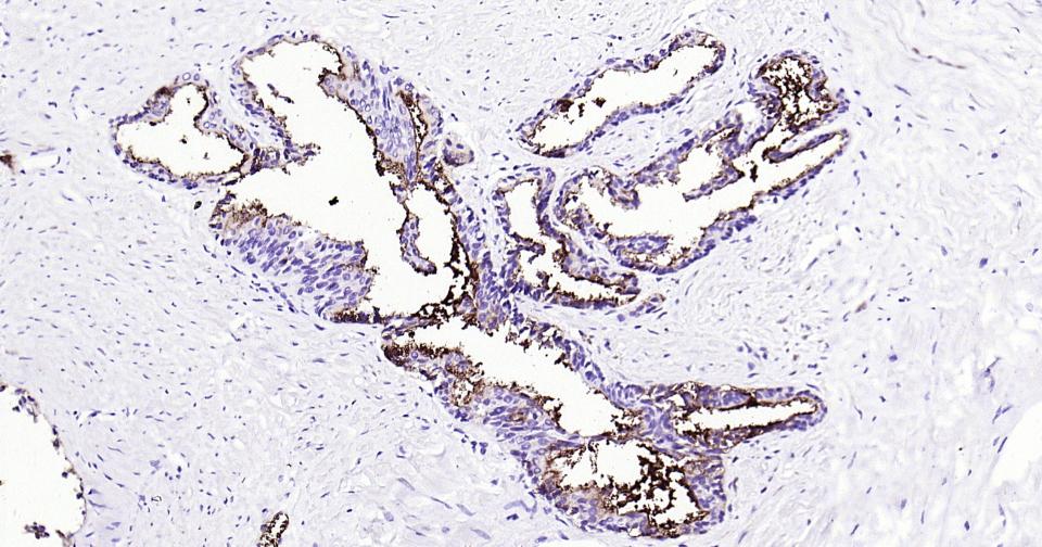
Figure 4. Paraformaldehyde-fixed, paraffin embedded Human Prostate; Antigen retrieval by boiling in sodium citrate buffer (pH6.0) for 15 min; Antibody incubation with CD13 Monoclonal Antibody, Unconjugated(SLM-60371R) at 1:200 overnight at 4°C, followed by conjugation to the SP Kit (Rabbit, SP-0023) and DAB (C-0010) staining
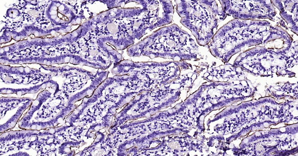
Figure 5. Paraformaldehyde-fixed, paraffin embedded Human Duodenum; Antigen retrieval by boiling in sodium citrate buffer (pH6.0) for 15 min; Antibody incubation with CD13 Monoclonal Antibody, Unconjugated(SLM-60371R) at 1:200 overnight at 4°C, followed by conjugation to the SP Kit (Rabbit, SP-0023) and DAB (C-0010) staining
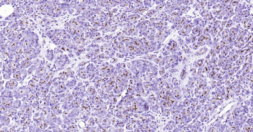
Figure 6. Paraformaldehyde-fixed, paraffin embedded Human Pancreatic Cancer; Antigen retrieval by boiling in sodium citrate buffer (pH6.0) for 15 min; Antibody incubation with CD13 Monoclonal Antibody, Unconjugated(SLM-60371R) at 1:200 overnight at 4°C, followed by conjugation to the SP Kit (Rabbit, SP-0023) and DAB (C-0010) staining
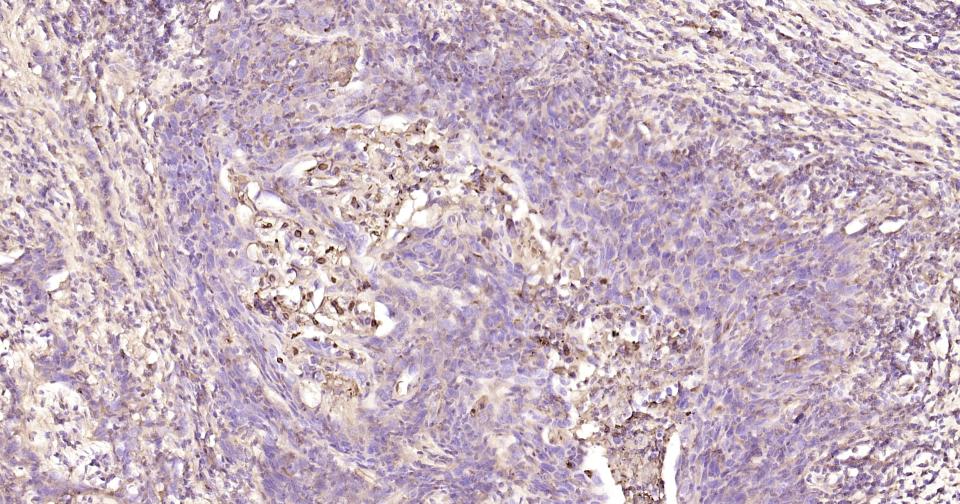
Figure 7. Paraformaldehyde-fixed, paraffin embedded Human Cervical Cancer; Antigen retrieval by boiling in sodium citrate buffer (pH6.0) for 15 min; Antibody incubation with CD13 Monoclonal Antibody, Unconjugated(SLM-60371R) at 1:200 overnight at 4°C, followed by conjugation to the SP Kit (Rabbit, SP-0023) and DAB (C-0010) staining
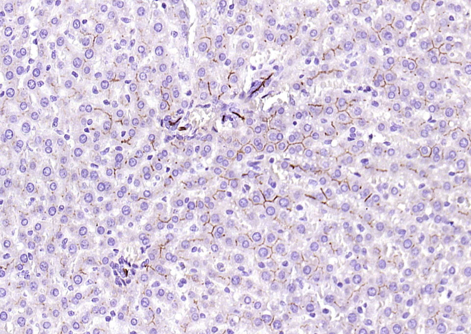
Figure 8. Paraformaldehyde-fixed, paraffin embedded (mouse liver); Antigen retrieval by boiling in sodium citrate buffer (pH6.0) for 15min; Block endogenous peroxidase by 3% hydrogen peroxide for 20 minutes; Blocking buffer (normal goat serum) at 37°C for 30min; Antibody incubation with (CD13) Monoclonal Antibody, Unconjugated (SLM-60371R) at 1:200 overnight at 4°C, followed by operating according to SP Kit(Rabbit) (sp-0023) instructionsand DAB staining
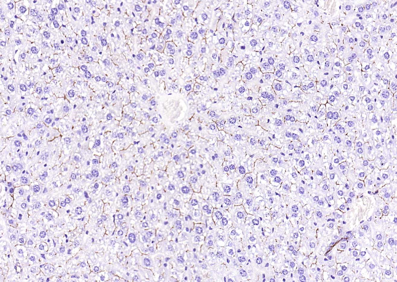
Figure 9. Paraformaldehyde-fixed, paraffin embedded (rat liver); Antigen retrieval by boiling in sodium citrate buffer (pH6.0) for 15min; Block endogenous peroxidase by 3% hydrogen peroxide for 20 minutes; Blocking buffer (normal goat serum) at 37°C for 30min; Antibody incubation with (CD13) Monoclonal Antibody, Unconjugated (SLM-60371R) at 1:200 overnight at 4°C, followed by operating according to SP Kit(Rabbit) (sp-0023) instructionsand DAB staining
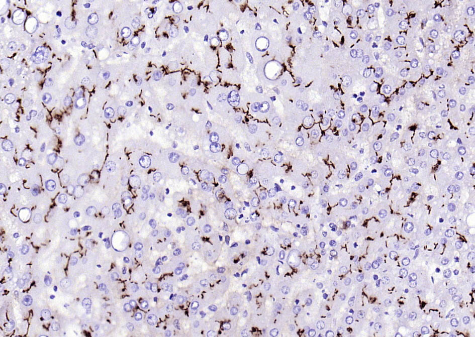
Figure 10. Paraformaldehyde-fixed, paraffin embedded (human liver); Antigen retrieval by boiling in sodium citrate buffer (pH6.0) for 15min; Block endogenous peroxidase by 3% hydrogen peroxide for 20 minutes; Blocking buffer (normal goat serum) at 37°C for 30min; Antibody incubation with (CD13) Monoclonal Antibody, Unconjugated (SLM-60371R) at 1:200 overnight at 4°C, followed by operating according to SP Kit(Rabbit) (sp-0023) instructionsand DAB staining
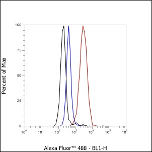
Figure 11. Cell line: THP-1 Fixative: 4% Paraformaldehyde Permeabilization: 90% methanol Primary ab dilution: 1:100 Secondary ab: Goat anti Rabbit IgG Unlabelled control: The cell without incubation with primary antibody and secondary antibody (Black line). Isotype control: Rabbit monoclonal IgG (Blue line). Comment: Line red is the positive signal for SLM-60371R
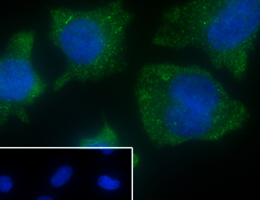
Figure 12. Cell line: A375 Fixative: 4% Paraformaldehyde Permeabilization: 0.1% TritonX-100 Primary ab dilution: 1:50 Primary incubation condition: 4°C overnight Secondary ab: Goat Anti-Rabbit IgG Nuclear counter stain: DAPI (Blue) Comment: Color green is the positive signal for SLM-60371R
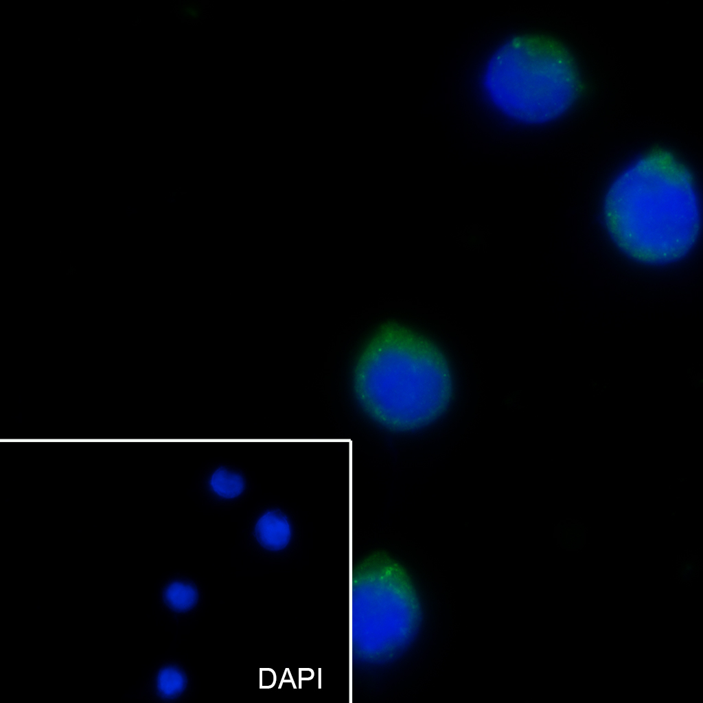
Figure 13. Cell line: THP-1 Fixative: 100% Ice-cold methanol Permeabilization: 0.1% TritonX-100 Primary ab dilution: 1:50 Primary incubation condition: 4°C overnight Secondary ab: Goat Anti-Rabbit IgG Nuclear counter stain: DAPI (Blue) Comment: Color green is the positive signal for SLM-60371R
Ordering & shipping
- Minimum order: 1 unit
- Lead time: varies with order quantity and specifications. Please consult your local distributor or our sales team for confirmation
- Payment: T/T 100% in advance; PayPal; Alibaba Trade Assurance
- Shipping: FedEx / DHL / UPS express; cold-chain packing available on request
- Price: USD, per unit (50ul / 100ul)
Partial purchase records(bought amounts latest4)
| Username | Quantity | bought time |
| Xa*** | 1 | 2026-08-27 |
| Ia*** | 1 | 2026-08-12 |
| Ol*** | 1 | 2026-05-13 |
| Ke*** | 1 | 2026-03-02 |
User Comment(Total0User Comment Num)
- No comment


 +86 571 56623320

 SUNLONG BIOTECH


