
Details
Product Name Recombinant Rabbit Anti-CD48 Monoclonal Antibody Cat No. SLA1778M Chinese Name CD48重组兔单抗 Alias BCM1; BLAST; BLAST1; MEM-102; SLAMF2; hCD48; mCD48; CD48_HUMAN; CD48; B-lymphocyte activation marker BLAST-1; BCM1 surface antigen; Leukocyte antigen MEM-102; SLAM family member 2 (SLAMF2); Signaling lymphocytic activation molecule 2; TCT.1; CD48 molecule; CD48 antigen (B-cell membrane protein) Host Species Rabbit Isotype IgG Clonality Monoclonal Clone No. React Species Human Applications WB (1:500-2000)
IHC-P (1:100-500)
IHC-F (1:100-500)
IF (1:100-500)
Flow-Cyt (1:50-100)
ICC/IF (1:50-200)
IP (1:20-50)
Not yet tested in other applications.
Optimal working dilutions must be determined by the end user.Immunogen A synthesized peptide derived from human CD48 Purification affinity purified by Protein A Predicted MW. 28 kDa Observed MW. 45-50 kDa Subcellular Location N/A Gene ID 962 Swiss-Prot P09326 Storage 0.01M TBS (pH7.4) with 1% BSA, 0.02% Proclin300 and 50% Glycerol.
Shipped at 4℃. Store at -20℃ for one year. Avoid repeated freeze/thaw cycles.Background Glycosylphosphatidylinositol (GPI)-anchored cell surface glycoprotein that interacts via its N-terminal immunoglobulin domain with cell surface receptors including 2B4/CD244 or CD2 to regulate immune cell function and activation. Participates in T-cell signaling transduction by associating with CD2 and efficiently bringing the Src family protein kinase LCK and LAT to the TCR/CD3 complex. Attention This product as supplied is intended for research use only, not for use in human, therapeutic or diagnostic applications.
Validation Images
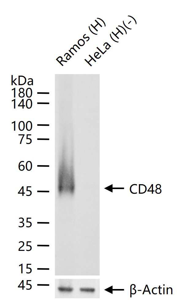
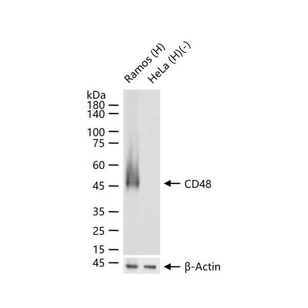
Figure 1. 25 ug total protein per lane of various lysates (see on figure) probed with CD48 monoclonal antibody, unconjugated (SLA1778M) at 1:1000 dilution and 4°C overnight incubation. Followed by conjugated secondary antibody incubation at r.t. for 60 min
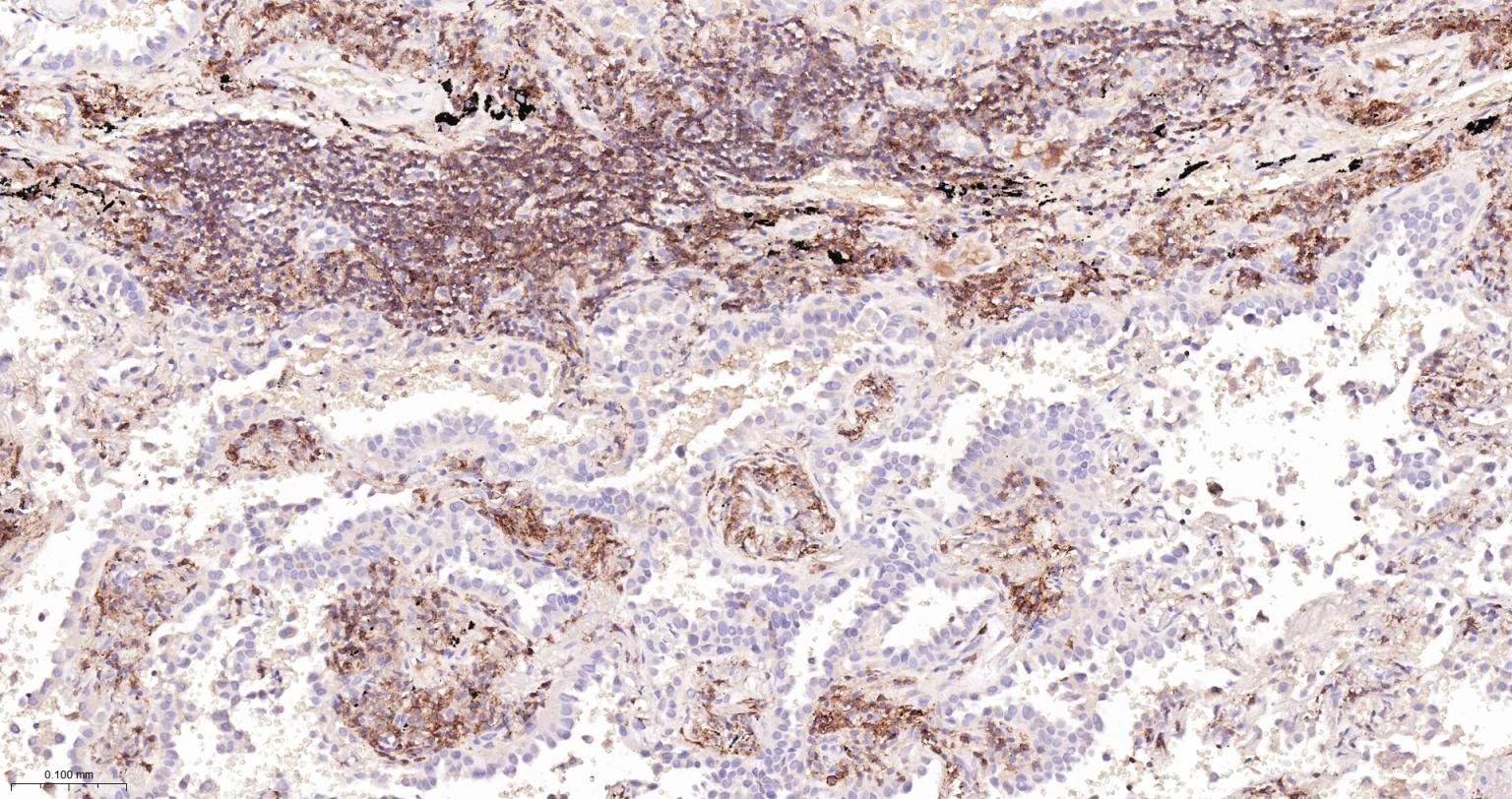
Figure 2. Paraformaldehyde-fixed, paraffin embedded Human Lung Cancer; Antigen retrieval by boiling in sodium citrate buffer (pH6.0) for 15 min; The section was incubated with CD48 Monoclonal Antibody, Unconjugated (SLA1778M) at 1:200 overnight at 4°C, followed by conjugation to the SL0295G-HRP and DAB (C-0010) staining
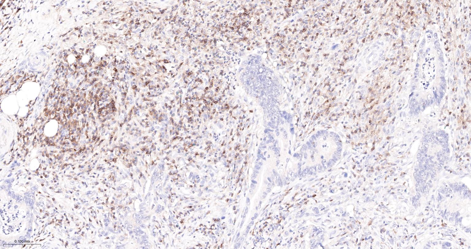
Figure 3. Paraformaldehyde-fixed, paraffin embedded Human Colon Cancer; Antigen retrieval by boiling in sodium citrate buffer (pH6.0) for 15 min; The section was incubated with CD48 Monoclonal Antibody, Unconjugated (SLA1778M) at 1:200 overnight at 4°C, followed by conjugation to the SL0295G-HRP and DAB (C-0010) staining
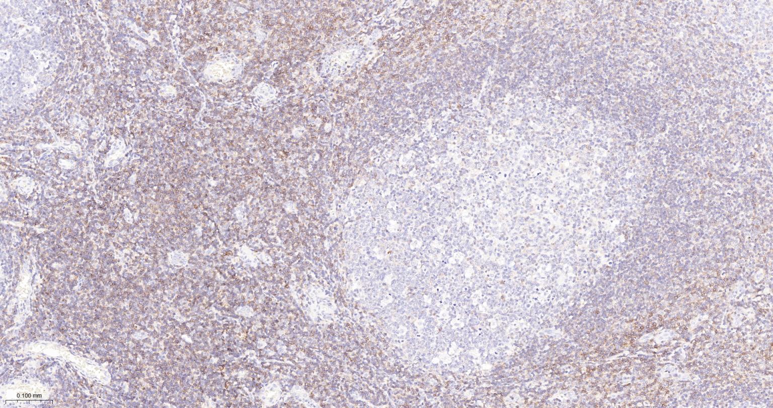
Figure 4. Paraformaldehyde-fixed, paraffin embedded Human Tonsil; Antigen retrieval by boiling in sodium citrate buffer (pH6.0) for 15 min; The section was incubated with CD48 Monoclonal Antibody, Unconjugated (SLA1778M) at 1:200 overnight at 4°C, followed by conjugation to the SL0295G-HRP and DAB (C-0010) staining
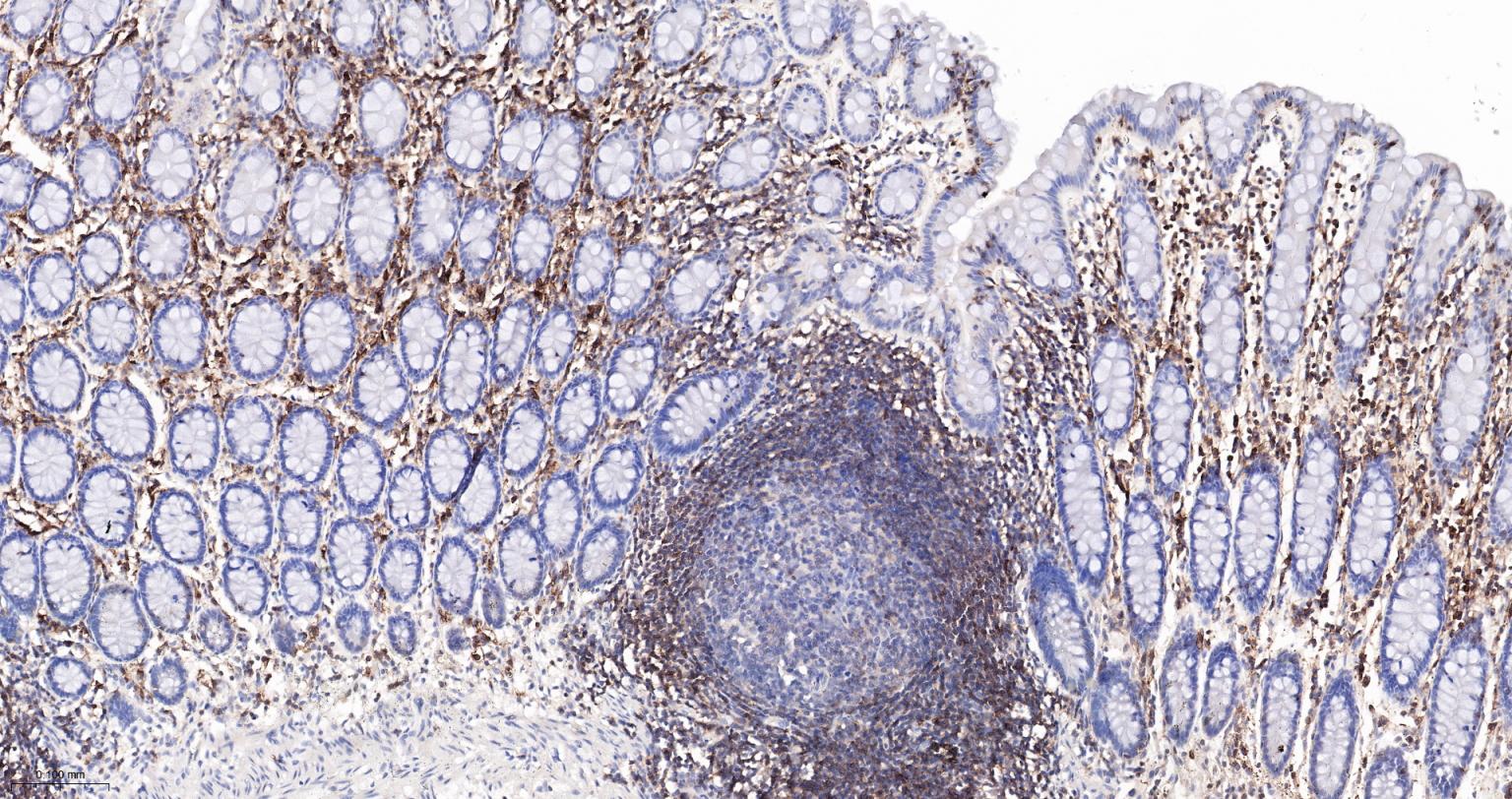
Figure 5. Paraformaldehyde-fixed, paraffin embedded Human Colon; Antigen retrieval by boiling in sodium citrate buffer (pH6.0) for 15 min; The section was incubated with CD48 Monoclonal Antibody, Unconjugated (SLA1778M) at 1:200 overnight at 4°C, followed by conjugation to the SL0295G-HRP and DAB (C-0010) staining
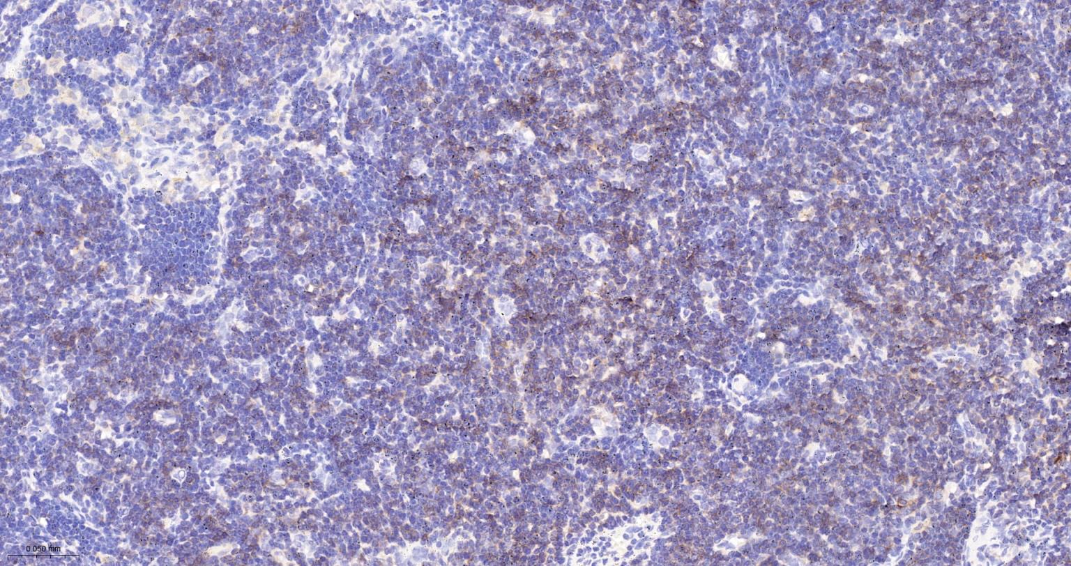
Figure 6. Paraformaldehyde-fixed, paraffin embedded Human Lymph; Antigen retrieval by boiling in sodium citrate buffer (pH6.0) for 15 min; The section was incubated with CD48 Monoclonal Antibody, Unconjugated (SLA1778M) at 1:200 overnight at 4°C, followed by conjugation to the SL0295G-HRP and DAB (C-0010) staining
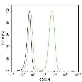
Figure 7. The Raji (H) cells were incubated in 5%BSA to block non-specific protein-protein interactions (30 min at r.t.), followed by secondary antibody incubation for 40 min at room temperature. Primary Antibody (green): Rabbit Anti-CD48 antibody (SLA1778M,1:100); Isotype Control (orange): Rabbit IgG (SL0295P). Blank control (black): PBS. Acquisition of 20,000 events was performed
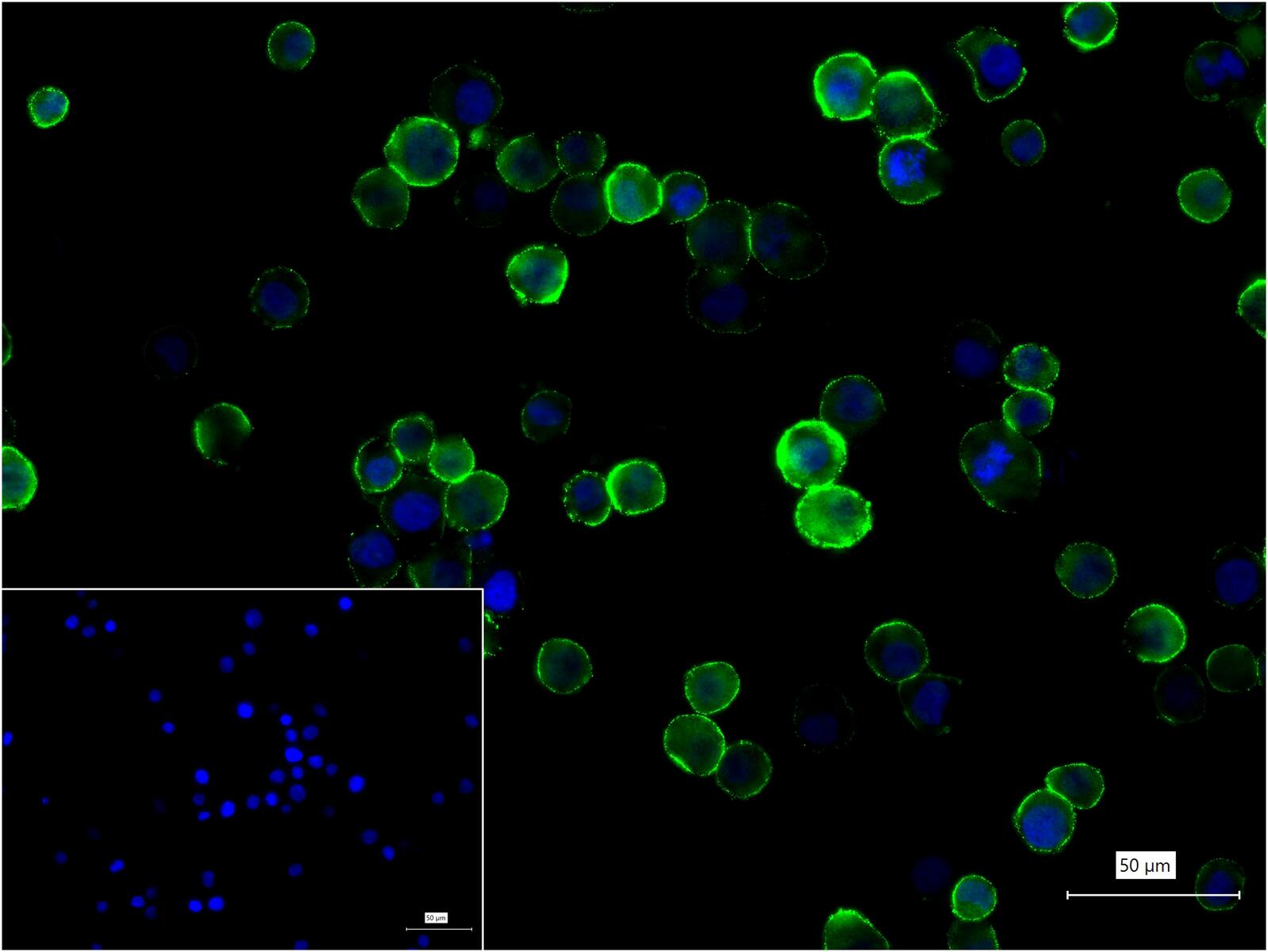
Figure 8. 4% Paraformaldehyde-fixed Raji (H) cell; Antibody incubation with (CD48) monoclonal Antibody, unconjugated (SLA1778M) 1:100, 90 min at 37°C; followed by BF488 conjugated Goat Anti-Rabbit IgG antibody (green, SL60295G-BF488) at 37°C for 90 min, DAPI (blue, C02-04002) was used to stain the cell nuclei. PBS instead of the primary antibody was used as the blank control
FOR RESEARCH USE ONLY. Not for use in diagnostic or therapeutic procedures.
Partial purchase records(bought amounts latest0)
User Comment(Total0User Comment Num)
- No comment


 +86 571 56623320

 SUNLONG BIOTECH


