Details
Product Name Recombinant Rabbit Anti-COL3A1 Monoclonal Antibody Cat No. SLA0904M Chinese Name Ⅲ型胶原蛋白重组兔单抗 Alias EDS4A; EDSVASC; PMGEDSV; Col3a-1; Tsk-2; Tsk2; CO3A1_BOVIN; COL3A1; CO3A1_HUMAN; CO3A1_MOUSE; CO3A1_RAT; collagen type III alpha 1 chain; Ehlers-Danlos syndrome type IV, autosomal dominant; collagen, type III, alpha 1 Host Species Rabbit Isotype IgG Clonality Monoclonal Clone No. 7E8 React Species Human Applications WB (1:500-2000)
IHC-P (1:100-500)
IHC-F (1:100-500)
IF (1:100-500)
Flow-Cyt (1:50-100)
ICC/IF (1:50-200)
Not yet tested in other applications.
Optimal working dilutions must be determined by the end user.Immunogen Recombinant human Collagen III protein Purification affinity purified by Protein A Concentration 1mg/ml Predicted MW. 139 kDa Observed MW. 150-200 kDa Subcellular Location Extracellular Gene ID 1281 Swiss-Prot P02461 Storage 0.01M TBS (pH7.4) with 1% BSA, 0.02% Proclin300 and 50% Glycerol.
Shipped at 4℃. Store at -20℃ for one year. Avoid repeated freeze/thaw cycles.Background The extensive family of COL gene products (collagens) is composed of several chain types, including fibril-forming interstitial collagens (types I, II, III and V) and basement membrane collagens (type IV), each type containing multiple isoforms. Collagens are fibrous, extracellular matrix proteins with high tensile strength and are the major components of connective tissue, such as tendons and cartilage. All collagens contain a triple helix domain and frequently show lateral self-association in order to form complex connective tissues. Several collagens also play a role in cell adhesion, important for maintaining normal tissue architecture and function. This gene encodes the pro-alpha1 chains of type III collagen, a fibrillar collagen that is found in extensible connective tissues such as skin, lung, uterus, intestine and the vascular system, frequently in association with type I collagen. Mutations in this gene are associated with Ehlers-Danlos syndrome types IV, and with aortic and arterial aneurysms. Two transcripts, resulting from the use of alternate polyadenylation signals, have been identified for this gene. Attention This product as supplied is intended for research use only, not for use in human, therapeutic or diagnostic applications.
Validation Images
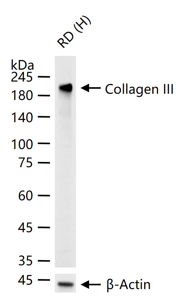
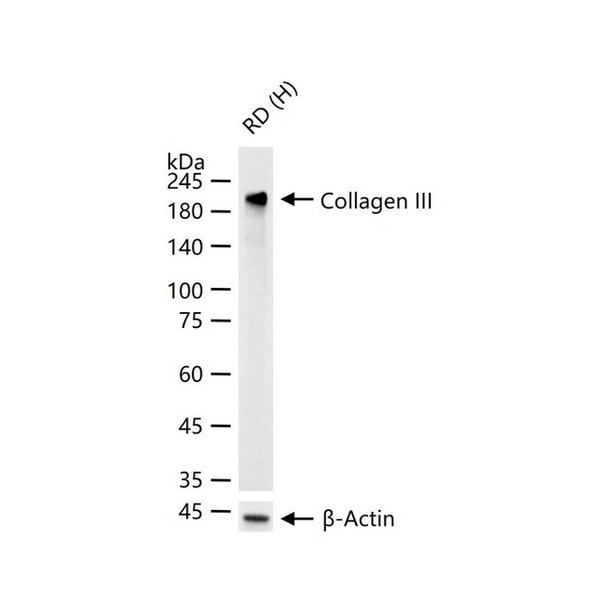
Figure 1. 25 ug total protein per lane of various lysates (see on figure) probed with Collagen III monoclonal antibody, unconjugated (SLA0904M) at 1:1000 dilution and 4°C overnight incubation. Followed by conjugated secondary antibody incubation at r.t. for 60 min
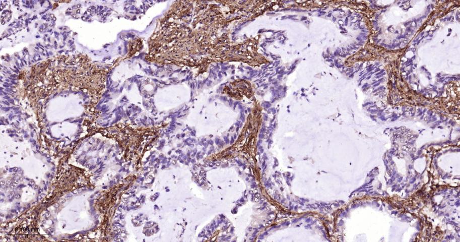
Figure 2. Paraformaldehyde-fixed, paraffin embedded Human Lung Cancer; Antigen retrieval by boiling in sodium citrate buffer (pH6.0) for 15 min; The section was incubated with Collagen III Monoclonal Antibody, Unconjugated (SLA0904M) at 1:500 overnight at 4°C, followed by conjugation to the SL0295G-HRP and DAB (C-0010) staining
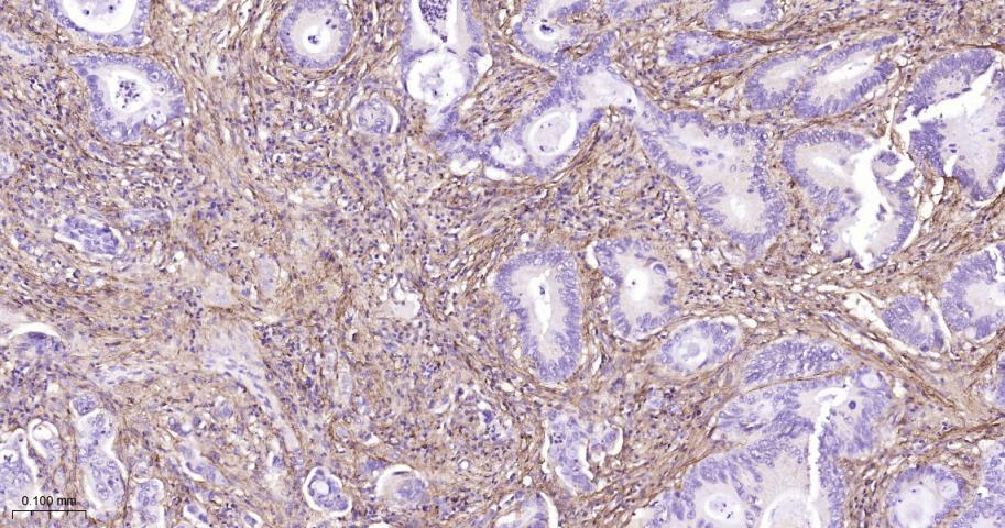
Figure 3. Paraformaldehyde-fixed, paraffin embedded Human Colon Cancer; Antigen retrieval by boiling in sodium citrate buffer (pH6.0) for 15 min; The section was incubated with Collagen III Monoclonal Antibody, Unconjugated (SLA0904M) at 1:500 overnight at 4°C, followed by conjugation to the SL0295G-HRP and DAB (C-0010) staining
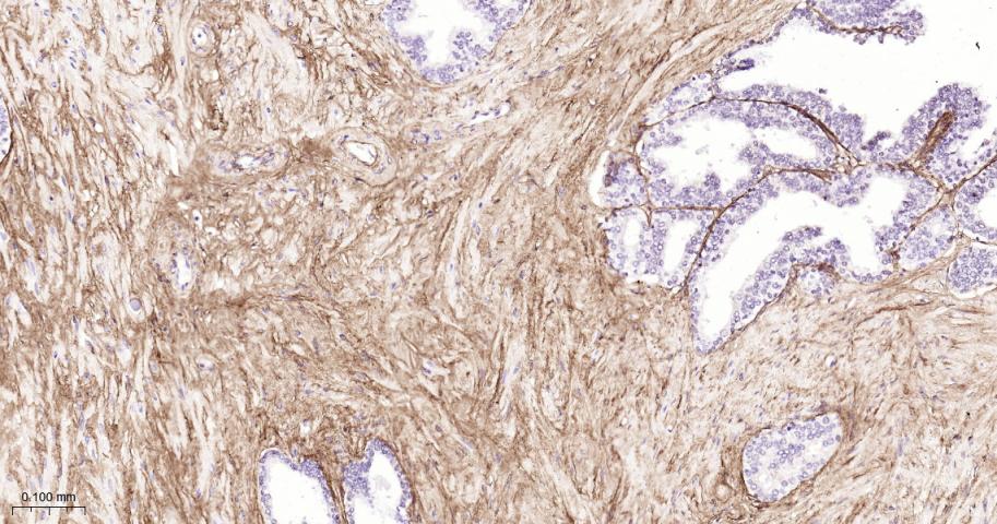
Figure 4. Paraformaldehyde-fixed, paraffin embedded Human Prostate Cancer; Antigen retrieval by boiling in sodium citrate buffer (pH6.0) for 15 min; The section was incubated with Collagen III Monoclonal Antibody, Unconjugated (SLA0904M) at 1:500 overnight at 4°C, followed by conjugation to the SL0295G-HRP and DAB (C-0010) staining
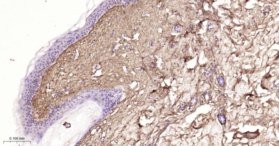
Figure 5. Paraformaldehyde-fixed, paraffin embedded Human Abdominal skin; Antigen retrieval by boiling in sodium citrate buffer (pH6.0) for 15 min; The section was incubated with Collagen III Monoclonal Antibody, Unconjugated (SLA0904M) at 1:500 overnight at 4°C, followed by conjugation to the SL0295G-HRP and DAB (C-0010) staining
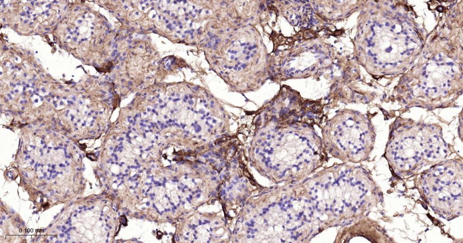
Figure 6. Paraformaldehyde-fixed, paraffin embedded Human Testicles; Antigen retrieval by boiling in sodium citrate buffer (pH6.0) for 15 min; The section was incubated with Collagen III Monoclonal Antibody, Unconjugated (SLA0904M) at 1:500 overnight at 4°C, followed by conjugation to the SL0295G-HRP and DAB (C-0010) staining
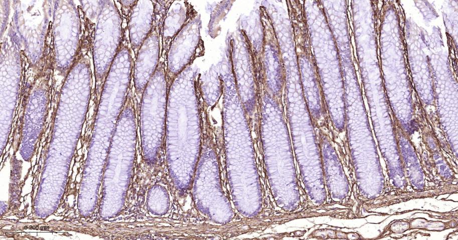
Figure 7. Paraformaldehyde-fixed, paraffin embedded Human Colon; Antigen retrieval by boiling in sodium citrate buffer (pH6.0) for 15 min; The section was incubated with Collagen III Monoclonal Antibody, Unconjugated (SLA0904M) at 1:500 overnight at 4°C, followed by conjugation to the SL0295G-HRP and DAB (C-0010) staining
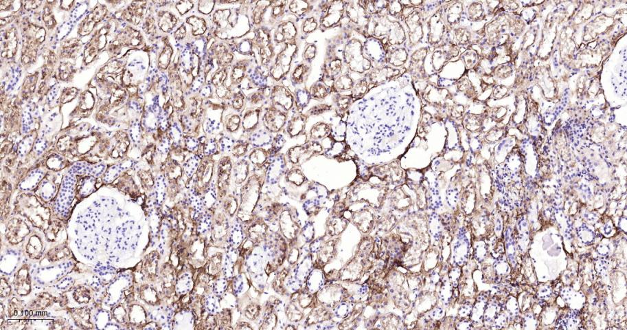
Figure 8. Paraformaldehyde-fixed, paraffin embedded Human Kidney; Antigen retrieval by boiling in sodium citrate buffer (pH6.0) for 15 min; The section was incubated with Collagen III Monoclonal Antibody, Unconjugated (SLA0904M) at 1:500 overnight at 4°C, followed by conjugation to the SL0295G-HRP and DAB (C-0010) staining
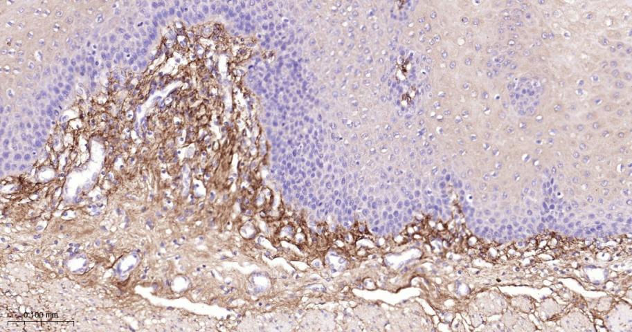
Figure 9. Paraformaldehyde-fixed, paraffin embedded Human Esophagus; Antigen retrieval by boiling in sodium citrate buffer (pH6.0) for 15 min; The section was incubated with Collagen III Monoclonal Antibody, Unconjugated (SLA0904M) at 1:500 overnight at 4°C, followed by conjugation to the SL0295G-HRP and DAB (C-0010) staining
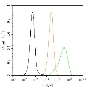
Figure 10. The RD (H) cells were fixed with 4% PFA (10 min at r.t.) and then permeabilized with 90% ice-cold methanol for 20 min at -20℃,the cells then were incubated in 5%BSA to block non-specific protein-protein interactions (30 min at r.t.).Primary Antibody (green):Rabbit Anti-Collagen III antibody (SLA0904M), dilution: 1:100; Isotype Control (orange): Rabbit IgG (SL0295P). Blank control (black): PBS. Acquisition of 20,000 events was performed
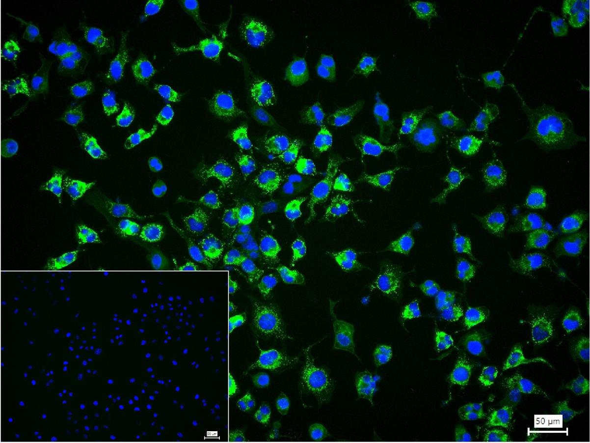
Figure 11. 4% Paraformaldehyde-fixed RD (H) cell; Triton X-100 at r.t. for 20 min; Antibody incubation with (Collagen III) monoclonal Antibody, unconjugated (SLA0904M) 1:100, 90 min at 37°C; followed by conjugated Goat Anti-Rabbit IgG antibody (green, SL60295G-BF488) at 37°C for 90 min, DAPI (blue, C02-04002) was used to stain the cell nuclei. PBS instead of the primary antibody was used as the blank control
FOR RESEARCH USE ONLY. Not for use in diagnostic or therapeutic procedures.
Partial purchase records(bought amounts latest0)
User Comment(Total0User Comment Num)
- No comment


 +86 571 56623320

 SUNLONG BIOTECH


