Details
Product Name Recombinant Rabbit Anti-H3-3A Monoclonal Antibody Cat No. SLA2924M Chinese Name 磷酸化组蛋白H3.3重组兔单抗 Alias BRYLIB1; H3.3A; H3F3; H3F3A; BRYLIB2; H3.3B; H3F3B; H3-3b; H33_HUMAN; H3-3A; H33_RAT; H3.3 histone A; H3 histone, family 3A; H3 histone family member 3A; H3-3A | Histone H3.3 (phospho-S22); H3.3S22P; p-Histone H3.3; phospho-Histone H3.3; p-H3.3; H3-3A | Histone H3.3 (phospho-Ser22) Host Species Rabbit Isotype IgG Clonality Monoclonal Clone No. 5D12 React Species Human,Mouse,Rat Applications WB (1:500-2000)
IHC-P (1:100-500)
IHC-F (1:100-500)
IF (1:100-500)
Flow-Cyt (1:50-100)
ICC/IF (1:50-200)
Not yet tested in other applications.
Optimal working dilutions must be determined by the end user.Immunogen A synthesized peptide derived from human Histone H3.3 around the phosphorylation site of S22 Purification affinity purified by Protein A Predicted MW. 15 kDa Observed MW. 15 kDa Subcellular Location N/A Gene ID 3020 Swiss-Prot P84243 Storage 0.01M TBS (pH7.4) with 1% BSA, 0.02% Proclin300 and 50% Glycerol.
Shipped at 4℃. Store at -20℃ for one year. Avoid repeated freeze/thaw cycles.Background Variant histone H3 which replaces conventional H3 in a wide range of nucleosomes in active genes. Constitutes the predominant form of histone H3 in non-dividing cells and is incorporated into chromatin independently of DNA synthesis. Deposited at sites of nucleosomal displacement throughout transcribed genes, suggesting that it represents an epigenetic imprint of transcriptionally active chromatin. Attention This product as supplied is intended for research use only, not for use in human, therapeutic or diagnostic applications.
Validation Images
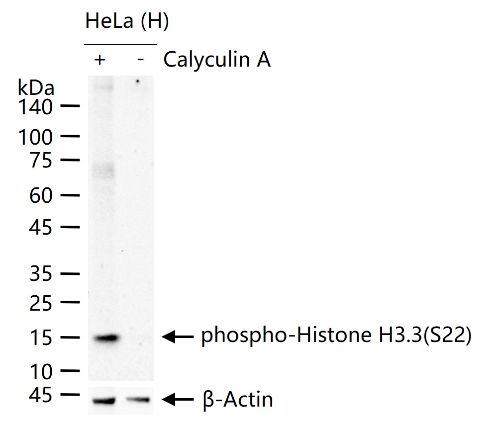
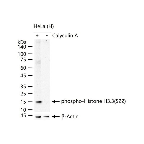
Figure 1. HeLa (H) cells were treated with or without Calyculin A (100nM) for 30 min, 25 μg total protein per lane of cell lysates (see on figure) probed with phospho-Histone H3.3(S22) monoclonal antibody, unconjugated (SLA2924M) at 1:1000 dilution and 4°C overnight incubation. Followed by conjugated secondary antibody incubation at r.t. for 60 min
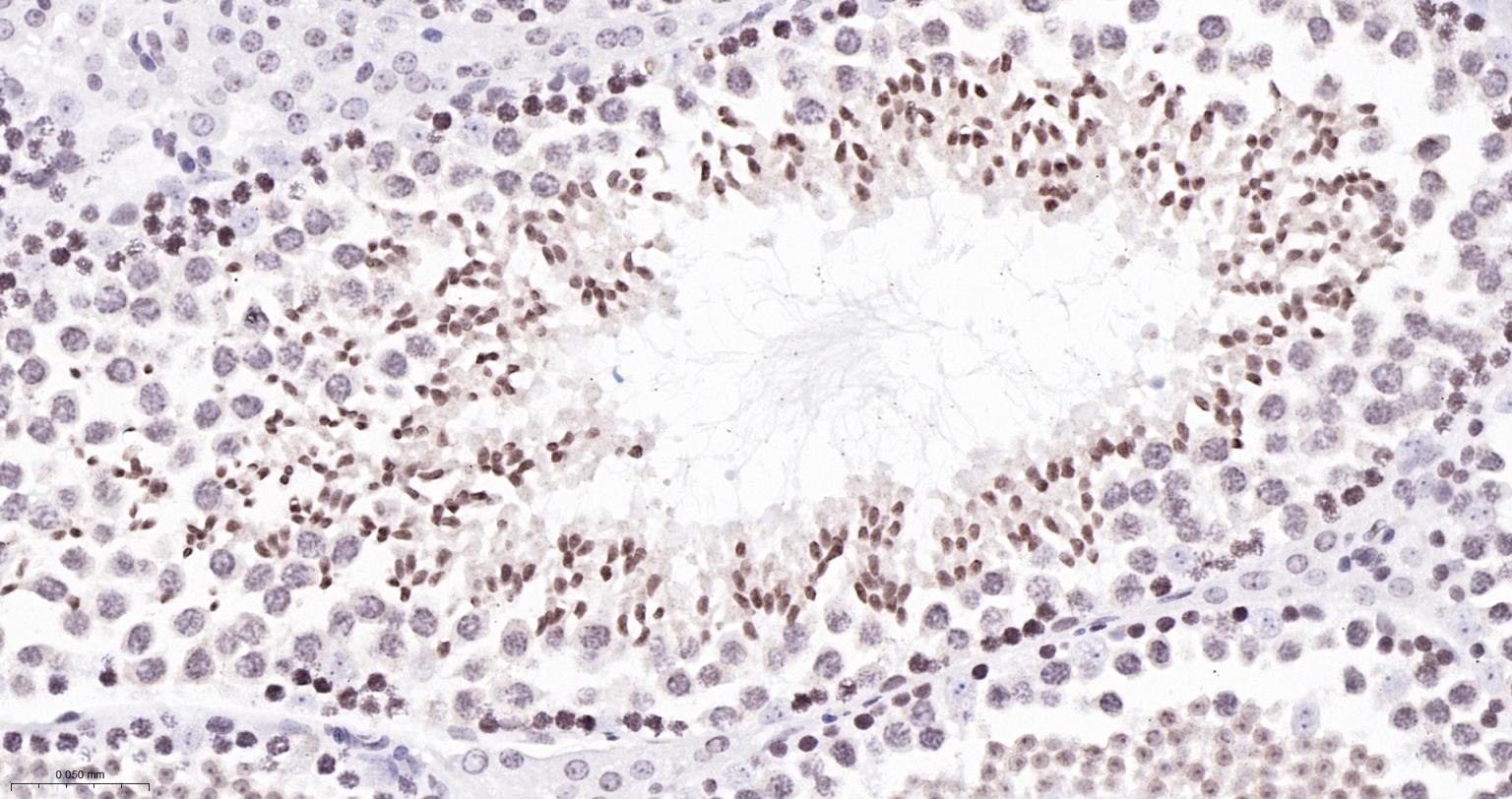
Figure 2. Paraformaldehyde-fixed, paraffin embedded Mouse Testicles; Antigen retrieval by boiling in sodium citrate buffer (pH6.0) for 15 min; The section was incubated with phospho-Histone H3.3(S22) Monoclonal Antibody, Unconjugated (SLA2924M) at 1:200 overnight at 4°C, followed by conjugation to the SL0295G-HRP and DAB (C-0010) staining
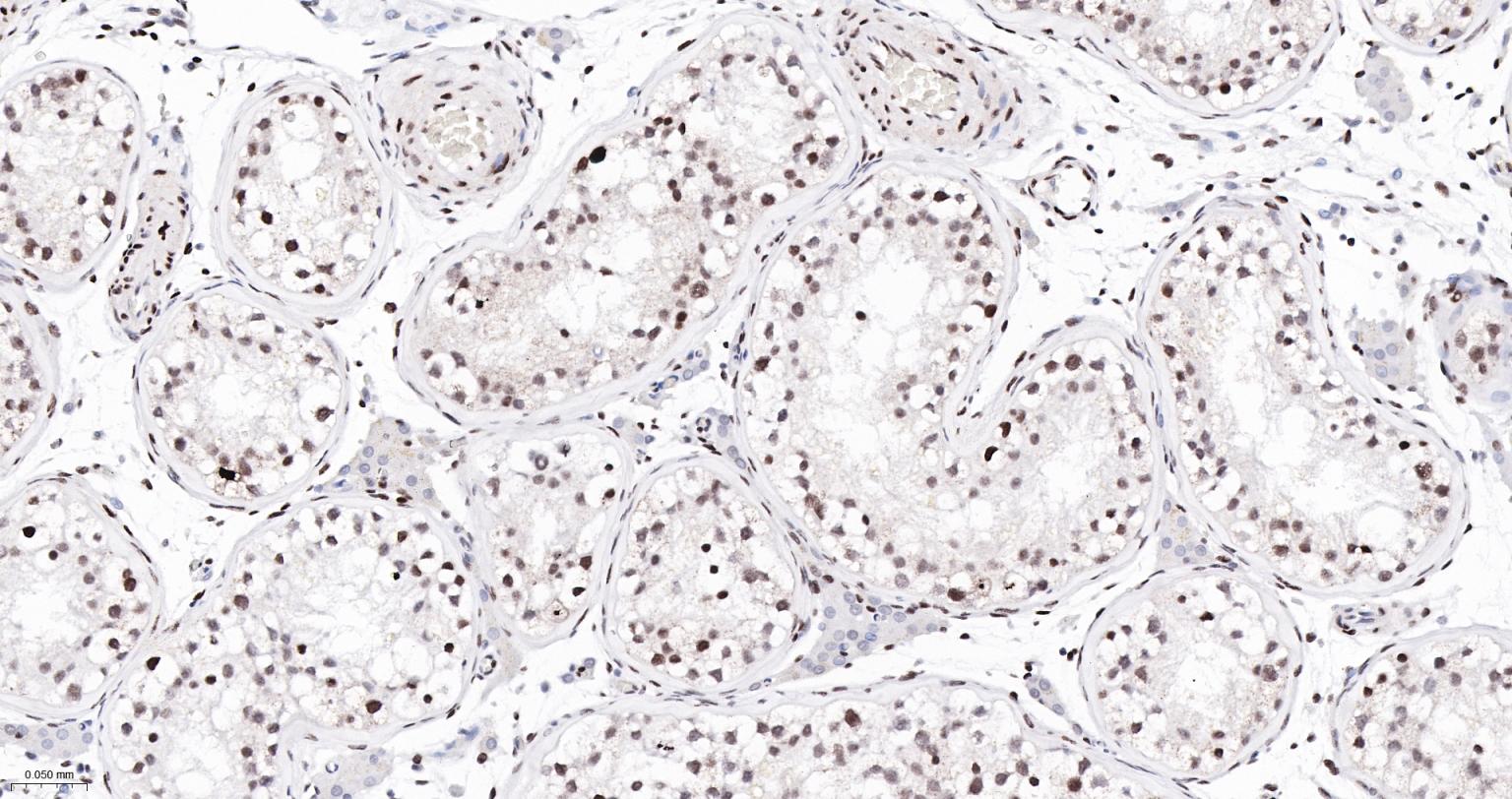
Figure 3. Paraformaldehyde-fixed, paraffin embedded Human Testicles; Antigen retrieval by boiling in sodium citrate buffer (pH6.0) for 15 min; The section was incubated with phospho-Histone H3.3(S22) Monoclonal Antibody, Unconjugated (SLA2924M) at 1:200 overnight at 4°C, followed by conjugation to the SL0295G-HRP and DAB (C-0010) staining
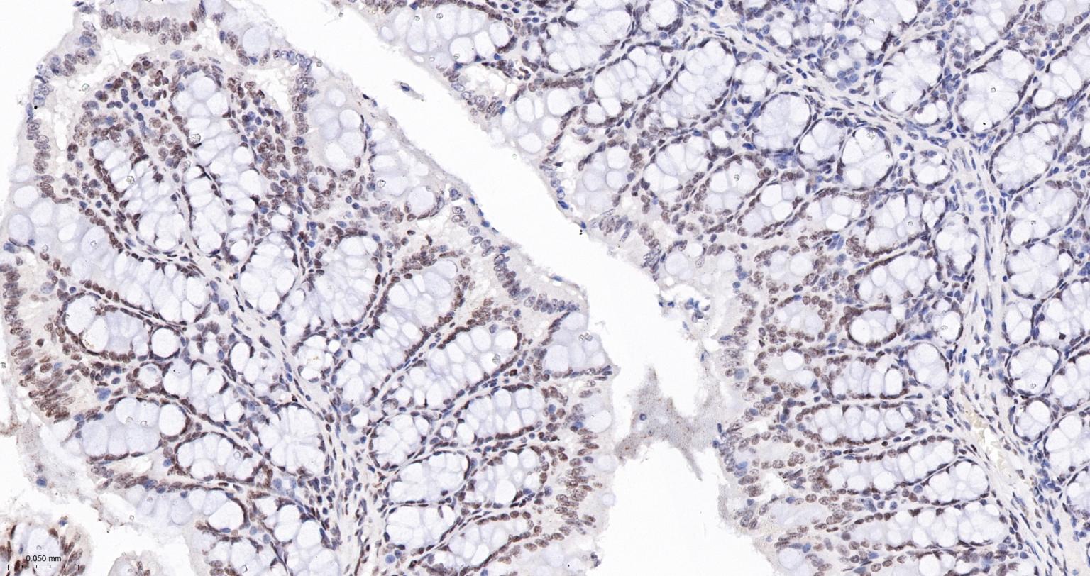
Figure 4. Paraformaldehyde-fixed, paraffin embedded Mouse Colon; Antigen retrieval by boiling in sodium citrate buffer (pH6.0) for 15 min; The section was incubated with phospho-Histone H3.3(S22) Monoclonal Antibody, Unconjugated (SLA2924M) at 1:200 overnight at 4°C, followed by conjugation to the SL0295G-HRP and DAB (C-0010) staining
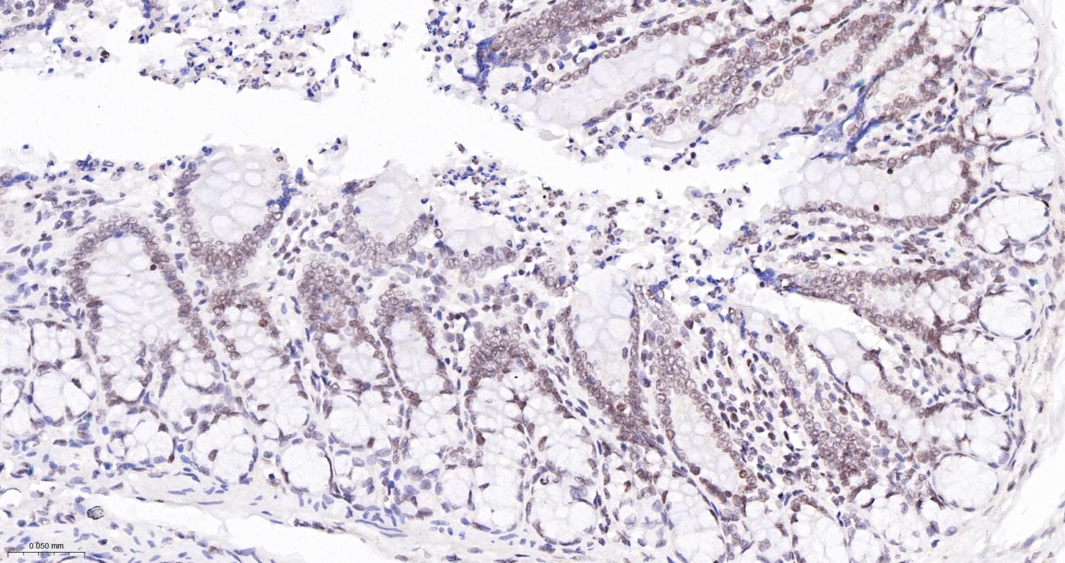
Figure 5. Paraformaldehyde-fixed, paraffin embedded Rat Colon; Antigen retrieval by boiling in sodium citrate buffer (pH6.0) for 15 min; The section was incubated with phospho-Histone H3.3(S22) Monoclonal Antibody, Unconjugated (SLA2924M) at 1:200 overnight at 4°C, followed by conjugation to the SL0295G-HRP and DAB (C-0010) staining
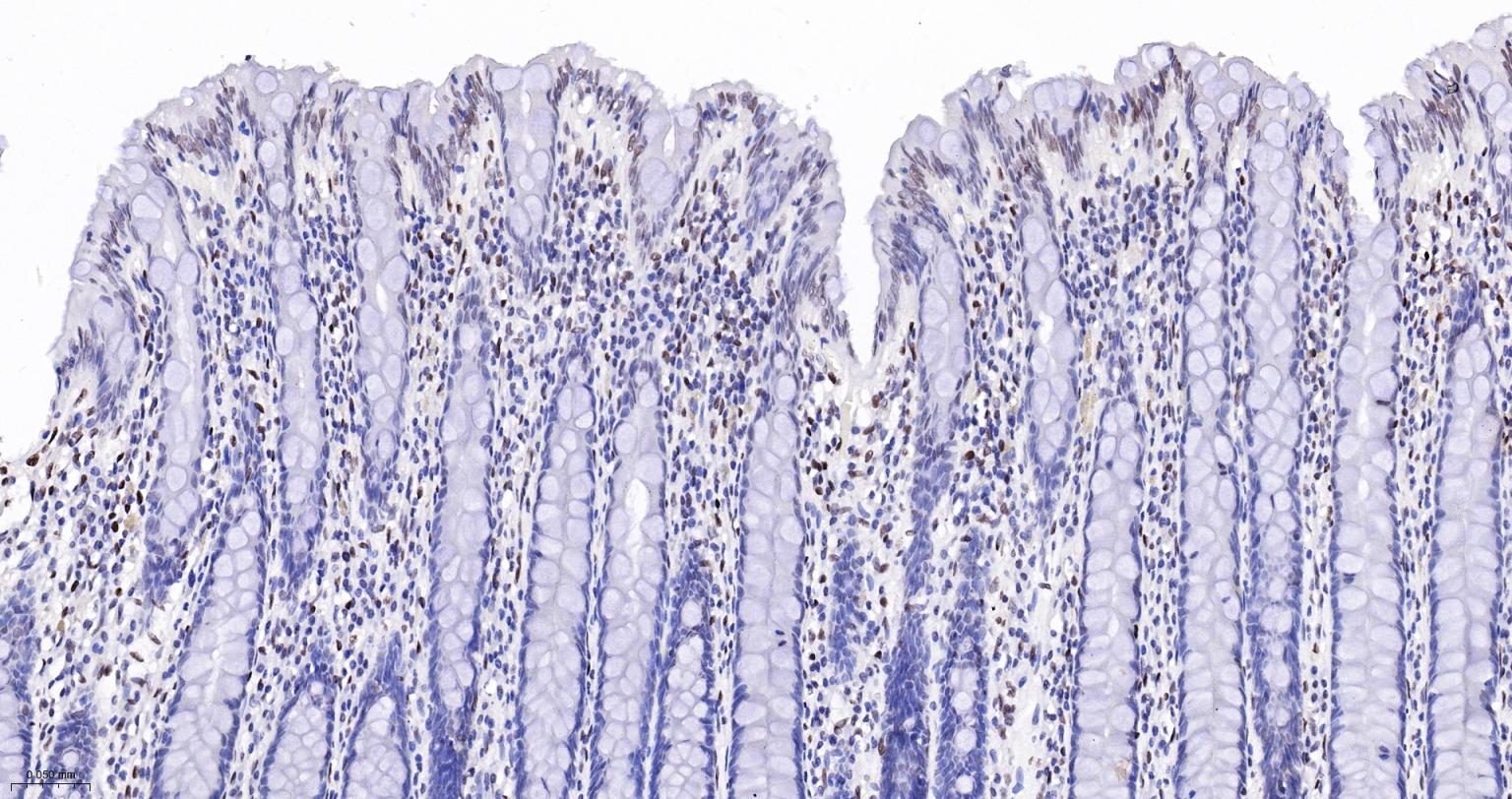
Figure 6. Paraformaldehyde-fixed, paraffin embedded Human Colon; Antigen retrieval by boiling in sodium citrate buffer (pH6.0) for 15 min; The section was incubated with phospho-Histone H3.3(S22) Monoclonal Antibody, Unconjugated (SLA2924M) at 1:200 overnight at 4°C, followed by conjugation to the SL0295G-HRP and DAB (C-0010) staining
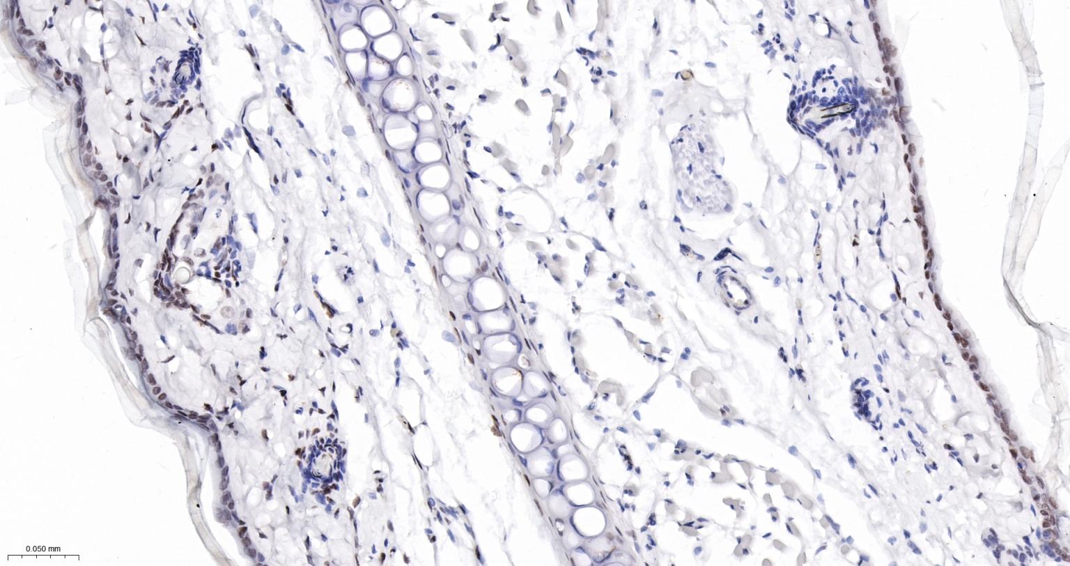
Figure 7. Paraformaldehyde-fixed, paraffin embedded Rat Skin; Antigen retrieval by boiling in sodium citrate buffer (pH6.0) for 15 min; The section was incubated with phospho-Histone H3.3(S22) Monoclonal Antibody, Unconjugated (SLA2924M) at 1:200 overnight at 4°C, followed by conjugation to the SL0295G-HRP and DAB (C-0010) staining
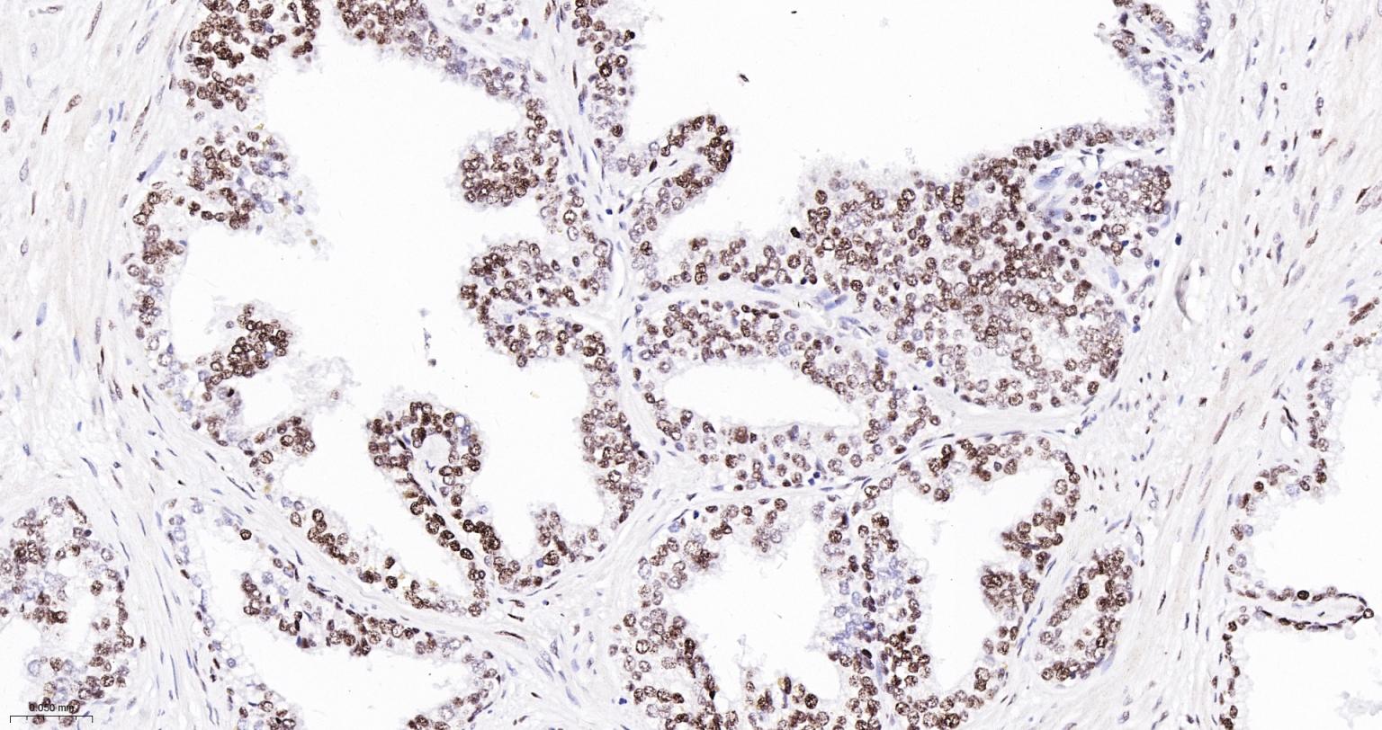
Figure 8. Paraformaldehyde-fixed, paraffin embedded Human Prostate Cancer; Antigen retrieval by boiling in sodium citrate buffer (pH6.0) for 15 min; The section was incubated with phospho-Histone H3.3(S22) Monoclonal Antibody, Unconjugated (SLA2924M) at 1:200 overnight at 4°C, followed by conjugation to the SL0295G-HRP and DAB (C-0010) staining
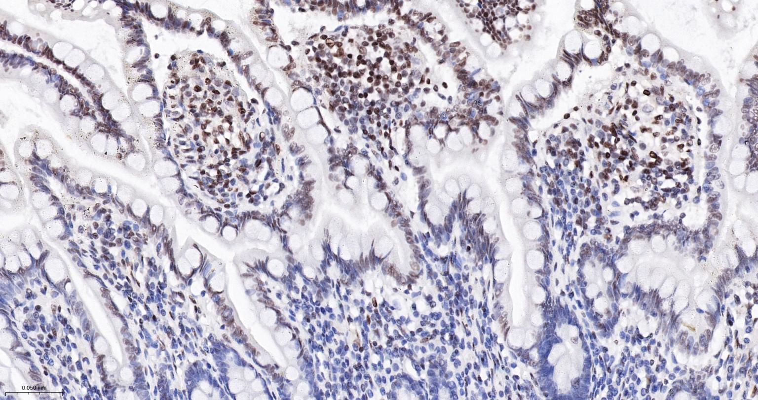
Figure 9. Paraformaldehyde-fixed, paraffin embedded Human Small Intestine; Antigen retrieval by boiling in sodium citrate buffer (pH6.0) for 15 min; The section was incubated with phospho-Histone H3.3(S22) Monoclonal Antibody, Unconjugated (SLA2924M) at 1:200 overnight at 4°C, followed by conjugation to the SL0295G-HRP and DAB (C-0010) staining
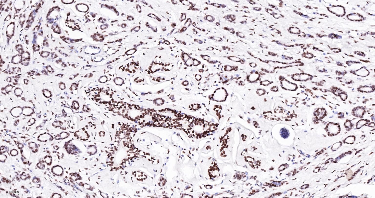
Figure 10. Paraformaldehyde-fixed, paraffin embedded Human Breast Cancer; Antigen retrieval by boiling in sodium citrate buffer (pH6.0) for 15 min; The section was incubated with phospho-Histone H3.3(S22) Monoclonal Antibody, Unconjugated (SLA2924M) at 1:200 overnight at 4°C, followed by conjugation to the SL0295G-HRP and DAB (C-0010) staining
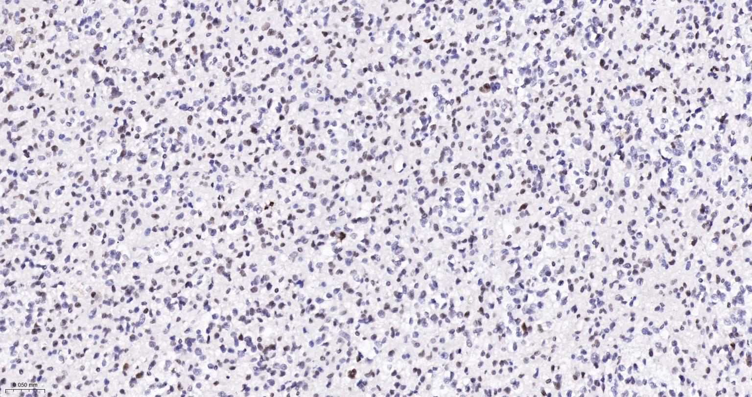
Figure 11. Paraformaldehyde-fixed, paraffin embedded Human Glioblastoma; Antigen retrieval by boiling in sodium citrate buffer (pH6.0) for 15 min; The section was incubated with phospho-Histone H3.3(S22) Monoclonal Antibody, Unconjugated (SLA2924M) at 1:200 overnight at 4°C, followed by conjugation to the SL0295G-HRP and DAB (C-0010) staining
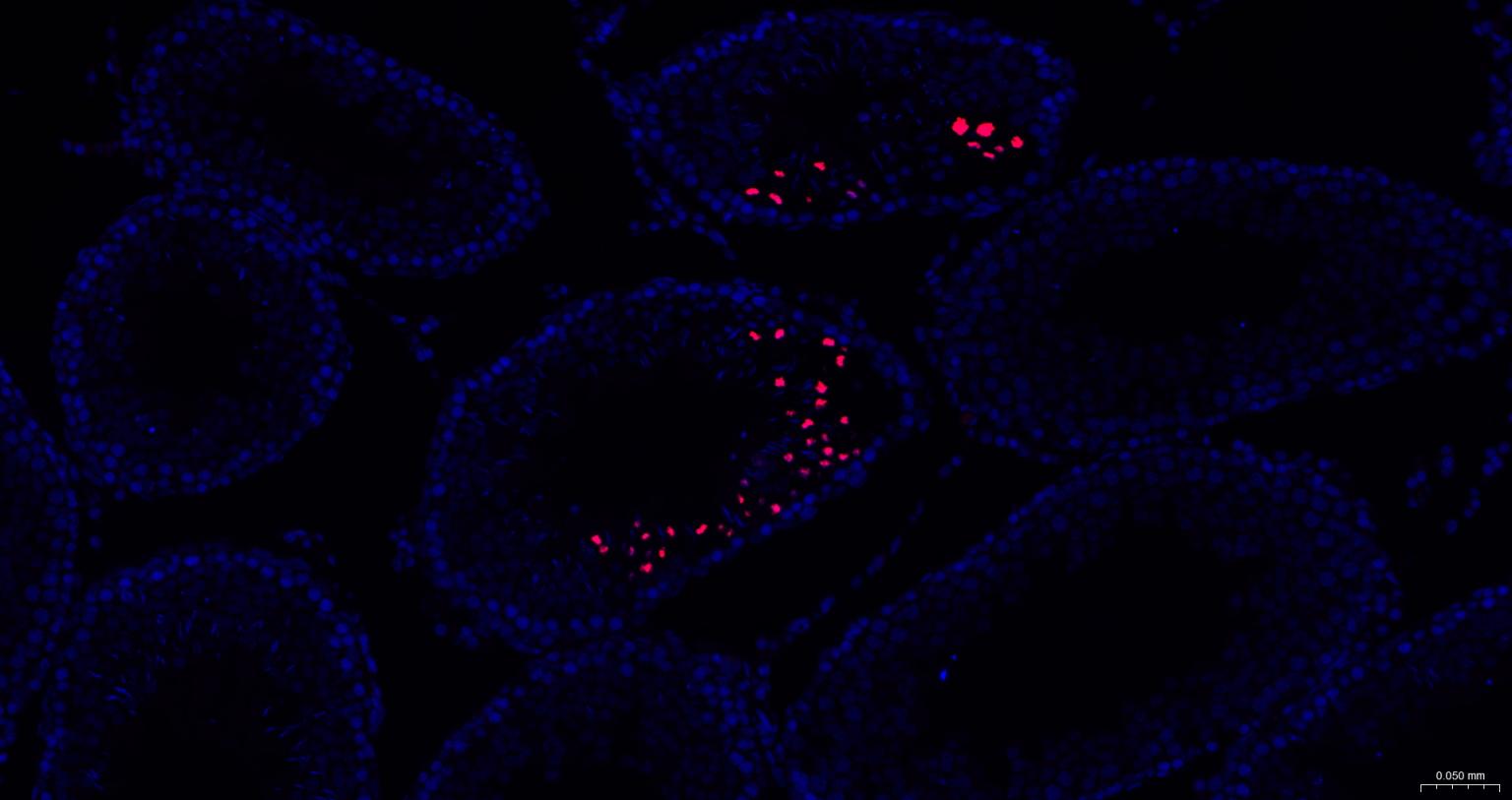
Figure 12. Paraformaldehyde-fixed, paraffin embedded Rat Testicles; Antigen retrieval by boiling in sodium citrate buffer (pH6.0) for 15 min; The section was incubated with H3-3A | Histone H3.3 (p-S22) Antibody, Unconjugated (SLA2924M) at 1:200 overnight at 4°C. Followed by conjugated Goat Anti-Rabbit IgG antibody (Red, SL0295G-BF594), DAPI (blue, C02-04002) was used to stain the cell nuclei
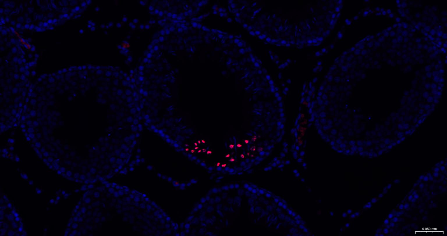
Figure 13. Paraformaldehyde-fixed, paraffin embedded Mouse Testicles; Antigen retrieval by boiling in sodium citrate buffer (pH6.0) for 15 min; The section was incubated with H3-3A | Histone H3.3 (p-S22) Antibody, Unconjugated (SLA2924M) at 1:200 overnight at 4°C. Followed by conjugated Goat Anti-Rabbit IgG antibody (Red, SL0295G-BF594), DAPI (blue, C02-04002) was used to stain the cell nuclei
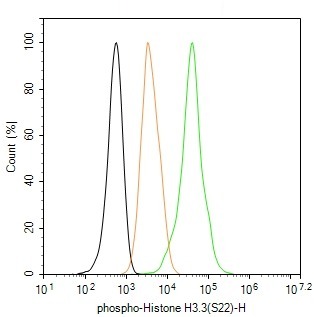
Figure 14. The HeLa (treated with 100ng/ml of nocodazole for 18 h) (H) cells were fixed with 4% PFA (10 min at r.t.) and then permeabilized with 90% ice-cold methanol for 20 min at -20℃,the cells then were incubated in 5%BSA to block non-specific protein-protein interactions (30 min at r.t.). Cells stained with Primary Antibody for 30 min at room temperature,followed by secondary antibody incubation for 40 min at room temperature. Primary Antibody (green):Rabbit Anti-phospho-Histone H3.3(S22) antibody (SLA2924M,1:100); Isotype Control (orange): Rabbit IgG (SL0295P). Blank control (black): PBS. Acquisition of 20,000 events was performed
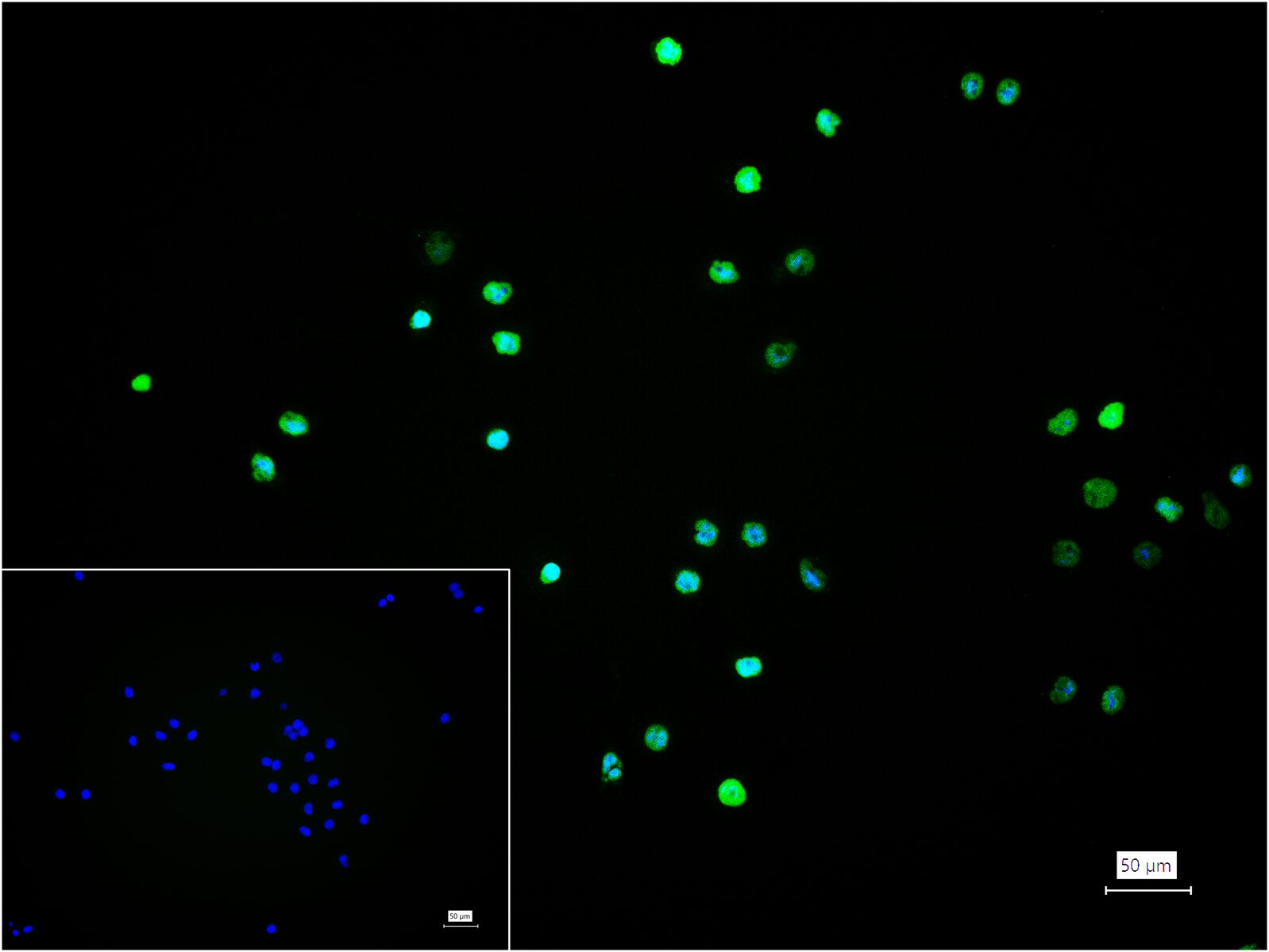
Figure 15. 4% Paraformaldehyde-fixed HeLa (treated with 100ng/ml of nocodazole for 18 h) (H) cell; Triton X-100 at r.t. for 20 min; Antibody incubation with (phospho-Histone H3.3(S22)) monoclonal Antibody, unconjugated (SLA2924M) 1:100, 90 min at 37°C; followed by BF488 conjugated Goat Anti-Rabbit IgG antibody (green, SL60295G-BF488) at 37°C for 90 min, DAPI (blue, C02-04002) was used to stain the cell nuclei. PBS instead of the primary antibody was used as the blank control
FOR RESEARCH USE ONLY. Not for use in diagnostic or therapeutic procedures.
Partial purchase records(bought amounts latest0)
User Comment(Total0User Comment Num)
- No comment


 +86 571 56623320

 SUNLONG BIOTECH


