Details
Product Name Recombinant Rabbit Anti-RPS19 Monoclonal Antibody Cat No. SLA0882M Chinese Name S19核糖体蛋白重组兔单抗 Alias DBA; DBA1; LOH19CR1; S19; eS19; ribosomal protein S19; loss of heterozygosity on chromosome 19, region 1; loss of heterozygosity, 19, chromosomal region 1; Diamond-Blackfan anemia Host Species Rabbit Isotype IgG Clonality Monoclonal Clone No. 13D8 React Species Human,Mouse,Rat Applications WB (1:500-1000)
IHC-P (1:100-500)
IHC-F (1:100-500)
IF (1:100-500)
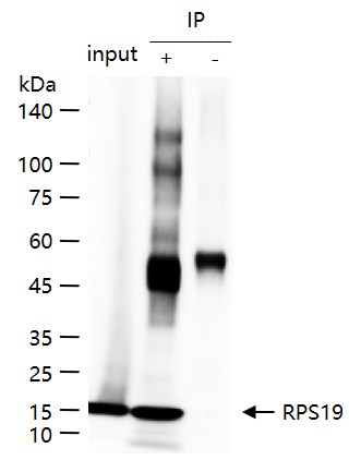
IP (1:20-50)
Not yet tested in other applications.
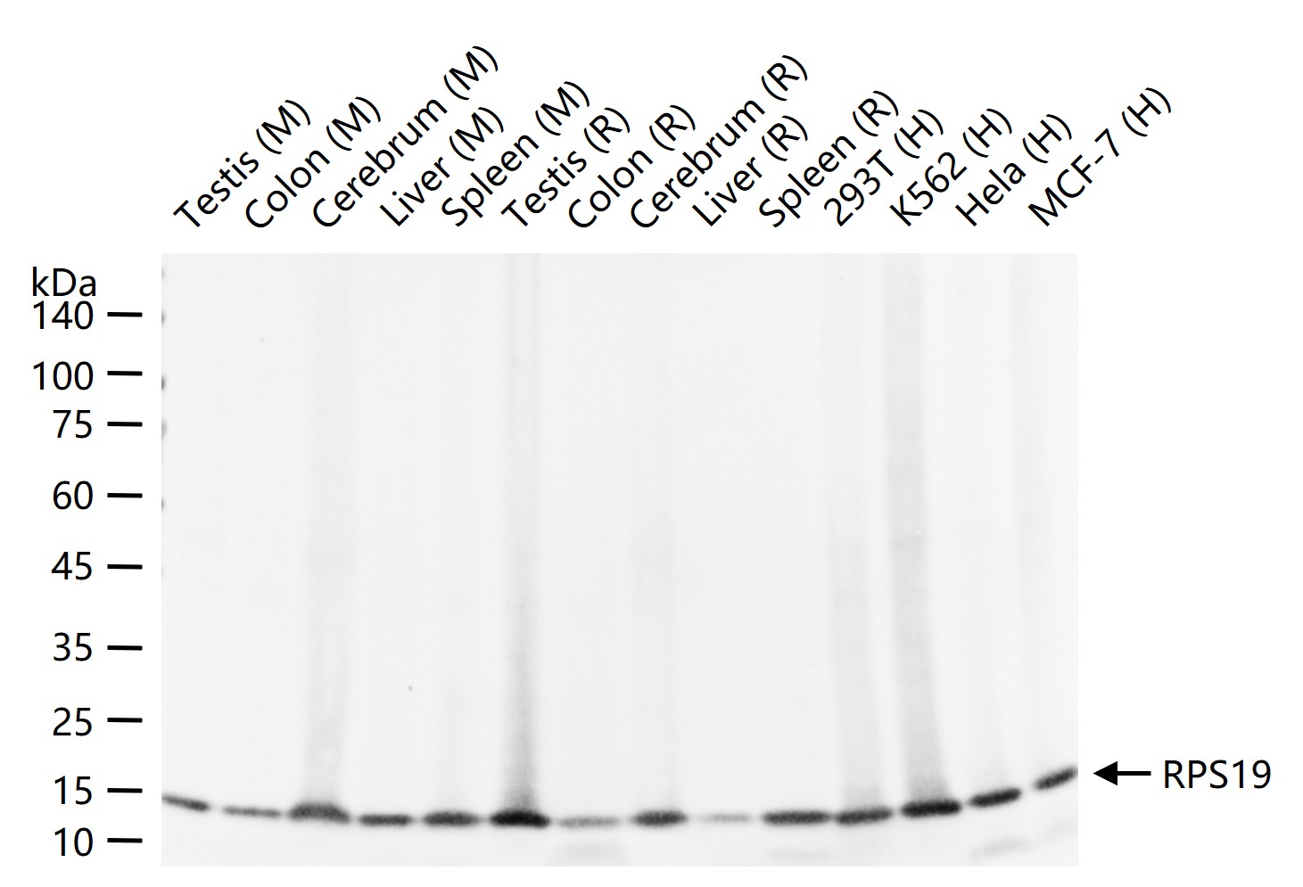
Optimal working dilutions must be determined by the end user.Immunogen A synthesized peptide derived from human RPS19 Purification affinity purified by Protein A Concentration 1mg/ml Predicted MW. 16 kDa Observed MW. 16 kDa Subcellular Location Nucleus Gene ID 6223 Storage 0.01M TBS (pH7.4) with 1% BSA, 0.02% Proclin300 and 50% Glycerol.
Shipped at 4℃. Store at -20℃ for one year. Avoid repeated freeze/thaw cycles.Background Ribosomes, the organelles that catalyze protein synthesis, consist of a small 40S subunit and a large 60S subunit. Together these subunits are composed of 4 RNA species and approximately 80 structurally distinct proteins. This gene encodes a ribosomal protein that is a component of the 40S subunit. The protein belongs to the S19E family of ribosomal proteins. It is located in the cytoplasm. Mutations in this gene cause Diamond-Blackfan anemia (DBA), a constitutional erythroblastopenia characterized by absent or decreased erythroid precursors, in a subset of patients. This suggests a possible extra-ribosomal function for this gene in erythropoietic differentiation and proliferation, in addition to its ribosomal function. Higher expression levels of this gene in some primary colon carcinomas compared to matched normal colon tissues has been observed. As is typical for genes encoding ribosomal proteins, there are multiple processed pseudogenes of this gene dispersed through the genome. [provided by RefSeq, Jul 2008]
Attention This product as supplied is intended for research use only, not for use in human, therapeutic or diagnostic applications.
Validation Images
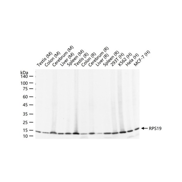
Figure 1. 25 ug total protein per lane of various lysates (see on figure) probed with RPS19 monoclonal antibody, unconjugated (SLA0882M) at 1:1000 dilution and 4°C overnight incubation. Followed by conjugated secondary antibody incubation at r.t. for 60 min.
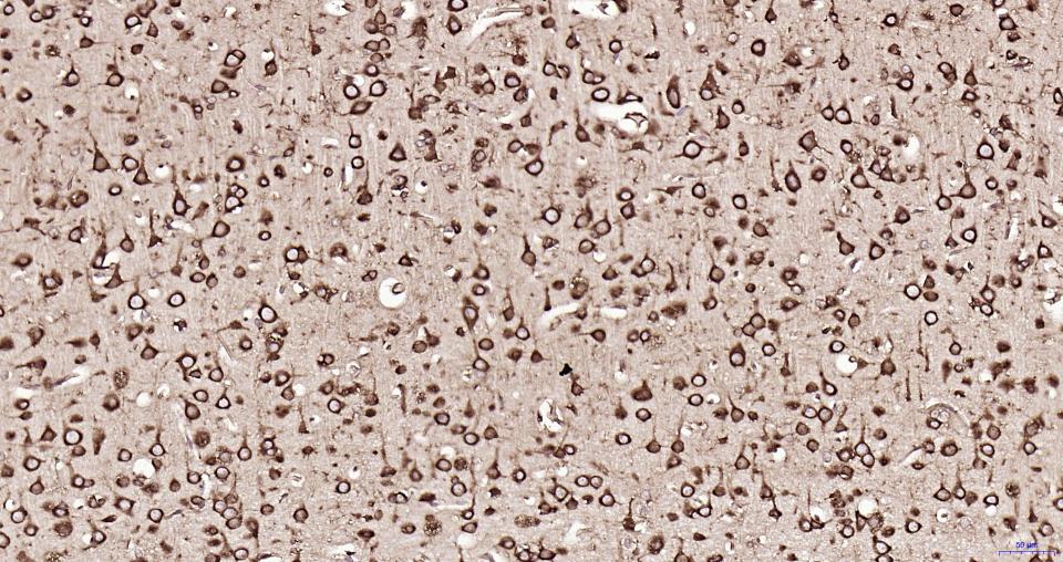
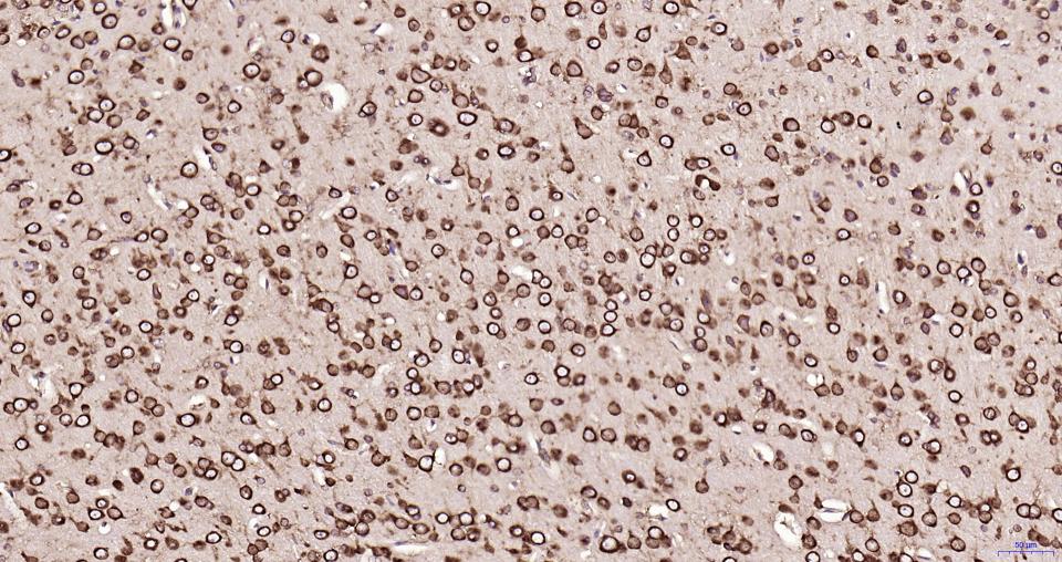
Figure 2. Paraformaldehyde-fixed, paraffin embedded Rat Cerebrum; Antigen retrieval by boiling in sodium citrate buffer (pH6.0) for 15 min; Antibody incubation with RPS19 Monoclonal Antibody, Unconjugated(SLA0882M) at 1:200 overnight at 4°C, followed by conjugation to the SP Kit(Rabbit, SP-0023) and DAB (C-0010) staining.
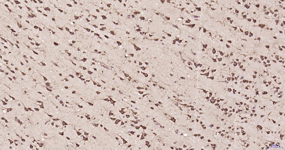
Figure 3. Paraformaldehyde-fixed, paraffin embedded Mouse Cerebrum; Antigen retrieval by boiling in sodium citrate buffer (pH6.0) for 15 min; Antibody incubation with RPS19 Monoclonal Antibody, Unconjugated(SLA0882M) at 1:200 overnight at 4°C, followed by conjugation to the SP Kit(Rabbit, SP-0023) and DAB (C-0010) staining.
Figure 4. Paraformaldehyde-fixed, paraffin embedded Human Cerebrum; Antigen retrieval by boiling in sodium citrate buffer (pH6.0) for 15 min; Antibody incubation with RPS19 Monoclonal Antibody, Unconjugated(SLA0882M) at 1:200 overnight at 4°C, followed by conjugation to the SP Kit(Rabbit, SP-0023) and DAB (C-0010) staining.
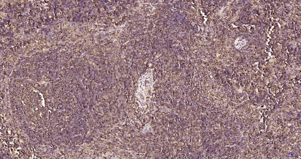
Figure 5. Paraformaldehyde-fixed, paraffin embedded Rat Spleen; Antigen retrieval by boiling in sodium citrate buffer (pH6.0) for 15 min; Antibody incubation with RPS19 Monoclonal Antibody, Unconjugated(SLA0882M) at 1:200 overnight at 4°C, followed by conjugation to the SP Kit(Rabbit, SP-0023) and DAB (C-0010) staining.
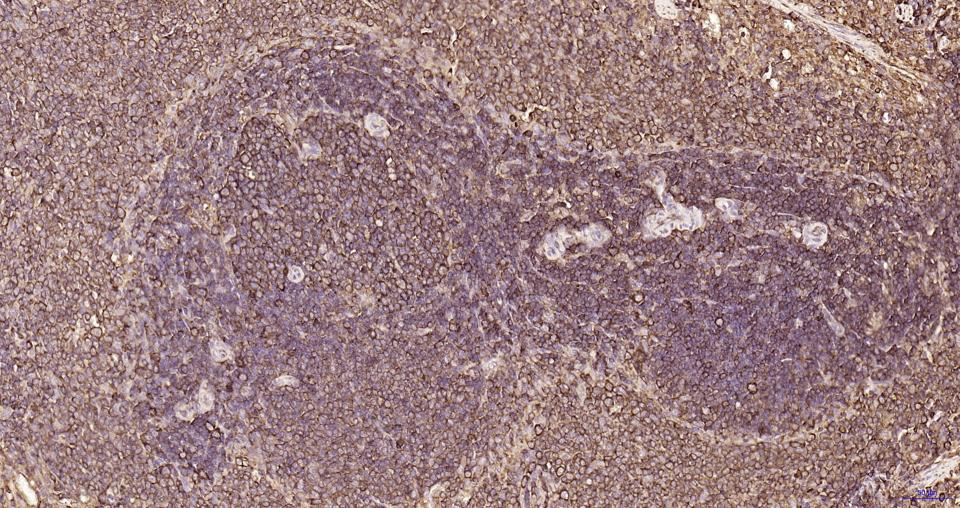
Figure 6. Paraformaldehyde-fixed, paraffin embedded Human Spleen; Antigen retrieval by boiling in sodium citrate buffer (pH6.0) for 15 min; Antibody incubation with RPS19 Monoclonal Antibody, Unconjugated(SLA0882M) at 1:200 overnight at 4°C, followed by conjugation to the SP Kit(Rabbit, SP-0023) and DAB (C-0010) staining.
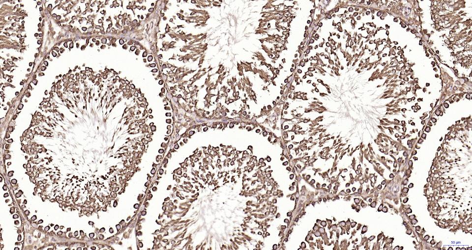
Figure 7. Paraformaldehyde-fixed, paraffin embedded Rat Testicles; Antigen retrieval by boiling in sodium citrate buffer (pH6.0) for 15 min; Antibody incubation with RPS19 Monoclonal Antibody, Unconjugated(SLA0882M) at 1:200 overnight at 4°C, followed by conjugation to the SP Kit(Rabbit, SP-0023) and DAB (C-0010) staining.
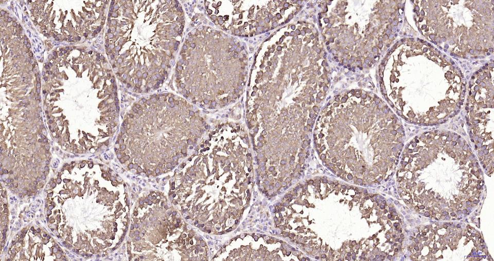
Figure 8. Paraformaldehyde-fixed, paraffin embedded Mouse Testicles; Antigen retrieval by boiling in sodium citrate buffer (pH6.0) for 15 min; Antibody incubation with RPS19 Monoclonal Antibody, Unconjugated(SLA0882M) at 1:200 overnight at 4°C, followed by conjugation to the SP Kit(Rabbit, SP-0023) and DAB (C-0010) staining.
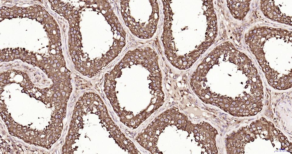
Figure 9. Paraformaldehyde-fixed, paraffin embedded Human Testicles; Antigen retrieval by boiling in sodium citrate buffer (pH6.0) for 15 min; Antibody incubation with RPS19 Monoclonal Antibody, Unconjugated(SLA0882M) at 1:200 overnight at 4°C, followed by conjugation to the SP Kit(Rabbit, SP-0023) and DAB (C-0010) staining.
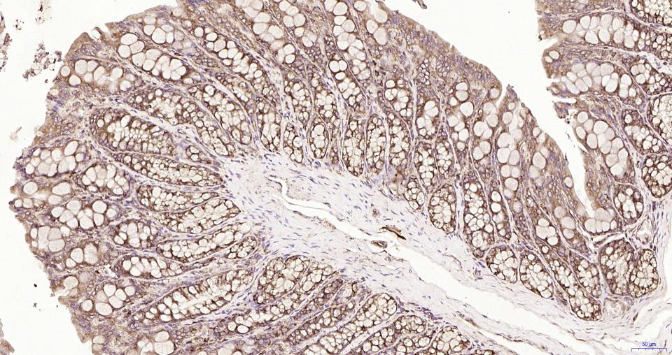
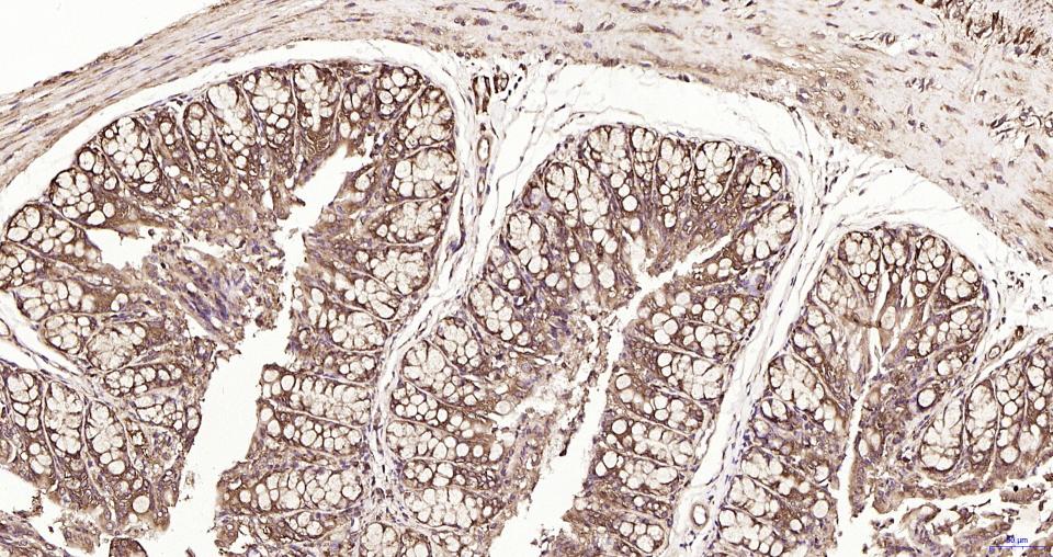
Figure 10. Paraformaldehyde-fixed, paraffin embedded Rat Colon; Antigen retrieval by boiling in sodium citrate buffer (pH6.0) for 15 min; Antibody incubation with RPS19 Monoclonal Antibody, Unconjugated(SLA0882M) at 1:200 overnight at 4°C, followed by conjugation to the SP Kit(Rabbit, SP-0023) and DAB (C-0010) staining.
Figure 11. Paraformaldehyde-fixed, paraffin embedded Mouse Colon; Antigen retrieval by boiling in sodium citrate buffer (pH6.0) for 15 min; Antibody incubation with RPS19 Monoclonal Antibody, Unconjugated(SLA0882M) at 1:200 overnight at 4°C, followed by conjugation to the SP Kit(Rabbit, SP-0023) and DAB (C-0010) staining.
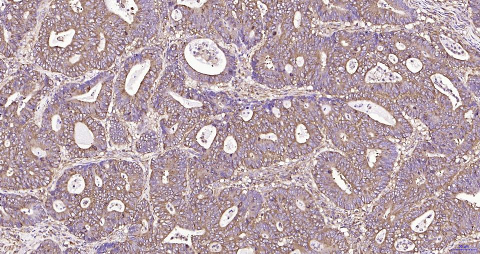
Figure 12. Paraformaldehyde-fixed, paraffin embedded Human Colon Cancer; Antigen retrieval by boiling in sodium citrate buffer (pH6.0) for 15 min; Antibody incubation with RPS19 Monoclonal Antibody, Unconjugated(SLA0882M) at 1:200 overnight at 4°C, followed by conjugation to the SP Kit(Rabbit, SP-0023) and DAB (C-0010) staining.
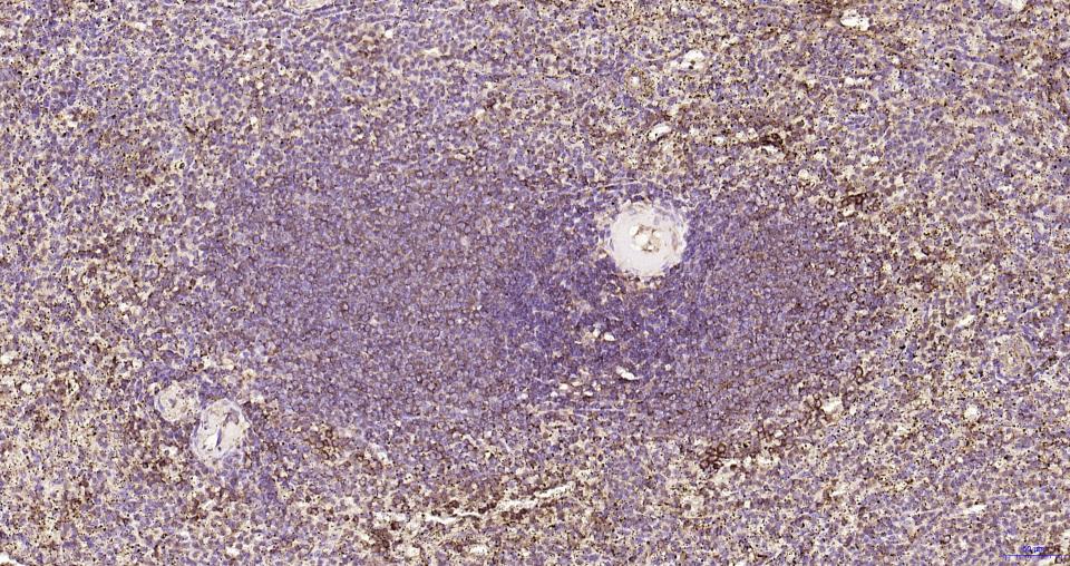
Figure 13. Paraformaldehyde-fixed, paraffin embedded MouseSpleen; Antigen retrieval by boiling in sodium citrate buffer (pH6.0) for 15 min; Antibody incubation with RPS19 Monoclonal Antibody, Unconjugated(SLA0882M) at 1:200 overnight at 4°C, followed by conjugation to the SP Kit(Rabbit, SP-0023) and DAB (C-0010) staining.
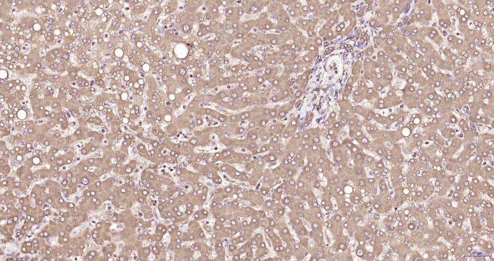
Figure 14. Paraformaldehyde-fixed, paraffin embedded Human Liver; Antigen retrieval by boiling in sodium citrate buffer (pH6.0) for 15 min; Antibody incubation with RPS19 Monoclonal Antibody, Unconjugated(SLA0882M) at 1:200 overnight at 4°C, followed by conjugation to the SP Kit(Rabbit, SP-0023) and DAB (C-0010) staining.
Figure 15. RPS19 was immunoprecipitated from HeLa cell lysate with SLA0882M at 1/100 dilution. Western blot was performed from the immunoprecipitate using protein A/G beads. HRP conjugated Goat Anti-Rabbit IgG was used as secondary antibody. Lane 1: HeLa cell lysate (Input). Lane 2: SLA0882M IP in HeLa cell lysate. Lane 3: native Rabbit IgG IP in HeLa cell lysate (negative control).
FOR RESEARCH USE ONLY. Not for use in diagnostic or therapeutic procedures.
Partial purchase records(bought amounts latest0)
User Comment(Total0User Comment Num)
- No comment


 +86 571 56623320

 SUNLONG BIOTECH


