Details
Product Name Rabbit Anti-GRIN2B Polyclonal Antibody Cat No. SLA0294 Chinese Name 磷酸化谷氨酸受体2B抗体 Alias DEE27; EIEE27; GluN2B; MRD6; NMDAR2B; NR2B; NR3; hNR3; GluRepsilon2; NMDE2_HUMAN; GRIN2B; Glutamate [NMDA] receptor subunit epsilon-2; N-methyl D-aspartate receptor subtype 2B (NMDAR2B | NR2B); N-methyl-D-aspartate receptor subunit 3 (NR3 | hNR3); NMDE2_MOUSE; NMDE2_RAT; glutamate ionotropic receptor NMDA type subunit 2B; glutamate receptor, ionotropic, N-methyl D-aspartate 2B; GRIN2B | NMDAR2B (phospho-Y1252); p-NMDAR2B; phospho-NMDAR2B; p-GRIN2B; GRIN2B | NMDAR2B (phospho-Tyr1252) Host Species Rabbit Isotype IgG Clonality Polyclonal React Species Human,Mouse,Rat Applications IHC-P (1:100-500)
IHC-F (1:100-500)
IF (1:100-500)
ICC/IF (1:50-200)
Not yet tested in other applications.
Optimal working dilutions must be determined by the end user.Immunogen KLH conjugated synthesised phosphopeptide derived from human NMDAR2B around the phosphorylation site of Tyr1252 Purification affinity purified by Protein A Concentration 1mg/ml Predicted MW. 164 kDa Gene ID 2904 Swiss-Prot Q13224 Storage 0.01M TBS (pH7.4) with 1% BSA, 0.02% Proclin300 and 50% Glycerol.
Shipped at 4℃. Store at -20℃ for one year. Avoid repeated freeze/thaw cycles.Background N-methyl-D-aspartate (NMDA) receptors are a class of ionotropic glutamate receptors. NMDA receptor channel has been shown to be involved in long-term potentiation, an activity-dependent increase in the efficiency of synaptic transmission thought to underlie certain kinds of memory and learning. NMDA receptor channels are heteromers composed of three different subunits: NR1 (GRIN1), NR2 (GRIN2A, GRIN2B, GRIN2C, or GRIN2D) and NR3 (GRIN3A or GRIN3B). The NR2 subunit acts as the agonist binding site for glutamate. This receptor is the predominant excitatory neurotransmitter receptor in the mammalian brain. [provided by RefSeq, Jul 2008]. Attention This product as supplied is intended for research use only, not for use in human, therapeutic or diagnostic applications.
Validation Images
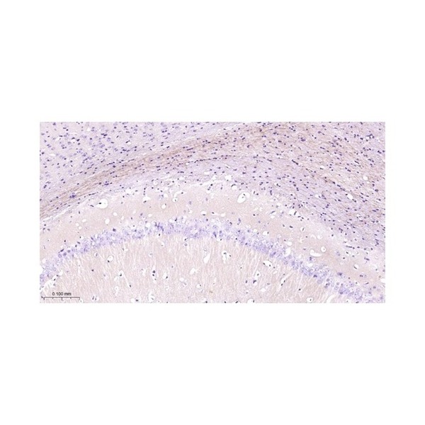
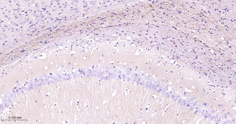
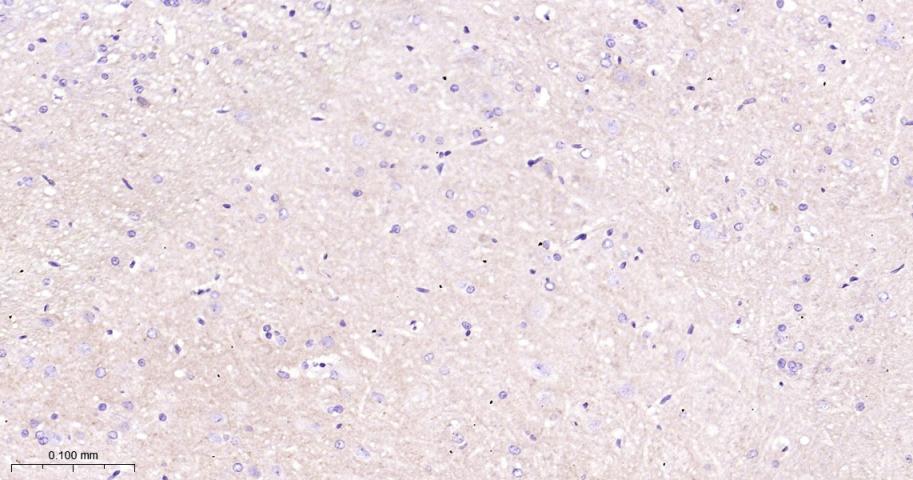
Figure 1. Paraformaldehyde-fixed, paraffin embedded Mouse seahorse; Antigen retrieval by boiling in sodium citrate buffer (pH6.0) for 15 min; The section was incubated with Phospho-NMDAR2B (Tyr1252) Polyclonal Antibody, Unconjugated (SLA0294) at 1:200 overnight at 4°C, followed by conjugation to the SL0295G-HRP and DAB (C-0010) staining.
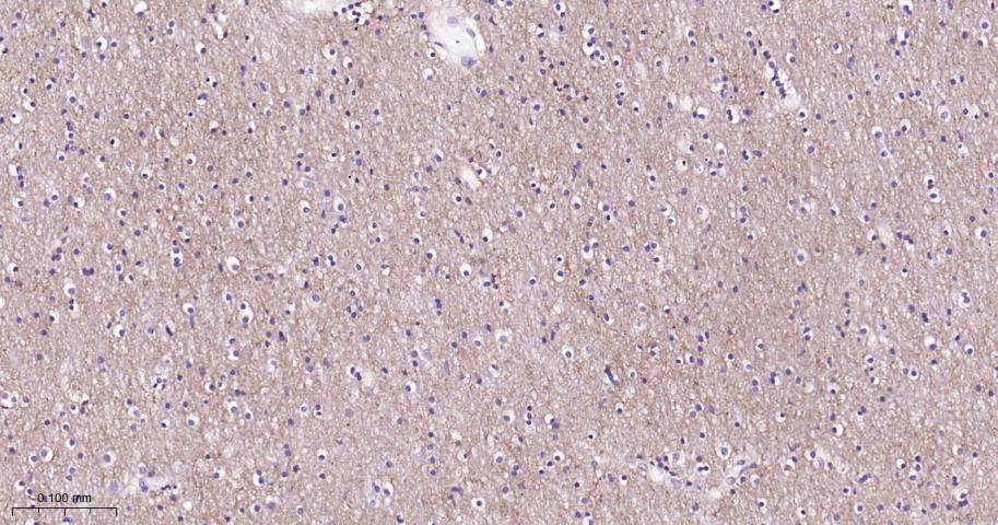
Figure 2. Paraformaldehyde-fixed, paraffin embedded Rat Cerebrum; Antigen retrieval by boiling in sodium citrate buffer (pH6.0) for 15 min; The section was incubated with Phospho-NMDAR2B (Tyr1252) Polyclonal Antibody, Unconjugated (SLA0294) at 1:200 overnight at 4°C, followed by conjugation to the SL0295G-HRP and DAB (C-0010) staining.
Figure 3. Paraformaldehyde-fixed, paraffin embedded Human Cerebrum; Antigen retrieval by boiling in sodium citrate buffer (pH6.0) for 15 min; The section was incubated with Phospho-NMDAR2B (Tyr1252) Polyclonal Antibody, Unconjugated (SLA0294) at 1:200 overnight at 4°C, followed by conjugation to the SL0295G-HRP and DAB (C-0010) staining.
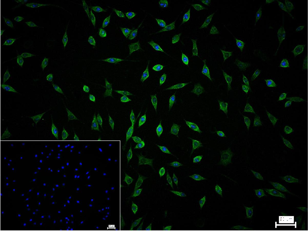
Figure 4. 4% Paraformaldehyde-fixed SH-SY5Y (H) cell; Triton X-100 at r.t. for 20 min; Antibody incubation with (Phospho-NMDAR2B (Tyr1252)) polyclonal Antibody, unconjugated (SLA0294) 1:100, 90 min at 37°C; followed by conjugated Goat Anti-Rabbit IgG antibody (green, SL40295G-FITC) at 37°C for 90 min, DAPI (blue, C02-04002) was used to stain the cell nuclei. PBS instead of the primary antibody was used as the blank control.
FOR RESEARCH USE ONLY. Not for use in diagnostic or therapeutic procedures.
Partial purchase records(bought amounts latest0)
User Comment(Total0User Comment Num)
- No comment


 +86 571 56623320

 SUNLONG BIOTECH


