Details
Product Name Recombinant Rabbit Anti-CASP3 Monoclonal Antibody Cat No. SLA0918M Chinese Name 半胱胺酸蛋白酶蛋白-3单克隆抗体 Alias CPP32; CPP32B; SCA-1; A830040C14Rik; AC-3; CASP-3; CC3; CPP-32; Caspase-3; Lice; Yama; mldy; CPP32-beta; CASP3_HUMAN; CASP3; Apopain; Cysteine protease CPP32 (CPP-32); Protein Yama; SREBP cleavage activity 1 (SCA-1); 3.4.22.56; CASP3_MOUSE; CASP3_PIG; CASP3_RABIT; Cysteine protease CPP32; CASP3_RAT; IRP; caspase 3; caspase 3, apoptosis-related cysteine protease; caspase 3, apoptosis-related cysteine peptidase Host Species Mouse Isotype IgG Clonality Monoclonal Clone No. 1B1 React Species Human Applications WB (1:1000-5000)
IHC-P (1:200-800)
IHC-F (1:200-800)
IF (1:200-800)
Flow-Cyt (1μg/Test)
ICC/IF (1:50-200)
Not yet tested in other applications.
Optimal working dilutions must be determined by the end user.Immunogen Recombinant human Caspase-3 protein Purification affinity purified by Protein A Concentration 1mg/ml Predicted MW. 32 kDa Observed MW. 35(pro-Caspase3),15-17(Cleaved Caspase-3) kDa Subcellular Location Cytoplasm Gene ID 836 Swiss-Prot P42574 Storage 0.01M TBS (pH7.4) with 1% BSA, 0.02% Proclin300 and 50% Glycerol.
Shipped at 4℃. Store at -20℃ for one year. Avoid repeated freeze/thaw cycles.Background The caspase family of cysteine proteases play a key role in apoptosis. Caspase 3 is the most extensively studied apoptotic protein among caspase family members. Caspase 3 is synthesized as inactive pro enzyme that is processed in cells undergoing apoptosis by self proteolysis and/or cleavage by other upstream proteases (e.g. Caspases 8, 9 and 10). The processed form of Caspase 3 consists of large (17kDa) and small (12kDa) subunits which associate to form an active enzyme. Caspase 3 is cleaved at Asp28 Ser29 and Asp175 Ser176. The active Caspase 3 proteolytically cleaves and activates other caspases (e.g. Caspases 6, 7 and 9), as well as relevant targets in the cells (e.g. PARP and DFF). Alternative splicing of this gene results in two transcript variants which encode the same protein. In immunohistochemical studies Caspase 3 expression has been shown to be widespread but not present in all cell types (e.g. commonly reported in epithelial cells of skin, renal proximal tubules and collecting ducts). Differences in the level of Caspase 3 have been reported in cells of short lived nature (eg germinal centre B cells) and those that are long lived (eg mantle zone B cells). Caspase 3 is the predominant caspase involved in the cleavage of amyloid beta 4A precursor protein, which is associated with neuronal death in Alzheimer's disease. Attention This product as supplied is intended for research use only, not for use in human, therapeutic or diagnostic applications.
Validation Images
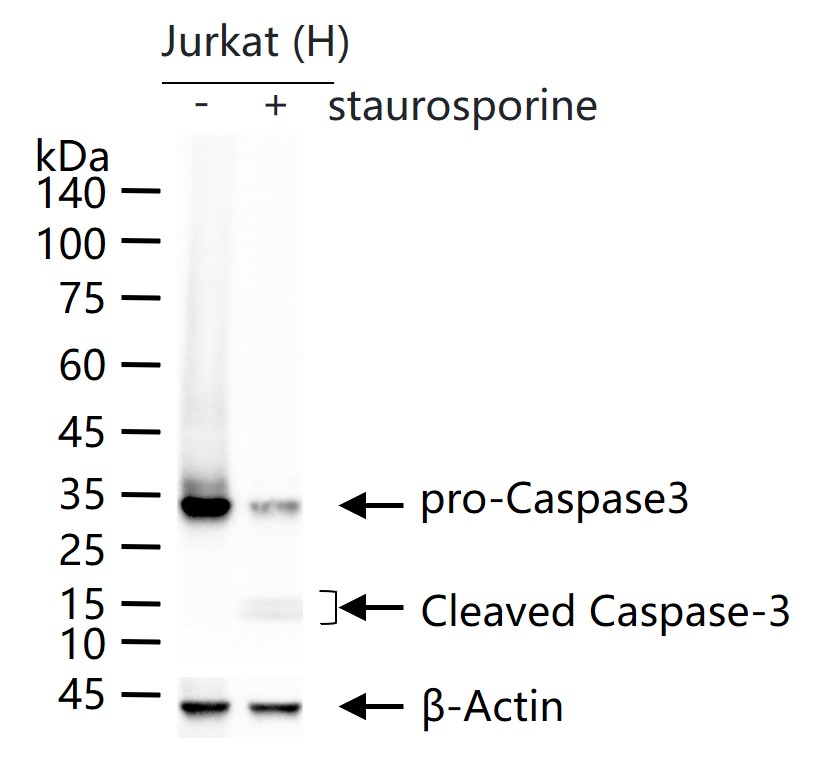
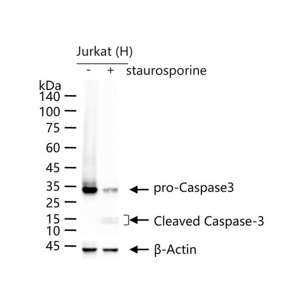
Figure 1. Jurkat (H) cells were treated with or without staurosporine treated (1uM) for 4 h, 25 μg total protein per lane of cell lysates (see on figure) probed with Caspase-3 monoclonal antibody, unconjugated (SLA0918M) at 1:1000 dilution and 4°C overnight incubation. Followed by conjugated secondary antibody incubation at r.t. for 60 min
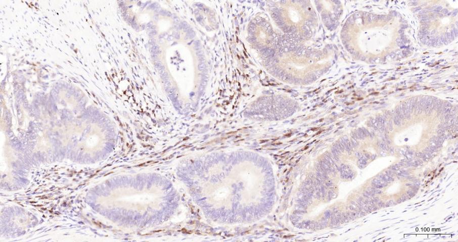
Figure 2. Paraformaldehyde-fixed, paraffin embedded Human Colon Cancer; Antigen retrieval by boiling in sodium citrate buffer (pH6.0) for 15 min; The section was incubated with Caspase-3 Monoclonal Antibody, Unconjugated (SLA0918M) at 1:200 overnight at 4°C, followed by conjugation to the SL40296G-HRP and DAB (C-0010) staining
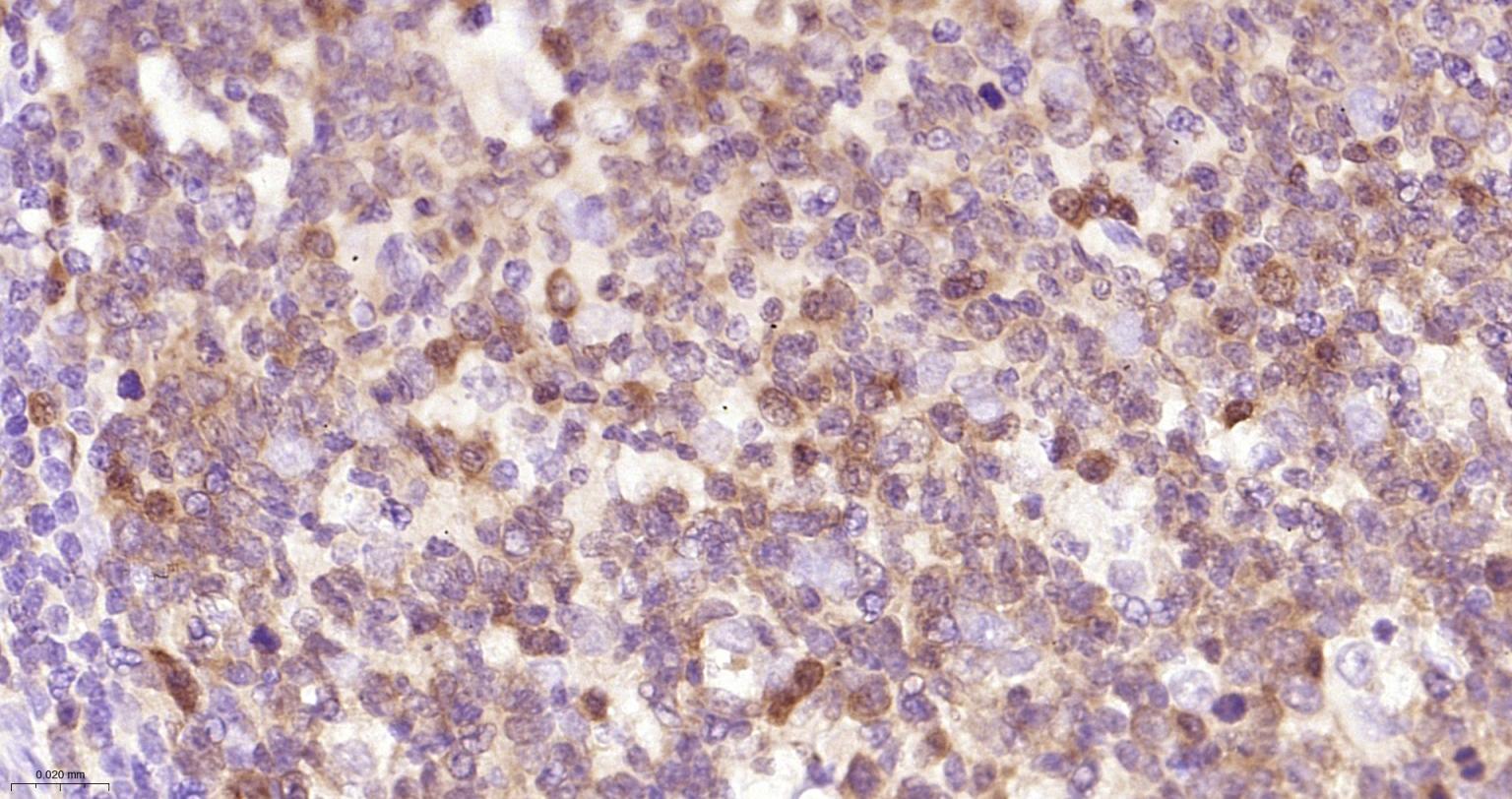
Figure 3. Paraformaldehyde-fixed, paraffin embedded Human Tonsil; Antigen retrieval by boiling in sodium citrate buffer (pH6.0) for 15 min; The section was incubated with Caspase-3 Monoclonal Antibody, Unconjugated (SLA0918M) at 1:200 overnight at 4°C, followed by conjugation to the SL40296G-HRP and DAB (C-0010) staining
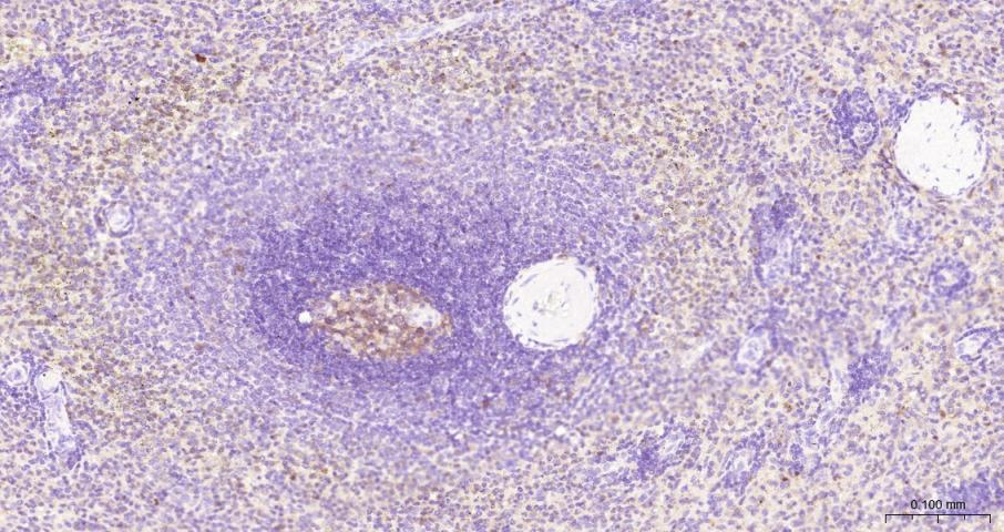
Figure 4. Paraformaldehyde-fixed, paraffin embedded Human Spleen; Antigen retrieval by boiling in sodium citrate buffer (pH6.0) for 15 min; The section was incubated with Caspase-3 Monoclonal Antibody, Unconjugated (SLA0918M) at 1:200 overnight at 4°C, followed by conjugation to the SL40296G-HRP and DAB (C-0010) staining
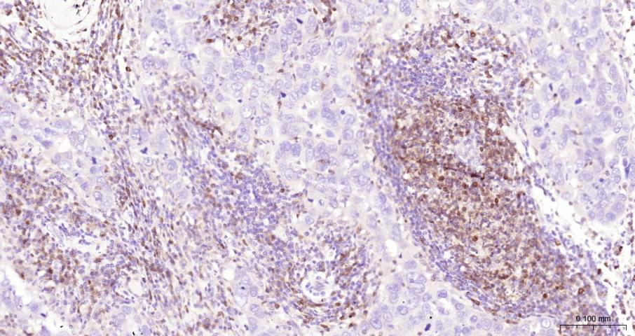
Figure 5. Paraformaldehyde-fixed, paraffin embedded Human Breast Cancer; Antigen retrieval by boiling in sodium citrate buffer (pH6.0) for 15 min; The section was incubated with Caspase-3 Monoclonal Antibody, Unconjugated (SLA0918M) at 1:200 overnight at 4°C, followed by conjugation to the SL40296G-HRP and DAB (C-0010) staining
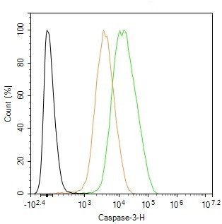
Figure 6. The Jurkat (H) cells were fixed with 4% PFA (10 min at r.t.) and then permeabilized with 90% ice-cold methanol for 20 min at -20℃,the cells then were incubated in 5%BSA to block non-specific protein-protein interactions (30 min at r.t.), followed by secondary antibody incubation for 40 min at room temperature. Primary Antibody (green):Mouse Anti-Caspase-3 antibody (SLA0918M): 1 μg/10^6 cells; Isotype Control (orange): Mouse IgG (SL0296P). Blank control (black): PBS. Acquisition of 20,000 events was performed
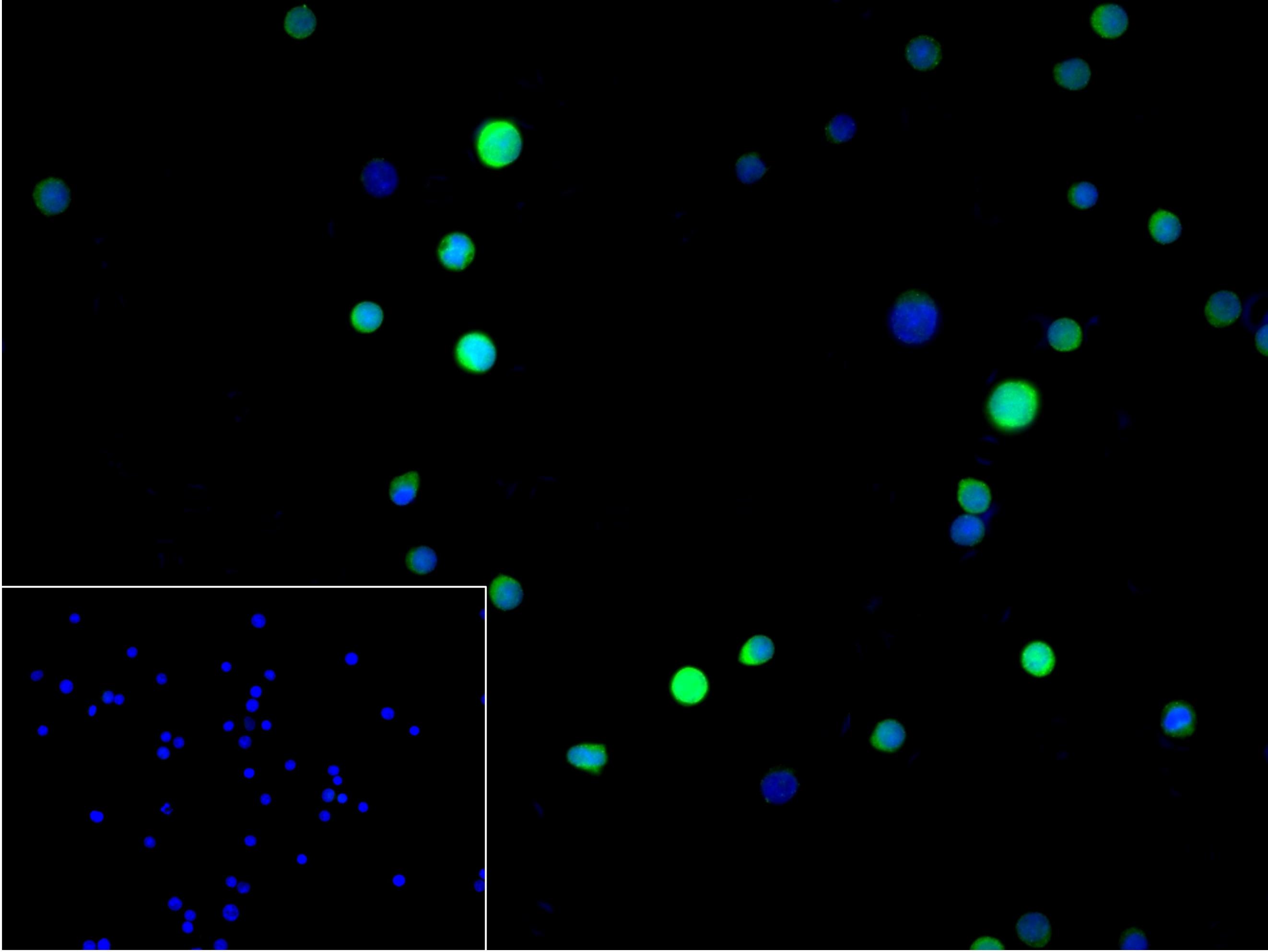
Figure 7. 4% Paraformaldehyde-fixed Jurkat (H) cell; Triton X-100 at r.t. for 20 min; Antibody incubation with (Caspase-3) monoclonal Antibody, unconjugated (SLA0918M) 1:100, 90 min at 37°C; followed by conjugated Goat Anti-Mouse IgG antibody (green, SL60296G-FITC) at 37°C for 90 min, DAPI (blue, C02-04002) was used to stain the cell nuclei. PBS instead of the primary antibody was used as the blank control
FOR RESEARCH USE ONLY. Not for use in diagnostic or therapeutic procedures.
Partial purchase records(bought amounts latest0)
User Comment(Total0User Comment Num)
- No comment


 +86 571 56623320

 SUNLONG BIOTECH


