Details
Product Name Recombinant Rabbit Anti-NCAM1 Monoclonal Antibody Cat No. SLA0598M Chinese Name 神经细胞粘附分子1单克隆抗体 Alias CD56; MSK39; NCAM; E-NCAM; NCAM-1; N-CAM; N-CAM-1; NCAM-C; NCAMC; NCAM1_HUMAN; NCAM1; NCAM1_MOUSE; NCAM1_RAT; neural cell adhesion molecule 1 Host Species Mouse Clonality Monoclonal Clone No. 5B7 React Species Human Applications IHC-P (1:100-500)
IHC-F (1:100-500)
IF (1:100-500)
Not yet tested in other applications.
Optimal working dilutions must be determined by the end user.Immunogen Recombinant human CD56 protein Purification affinity purified by Protein A Predicted MW. 92 kDa Subcellular Location Membrane Gene ID 4684 Swiss-Prot P13591 Storage 0.01M TBS (pH7.4) with 1% BSA, 0.02% Proclin300 and 50% Glycerol.
Shipped at 4℃. Store at -20℃ for one year. Avoid repeated freeze/thaw cycles.Background This gene encodes a cell adhesion protein which is a member of the immunoglobulin superfamily. The encoded protein is involved in cell-to-cell interactions as well as cell-matrix interactions during development and differentiation. The encoded protein has been shown to be involved in development of the nervous system, and for cells involved in the expansion of T cells and dendritic cells which play an important role in immune surveillance. Alternative splicing results in multiple transcript variants. [provided by RefSeq, Jun 2011]
Attention This product as supplied is intended for research use only, not for use in human, therapeutic or diagnostic applications.
Validation Images
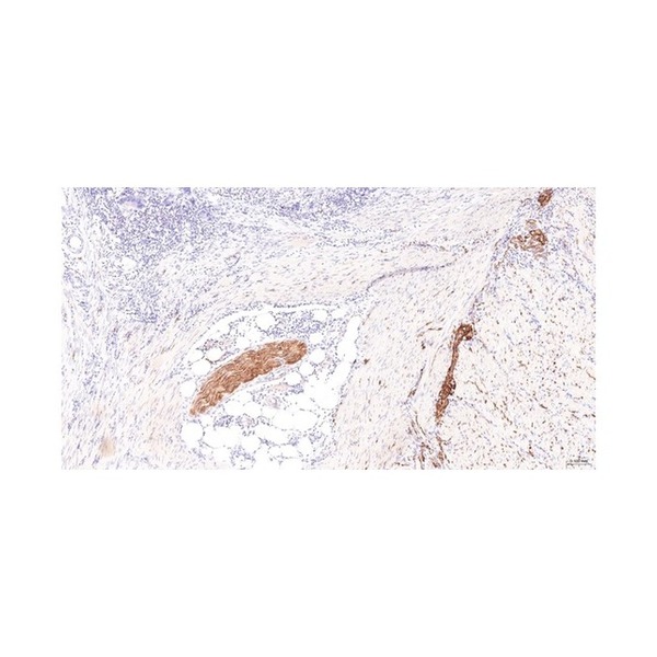
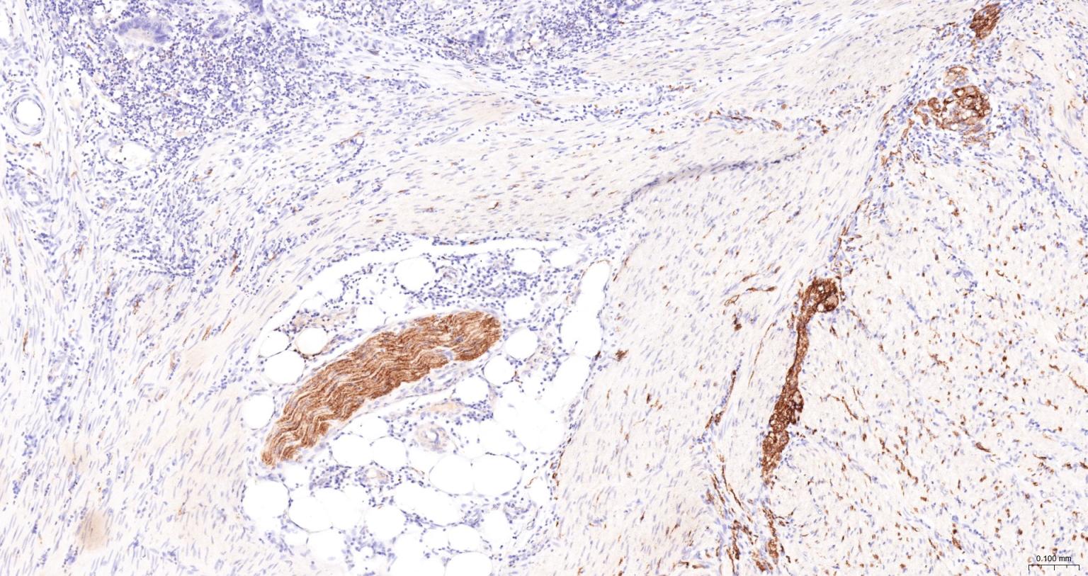
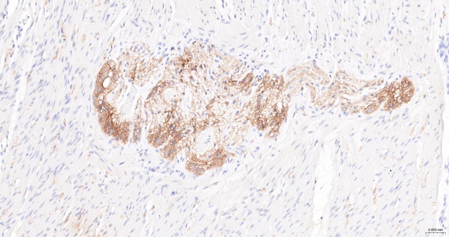
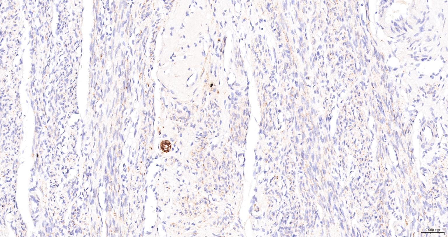
Figure 1. Paraformaldehyde-fixed, paraffin embedded Human Colon Cancer; Antigen retrieval by boiling in sodium citrate buffer (pH6.0) for 15 min; The section was incubated with CD56 Monoclonal Antibody, Unconjugated (SLA0598M) at 1:200 overnight at 4°C, followed by conjugation to the SL40296G-HRP and DAB (C-0010) staining.
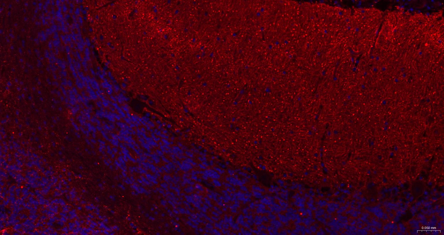
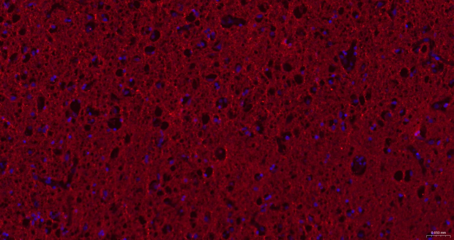
Figure 2. Paraformaldehyde-fixed, paraffin embedded Human Colon; Antigen retrieval by boiling in sodium citrate buffer (pH6.0) for 15 min; The section was incubated with CD56 Monoclonal Antibody, Unconjugated (SLA0598M) at 1:200 overnight at 4°C, followed by conjugation to the SL40296G-HRP and DAB (C-0010) staining.
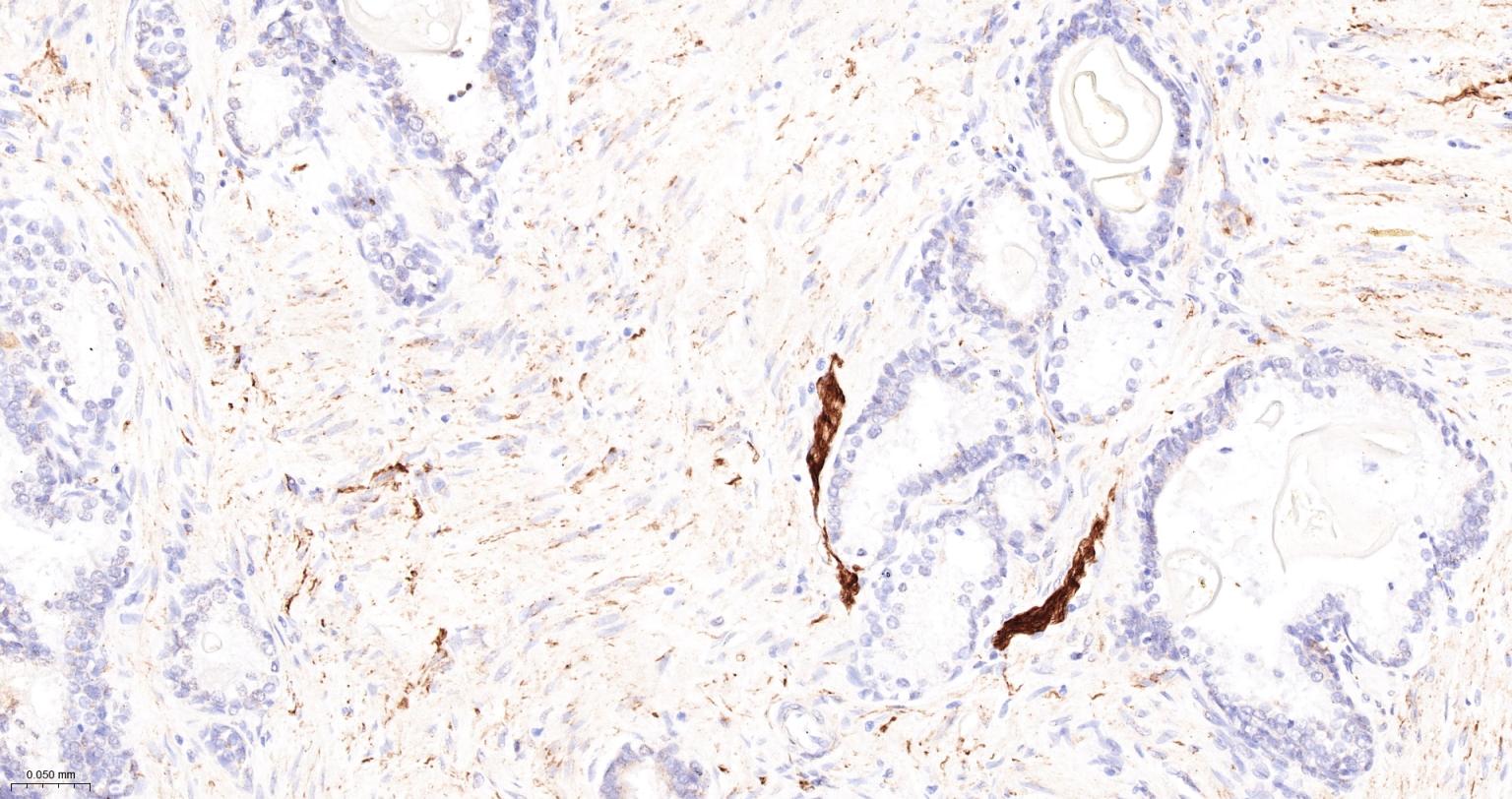
Figure 3. Paraformaldehyde-fixed, paraffin embedded Human Prostate; Antigen retrieval by boiling in sodium citrate buffer (pH6.0) for 15 min; The section was incubated with CD56 Monoclonal Antibody, Unconjugated (SLA0598M) at 1:200 overnight at 4°C, followed by conjugation to the SL40296G-HRP and DAB (C-0010) staining.
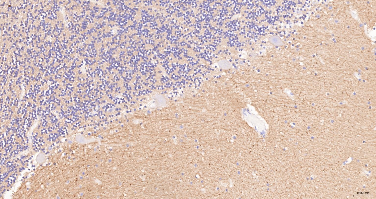
Figure 4. Paraformaldehyde-fixed, paraffin embedded Human Cerebellum; Antigen retrieval by boiling in sodium citrate buffer (pH6.0) for 15 min; The section was incubated with CD56 Monoclonal Antibody, Unconjugated (SLA0598M) at 1:200 overnight at 4°C, followed by conjugation to the SL40296G-HRP and DAB (C-0010) staining.
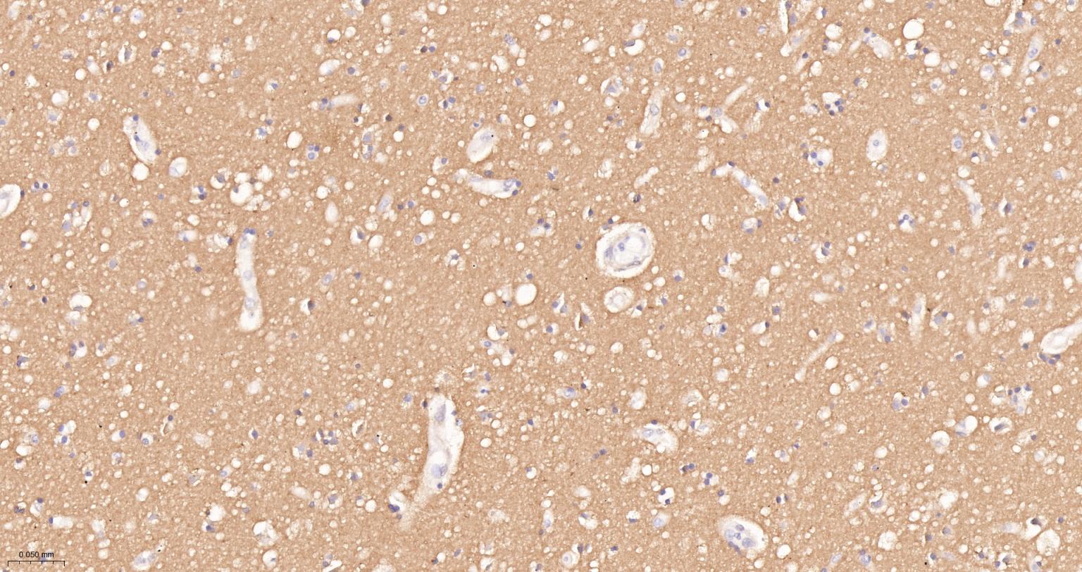
Figure 5. Paraformaldehyde-fixed, paraffin embedded Human Left Parietal Lobe; Antigen retrieval by boiling in sodium citrate buffer (pH6.0) for 15 min; The section was incubated with CD56 Monoclonal Antibody, Unconjugated (SLA0598M) at 1:200 overnight at 4°C, followed by conjugation to the SL40296G-HRP and DAB (C-0010) staining.
Figure 6. Paraformaldehyde-fixed, paraffin embedded Human Endometrial Cancer; Antigen retrieval by boiling in sodium citrate buffer (pH6.0) for 15 min; The section was incubated with CD56 Monoclonal Antibody, Unconjugated (SLA0598M) at 1:200 overnight at 4°C, followed by conjugation to the SL40296G-HRP and DAB (C-0010) staining.
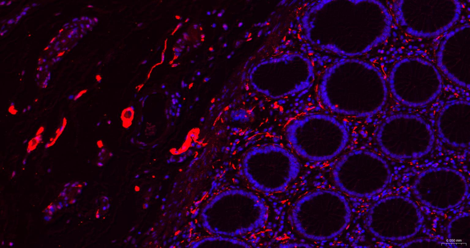
Figure 7. Paraformaldehyde-fixed, paraffin embedded Human Colon; Antigen retrieval by boiling in sodium citrate buffer (pH6.0) for 15 min; The section was incubated with CD56 Monoclonal Antibody, Unconjugated (SLA0598M) at 1:200 overnight at 4°C. Followed by conjugated Goat Anti-Mouse IgG antibody (Red, SL0296G-BF594), DAPI (blue, C02-04002) was used to stain the cell nuclei.
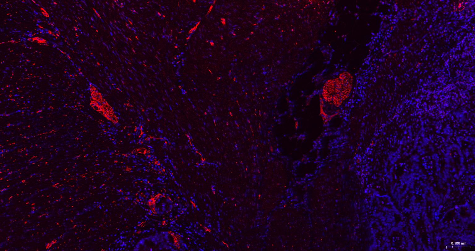
Figure 8. Paraformaldehyde-fixed, paraffin embedded Human Colon Cancer; Antigen retrieval by boiling in sodium citrate buffer (pH6.0) for 15 min; The section was incubated with CD56 Monoclonal Antibody, Unconjugated (SLA0598M) at 1:200 overnight at 4°C. Followed by conjugated Goat Anti-Mouse IgG antibody (Red, SL0296G-BF594), DAPI (blue, C02-04002) was used to stain the cell nuclei.
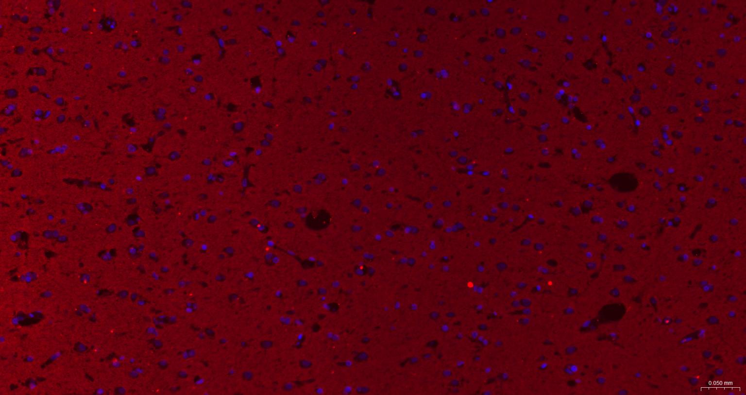
Figure 9. Paraformaldehyde-fixed, paraffin embedded Human Cerebrum; Antigen retrieval by boiling in sodium citrate buffer (pH6.0) for 15 min; The section was incubated with CD56 Monoclonal Antibody, Unconjugated (SLA0598M) at 1:200 overnight at 4°C. Followed by conjugated Goat Anti-Mouse IgG antibody (Red, SL0296G-BF594), DAPI (blue, C02-04002) was used to stain the cell nuclei.
Figure 10. Paraformaldehyde-fixed, paraffin embedded Human Cerebellum; Antigen retrieval by boiling in sodium citrate buffer (pH6.0) for 15 min; The section was incubated with CD56 Monoclonal Antibody, Unconjugated (SLA0598M) at 1:200 overnight at 4°C. Followed by conjugated Goat Anti-Mouse IgG antibody (Red, SL0296G-BF594), DAPI (blue, C02-04002) was used to stain the cell nuclei.
Figure 11. Paraformaldehyde-fixed, paraffin embedded Human Left parietal lobe; Antigen retrieval by boiling in sodium citrate buffer (pH6.0) for 15 min; The section was incubated with CD56 Monoclonal Antibody, Unconjugated (SLA0598M) at 1:200 overnight at 4°C. Followed by conjugated Goat Anti-Mouse IgG antibody (Red, SL0296G-BF594), DAPI (blue, C02-04002) was used to stain the cell nuclei.
FOR RESEARCH USE ONLY. Not for use in diagnostic or therapeutic procedures.
Partial purchase records(bought amounts latest0)
User Comment(Total0User Comment Num)
- No comment


 +86 571 56623320

 SUNLONG BIOTECH


