Details
Product Name Recombinant Rabbit Anti-MS4A1 Monoclonal Antibody Cat No. SLA3106M Chinese Name CD20重组兔单抗 Alias B1; Bp35; CD20; CVID5; FMC7; LEU-16; S7; CD20_HUMAN; MS4A1; B-lymphocyte surface antigen B1; Leukocyte surface antigen Leu-16; Membrane-spanning 4-domains subfamily A member 1; membrane spanning 4-domains A1; membrane-spanning 4-domains, subfamily A, member 1 Host Species Rabbit Isotype IgG Clonality Monoclonal Clone No. 1B9 React Species Human Applications WB (1:1000-5000)
IHC-P (1:200-2000)
IHC-F (1:200-2000)
IF (1:200-2000)
Flow-Cyt (1:100-200)
Not yet tested in other applications.
Optimal working dilutions must be determined by the end user.Immunogen A synthesized peptide derived from human CD20 Purification affinity purified by Protein A Concentration 1mg/ml Predicted MW. 33 kDa Observed MW. 36 kDa Subcellular Location Membrane Gene ID 931 Swiss-Prot P11836 Storage 0.01M TBS (pH7.4) with 1% BSA, 0.02% Proclin300 and 50% Glycerol.
Shipped at 4℃. Store at -20℃ for one year. Avoid repeated freeze/thaw cycles.Background CD20 is a non glycosylated protein with a molecular weight of 35 or 37 kDa depending on the degree of phosphorylation. Although not a member of the tetraspanin superfamily of cell surface receptors, it crosses the cell membrane four times. The CD20 antigen is present on human pre B lymphocytes and on B lymphocytes at all stages of maturation, except on plasma cells. Low level expression of the CD20 antigen has been detected on normal T lymphocytes. The CD20 molecule is involved in regulation of B cell differentiation, presumably via its reported function as a Ca++ channel subunit. CD20 is a non-glycosylated surface phosphoprotein that has a molecular weight range of 33-37 kDa depending on the degree of phosphorylation. CD20 is expressed on mature and most malignant B cells, in a subpopulation of T lymphocytes and follicular dendritic cells. CD20 expression on B cells is synchronous with the expression of surface IgM and it regulates transmembrane calcium conductance, cell cycle progression and B-cell proliferation. CD20 is also associated with lipid rafts, but the intensity of this association depends on extracellular triggering, employing CD20 conformational change, and/or BCR (B cell antigen receptor) aggregation. After the receptor ligation, BCR and CD20 colocalize and then rapidly dissociate before BCR endocytosis, whereas CD20 remains at the cell surface. CD20 serves as a useful target for antibody-mediated therapeutic depletion of B cells, as it is expressed at high levels on most B-cell malignancies, but does not become internalized or shed from the plasma membrane following monoclonal antibody treatment. Diseases associated with CD20 dysfunction include Ms4a1-related common variable immune deficiency. Attention This product as supplied is intended for research use only, not for use in human, therapeutic or diagnostic applications.
Validation Images
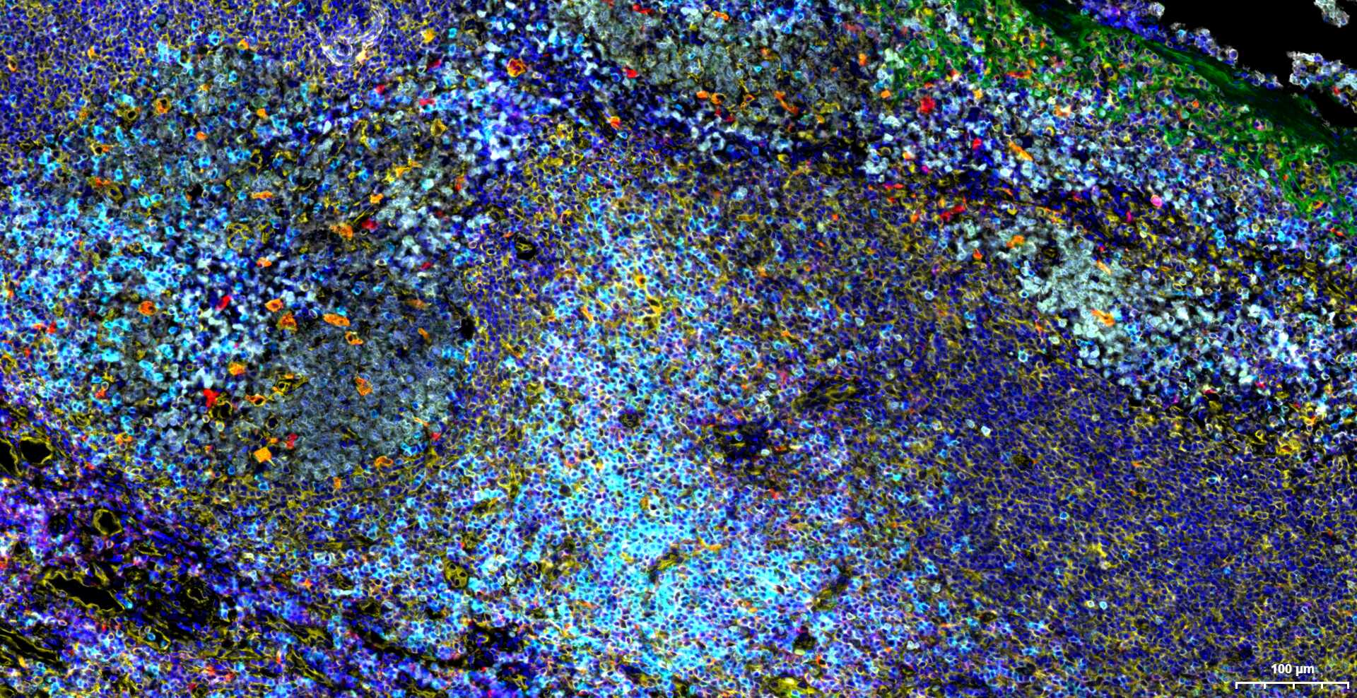
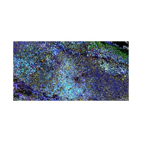
Figure 1. Paraformaldehyde-fixed, paraffin embedded Human Tonsil. Merged staining of anti-CD68 (SLM-60634R; 1:200; red) anti- CD20 (SLA3106M; 1:200; white) anti-CD3E (SLM-60002R; 1:200; light blue) anti-CD4 (SLM-52469R; 1:200; rose red) anti-Ki67 (SLM-52455R; 1:200; bean green) anti-Vimentin (SLM-33170M; 1:200; yellow) and anti-Pan Cytokeratin (SLM-34137M; 1:150; green) DAPI (dark blue) was used as a nuclear counter stain
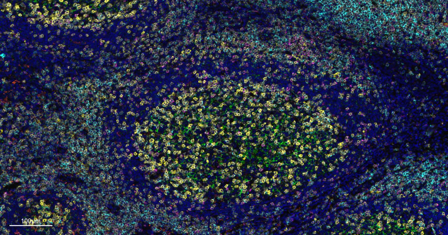
Figure 2. Paraformaldehyde-fixed, paraffin embedded Human Tonsil. Merged staining of anti-CD8A (SLA0485M; 1:200; pink) anti-CD4 (SLM-52469R; 1:200; yellow) anti-CD3E (SLM-60002R; 1:200; light blue) anti-CD68 (SLM-60634R; 1:200; white) anti-CD11c (SLM-61135R; 1:200; orange) and anti-CD20 (SLA3106M; 1:200; green) DAPI (dark blue) was used as a nuclear counter stain
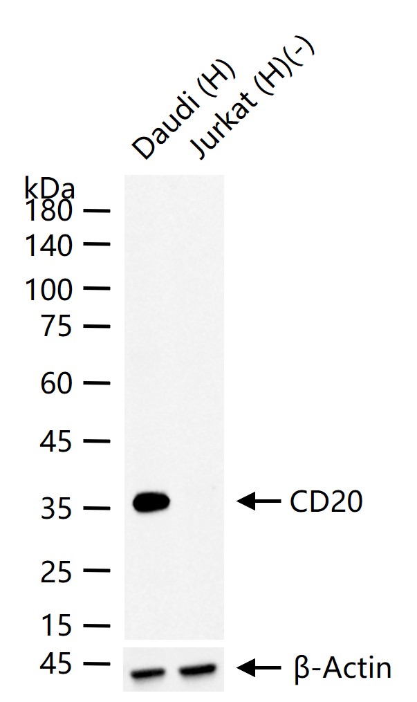
Figure 3. 25 ug total protein per lane of various lysates (see on figure) probed with CD20 monoclonal antibody, unconjugated (SLA3106M) at 1:1000 dilution and 4°C overnight incubation. Followed by conjugated secondary antibody incubation at r.t. for 60 min
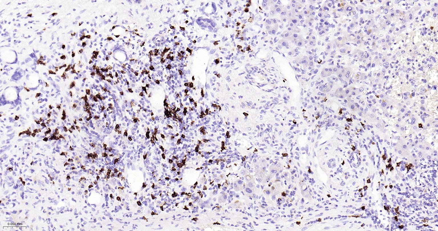
Figure 4. Paraformaldehyde-fixed, paraffin embedded Human Liver Cancer; Antigen retrieval by boiling in sodium citrate buffer (pH6.0) for 15 min; Antibody incubation with CD20 Monoclonal Antibody, Unconjugated(SLA3106M) at 1:200 overnight at 4°C, followed by conjugation to the SL0295G-HRP and DAB (C-0010) staining
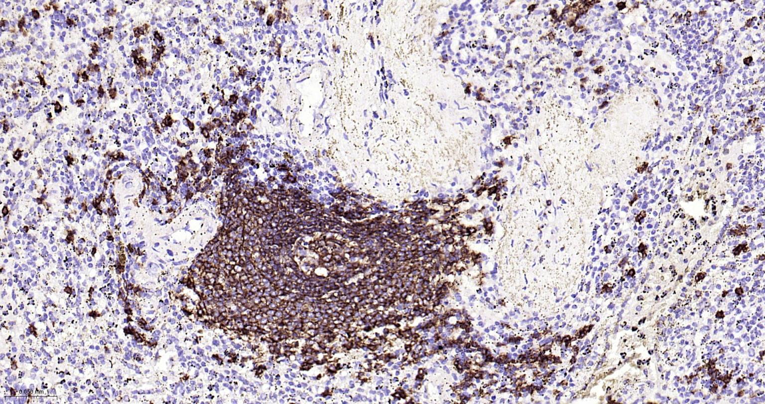
Figure 5. Paraformaldehyde-fixed, paraffin embedded Human Spleen; Antigen retrieval by boiling in sodium citrate buffer (pH6.0) for 15 min; Antibody incubation with CD20 Monoclonal Antibody, Unconjugated(SLA3106M) at 1:200 overnight at 4°C, followed by conjugation to the SL0295G-HRP and DAB (C-0010) staining
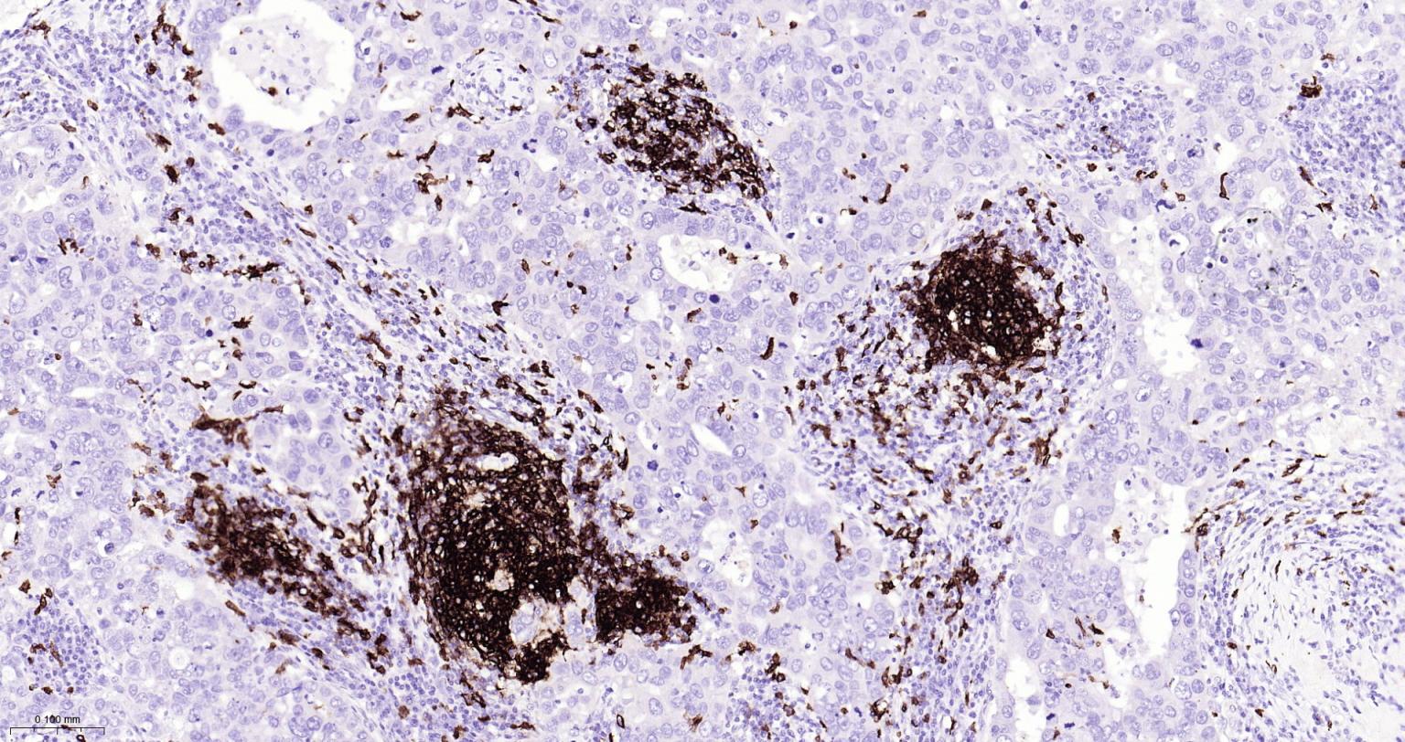
Figure 6. Paraformaldehyde-fixed, paraffin embedded Human Breast Cancer; Antigen retrieval by boiling in sodium citrate buffer (pH6.0) for 15 min; Antibody incubation with CD20 Monoclonal Antibody, Unconjugated(SLA3106M) at 1:200 overnight at 4°C, followed by conjugation to the SL0295G-HRP and DAB (C-0010) staining
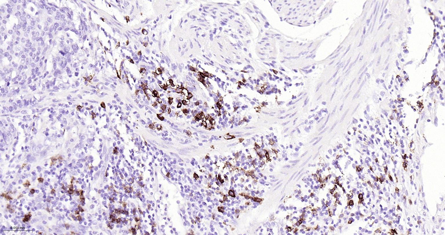
Figure 7. Paraformaldehyde-fixed, paraffin embedded Human Esophageal Cancer; Antigen retrieval by boiling in sodium citrate buffer (pH6.0) for 15 min; Antibody incubation with CD20 Monoclonal Antibody, Unconjugated(SLA3106M) at 1:200 overnight at 4°C, followed by conjugation to the SL0295G-HRP and DAB (C-0010) staining
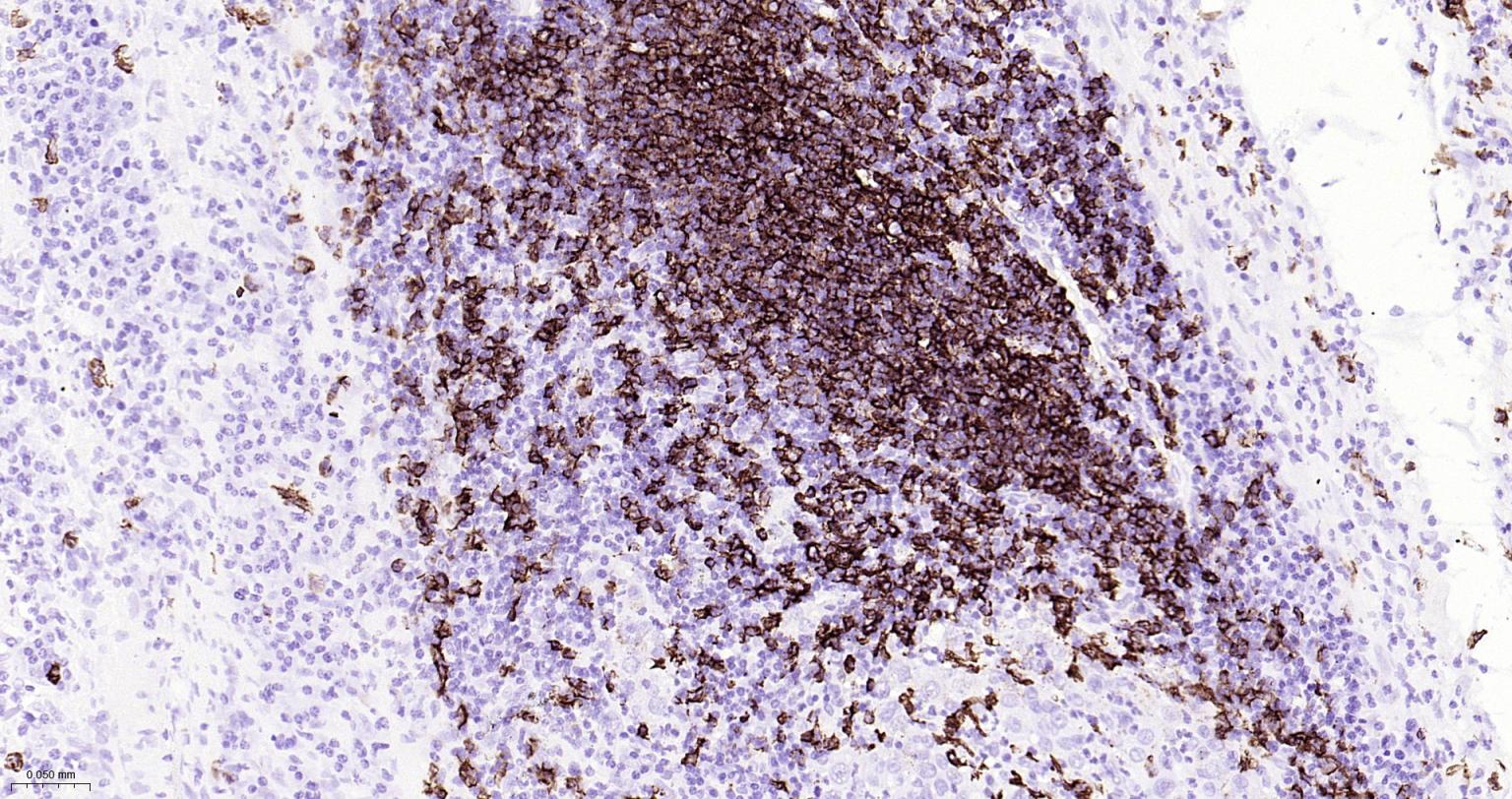
Figure 8. Paraformaldehyde-fixed, paraffin embedded Human Gastric Cancer; Antigen retrieval by boiling in sodium citrate buffer (pH6.0) for 15 min; Antibody incubation with CD20 Monoclonal Antibody, Unconjugated(SLA3106M) at 1:200 overnight at 4°C, followed by conjugation to the SL0295G-HRP and DAB (C-0010) staining
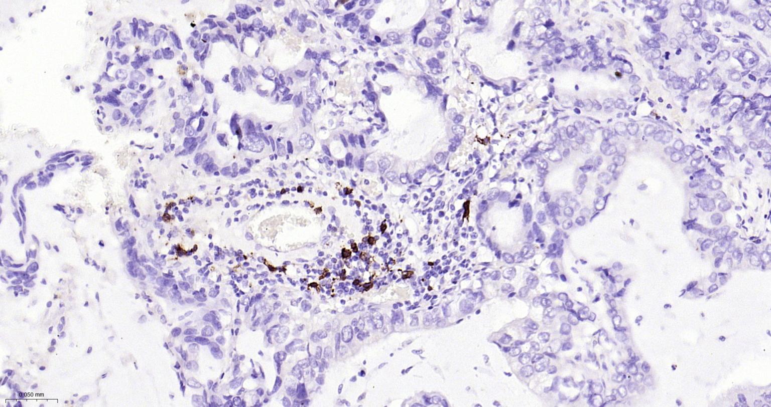
Figure 9. Paraformaldehyde-fixed, paraffin embedded Human Lung Cancer; Antigen retrieval by boiling in sodium citrate buffer (pH6.0) for 15 min; Antibody incubation with CD20 Monoclonal Antibody, Unconjugated(SLA3106M) at 1:200 overnight at 4°C, followed by conjugation to the SL0295G-HRP and DAB (C-0010) staining
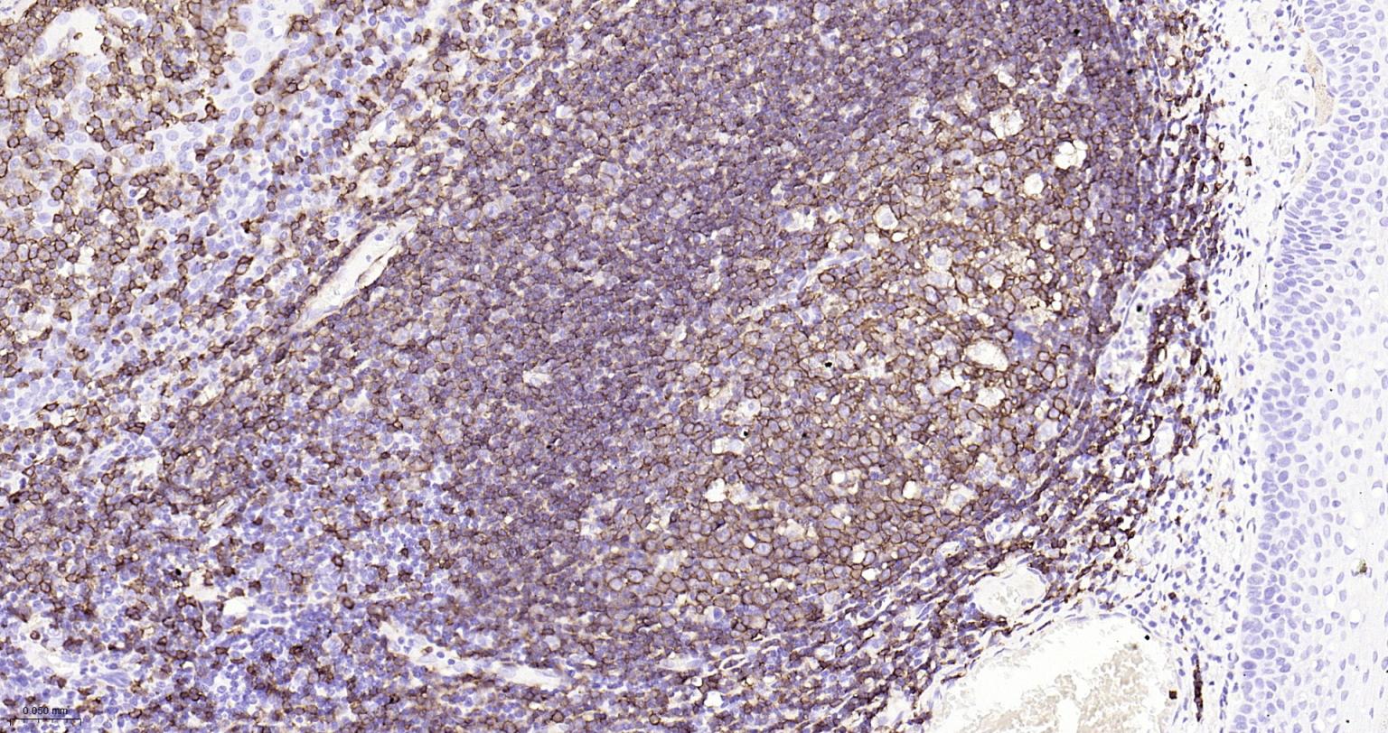
Figure 10. Paraformaldehyde-fixed, paraffin embedded Human Tonsil; Antigen retrieval by boiling in sodium citrate buffer (pH6.0) for 15 min; Antibody incubation with CD20 Monoclonal Antibody, Unconjugated(SLA3106M) at 1:200 overnight at 4°C, followed by conjugation to the SL0295G-HRP and DAB (C-0010) staining
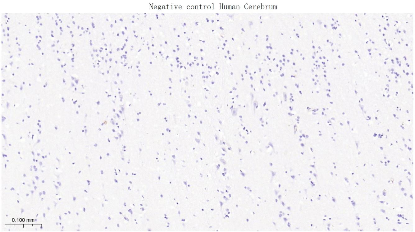
Figure 11. (Negative control)Paraformaldehyde-fixed, paraffin embedded Human Cerebrum; Antigen retrieval by boiling in sodium citrate buffer (pH6.0) for 15 min; Antibody incubation with CD20 Monoclonal Antibody, Unconjugated(SLA3106M) at 1:200 overnight at 4°C, followed by conjugation to the SL0295G-HRP and DAB (C-0010) staining
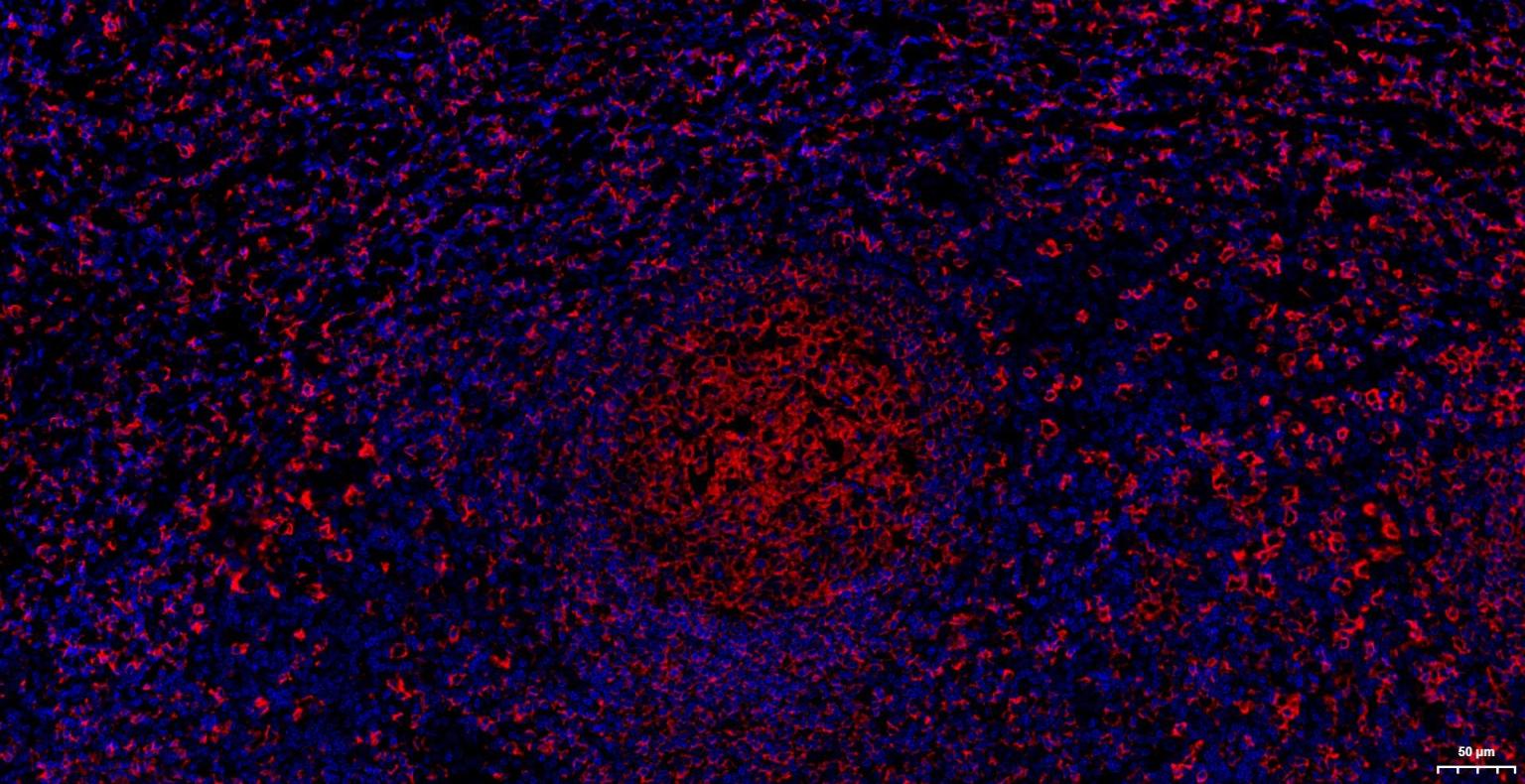
Figure 12. Paraformaldehyde-fixed, paraffin embedded Human Tonsil; Antigen retrieval by boiling in sodium citrate buffer (pH6.0) for 15 min; The section was incubated with CD20 Monoclonal Antibody, Unconjugated (SLA3106M) at 1:200 overnight at 4°C. Followed by conjugated Goat Anti-Rabbit IgG antibody (Red, SL0295G-BF594), DAPI (blue, C02-04002) was used to stain the cell nuclei
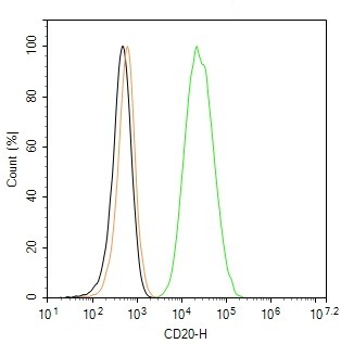
Figure 13. The Raji (H) cells were incubated in 5%BSA to block non-specific protein-protein interactions (30 min at r.t.), followed by secondary antibody incubation for 40 min at room temperature. Primary Antibody (green): Rabbit Anti-CD20 antibody (SLA3106M,1:100); Isotype Control (orange): Rabbit IgG (SL0295P). Blank control (black): PBS. Acquisition of 20,000 events was performed
FOR RESEARCH USE ONLY. Not for use in diagnostic or therapeutic procedures.
Partial purchase records(bought amounts latest0)
User Comment(Total0User Comment Num)
- No comment


 +86 571 56623320

 SUNLONG BIOTECH


