For general and specific antibody protocols please visit our website. Read all instructions before using this product.
Flow cytometric analysis of JNK3 expression in HT-1080 cells using JNK3 antibody 1:2,000. Green, isotype control; red, JNK3.
Immunocytochemical staining of HT-1080 cells with anti-JNK3 antibody 1:1,000. Nuclei were stained blue with DAPI; JNK3 was stained magenta with Alexa Fluor® 647. Images were taken using Leica stellaris 5. Protein abundance based on laser Intensity and smart gain: Medium. Scale bar: 20 μm.
Western blotting analysis using anti-JNK3 antibody 1:5,000 and HRP-conjugated goat anti-rabbit secondary antibody 1:20,000 respectively.
Western blotting analysis using anti-JNK3 antibody 1:5,000 and HRP-conjugated goat anti-rabbit secondary antibody 1:20,000 respectively. Image was developed using NaQ™ ECL Substrate Kit .
![[KD-Validated] Anti-MAPK10 Rabbit Monoclonal Antibody](images/default_images/52.jpg)
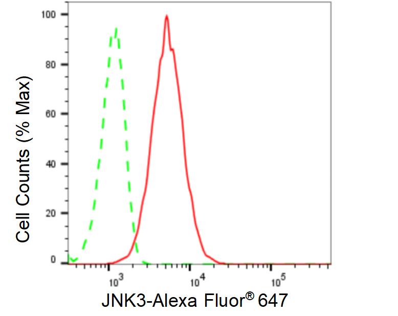 Flow cytometric analysis of JNK3 expression in HT-1080 cells using JNK3 antibody 1:2,000. Green, isotype control; red, JNK3.
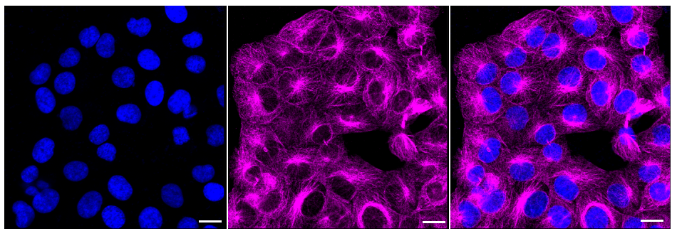 Immunocytochemical staining of HT-1080 cells with anti-JNK3 antibody 1:1,000. Nuclei were stained blue with DAPI; JNK3 was stained magenta with Alexa Fluor® 647. Images were taken using Leica stellaris 5. Protein abundance based on laser Intensity and smart gain: Medium. Scale bar: 20 μm.
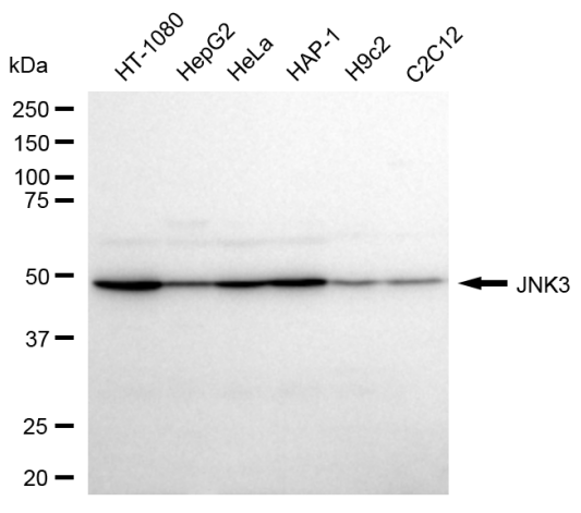 Western blotting analysis using anti-JNK3 antibody 1:5,000 and HRP-conjugated goat anti-rabbit secondary antibody 1:20,000 respectively.
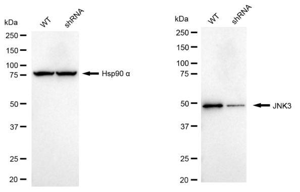 Western blotting analysis using anti-JNK3 antibody 1:5,000 and HRP-conjugated goat anti-rabbit secondary antibody 1:20,000 respectively. Image was developed using NaQ™ ECL Substrate Kit .

 +86 571 56623320
 [email protected]
 SUNLONG BIOTECH

