Details
Product Name MTA1 Chinese Name Tumour转移相关蛋白1抗体 Alias Metastasis-associated 1; Metastasis associated 1; Metastasis associated gene 1; Metastasis associated gene 1 protein; Metastasis associated protein; Metastasis associated protein MTA 1; Metastasis associated protein MTA1; MTA 1; MTA1_HUMAN. literatures Research Area Tumour Cell biology Epigenetics Immunogen Species Rabbit Clonality Polyclonal React Species Human,Mouse,Rat Applications WB=1:500-2000,IHC-P=1:100-500,IHC-F=1:100-500,IF=1:100-500,Flow-Cyt=1ug/Test (Paraffin sections need antigen repair)
not yet tested in other applications.
optimal dilutions/concentrations should be determined by the end user.Theoretical molecular weight 81kDa Cellular localization The nucleus cytoplasmic Form Liquid Concentration 1mg/ml immunogen KLH conjugated synthetic peptide derived from human MTA1: 601-715/715 Lsotype IgG Purification affinity purified by Protein A Buffer Solution 1M TBS(pH7.4) with 1% BSA, 3% Proclin300 and 50% Glycerol. Storage Shipped at 4℃. Store at -20 °C for one year. Avoid repeated freeze/thaw cycles. Attention This product as supplied is intended for research use only, not for use in human, therapeutic or diagnostic applications. PubMed PubMed Product Detail This protein was identified in metastatic cells, specifically, mammary adenocarcinoma cell lines. Expression of this protein has been correlated with the metastatic potential of at least two types of carcinomas although it is also expressed in many normal tissues. The role it plays in metastasis is unclear. It was initially thought to be the 70 kDa component of a nucleosome remodeling deacetylase complex, NuRD, but it is more likely that this component is a different but very similar protein. These two proteins are so closely related, though, that they share the same types of domains. These domains include two DNA binding domains, a dimerization domain, and a domain commonly found in proteins that methylate DNA. The profile and activity of this protein suggests that it is involved in regulating transcription and that this may be accomplished by chromatin remodeling.
Function:
May be involved in the regulation of gene expression by covalent modification of histone proteins. Isoform Long is a corepressor of estrogen receptor (ER). Isoform Short binds to ER and sequesters it in the cytoplasm and enhances non-genomic responses of ER.
Subunit:
Component of the nucleosome-remodeling and histone-deacetylase multiprotein complex (NuRD). Interacts with HDAC1 and ITGB3BP/CENPR. Binds to CSNK1G2 in the cytoplasm.
Subcellular Location:
Cytoplasm and Nucleus.
Tissue Specificity:
Widely expressed. High expression in brain, ovaries, adrenal glands and virgin mammary glands. Higher in tumors than in adjacent normal tissue from the same individual.
Similarity:
Contains 1 BAH domain. Contains 1 ELM2 domain.
Contains 1 GATA-type zinc finger.
Contains 1 SANT domain.
SWISS:
Q13330
Gene ID:
9112
Database links:Entrez Gene: 9112 Human
Entrez Gene: 116870 Mouse
Omim: 603526 Human
SwissProt: Q13330 Human
SwissProt: Q2KHS8 Mouse
SwissProt: Q8K4B0 Mouse
Unigene: 101448 Human
Unigene: 525629 Human
Unigene: 212577 Mouse
Unigene: 5840 Rat
transcriptional regulatory factor(Transcriptin Regulators)
MTA1(Metastasis-associated 1)The protein is a protein complex that possesses histone deacetylase activity and plays an important role in histone deacetylation, chromatin remodeling, and transcriptional control.。Product Picture
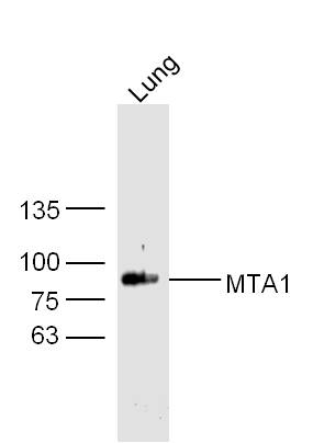
Figure 1. Sample: Lung(Mouse) Lysate at 30 ug Primary: Anti- MTA1 (SL1412R) at 1/300 dilution Secondary: IRDye800CW Goat Anti-Mouse IgG at 1/10000 dilution Predicted band size: 81 kD Observed band size: 85 kD
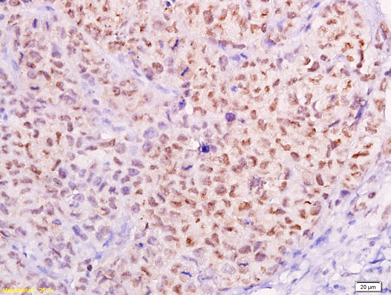
Figure 2. Tissue/cell: transplanted tumor of cervical carcinoma in nude mice; 4% Paraformaldehyde-fixed and paraffin-embedded; Antigen retrieval: citrate buffer ( 0.01M, pH 6.0 ), Boiling bathing for 15min; Block endogenous peroxidase by 3% Hydrogen peroxide for 30min; Blocking buffer (normal goat serum,C-0005) at 37℃ for 20 min; Incubation: Anti-MTA1 Polyclonal Antibody, Unconjugated(SL1412R) 1:300, overnight at 4°C, followed by conjugation to the secondary antibody(SP-0023) and DAB(C-0010) staining
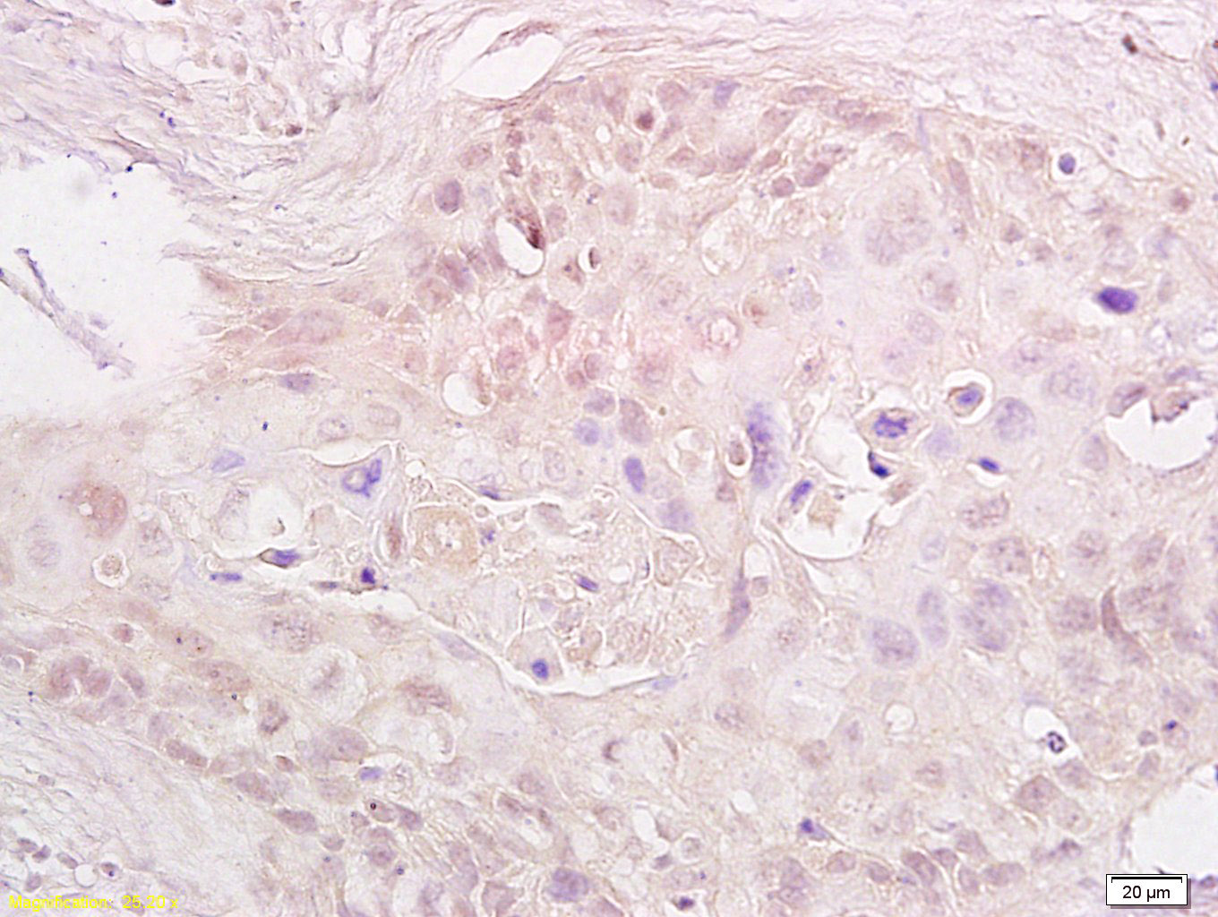
Figure 3. Tissue/cell: human cervical carcinoma; 4% Paraformaldehyde-fixed and paraffin-embedded; Antigen retrieval: citrate buffer ( 0.01M, pH 6.0 ), Boiling bathing for 15min; Block endogenous peroxidase by 3% Hydrogen peroxide for 30min; Blocking buffer (normal goat serum,C-0005) at 37℃ for 20 min; Incubation: Anti-MTA1 Polyclonal Antibody, Unconjugated(SL1412R) 1:600, overnight at 4°C, followed by conjugation to the secondary antibody(SP-0023) and DAB(C-0010) staining
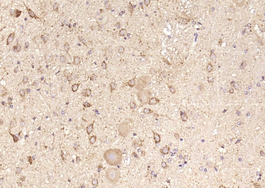
Figure 4. Paraformaldehyde-fixed, paraffin embedded (rat brain); Antigen retrieval by boiling in sodium citrate buffer (pH6.0) for 15min; Block endogenous peroxidase by 3% hydrogen peroxide for 20 minutes; Blocking buffer (normal goat serum) at 37°C for 30min; Antibody incubation with (MTA1) Polyclonal Antibody, Unconjugated (SL1412R) at 1:200 overnight at 4°C, followed by operating according to SP Kit(Rabbit) (sp-0023) instructionsand DAB staining
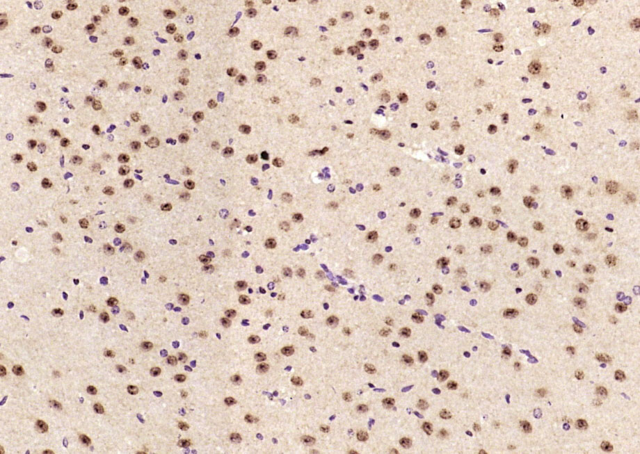
Figure 5. Paraformaldehyde-fixed, paraffin embedded (Mouse brain); Antigen retrieval by boiling in sodium citrate buffer (pH6.0) for 15min; Block endogenous peroxidase by 3% hydrogen peroxide for 20 minutes; Blocking buffer (normal goat serum) at 37°C for 30min; Antibody incubation with (MTA1) Polyclonal Antibody, Unconjugated (SL1412R) at 1:200 overnight at 4°C, followed by operating according to SP Kit(Rabbit) (sp-0023) instructionsand DAB staining
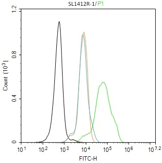
Figure 6. Blank control (black line) :SH-SY5Y. Primary Antibody (green line): Rabbit Anti-MTA1 antibody (SL1412R) Dilution:1ug/Test; Secondary Antibody (white blue line) : Goat anti-rabbit IgG-FITC Dilution: 0.5ug/Test. Isotype control (orange line) : Normal Rabbit IgG Protocol The cells were fixed with 4% PFA (10min at room temperature)and then permeabilized with 90% ice-cold methanol for 20 min at -20℃, The cells were then incubated in 5%BSA to block non-specific protein-protein interactions for 30 min at room temperature.Cells stained with Primary Antibody for 30 min at room temperature. The secondary antibody used for 40 min at room temperature. Acquisition of 20,000 events was performed
Ordering & shipping
- Minimum order: 1 unit
- Lead time: varies with order quantity and specifications. Please consult your local distributor or our sales team for confirmation
- Payment: T/T 100% in advance; PayPal; Alibaba Trade Assurance
- Shipping: FedEx / DHL / UPS express; cold-chain packing available on request
- Price: USD, per unit (50ul / 100ul)
Partial purchase records(bought amounts latest4)
| Username | Quantity | bought time |
| Vi*** | 1 | 2026-05-12 |
| Di*** | 1 | 2026-05-05 |
| Ia*** | 1 | 2026-05-01 |
| Ca*** | 1 | 2026-02-26 |
User Comment(Total0User Comment Num)
- No comment


 +86 571 56623320

 SUNLONG BIOTECH


