Details
Product Name Cathepsin L Chinese Name 组织蛋白酶L抗体 Alias Cathepsin L heavy chain; Cathepsin L light chain; CathepsinL; CATL; CTSL; CTSL protein; DKFZp686A18159; FLJ31037; Major excreted protein; MEP; cath-L; CL; CATL1_HUMAN; CathepsinL1; Cathepsin L1,Cathepsin-L1; CTSL 1; CTSL-1,CTS L1,CTS-L1. literatures Research Area Kinases and Phosphatases Synthesis and Degradation Immunogen Species Rabbit Clonality Polyclonal React Species Human, Rat, (predicted: Mouse, ) Applications WB=1:500-2000,IHC-P=1:100-500,IHC-F=1:100-500,IF=1:100-500,Flow-Cyt=1ug/Test (Paraffin sections need antigen repair)
not yet tested in other applications.
optimal dilutions/concentrations should be determined by the end user.Theoretical molecular weight 19/30/37kDa Cellular localization cytoplasmic Form Liquid Concentration 1mg/ml immunogen KLH conjugated synthetic peptide derived from human cathepsin L1 proprotein: 71-170/334 Lsotype IgG Purification affinity purified by Protein A Buffer Solution 1M TBS(pH7.4) with 1% BSA, 3% Proclin300 and 50% Glycerol. Storage Shipped at 4℃. Store at -20 °C for one year. Avoid repeated freeze/thaw cycles. Attention This product as supplied is intended for research use only, not for use in human, therapeutic or diagnostic applications. PubMed PubMed Product Detail The protein encoded by this gene is a lysosomal cysteine proteinase that plays a major role in intracellular protein catabolism. Its substrates include collagen and elastin, as well as alpha-1 protease inhibitor, a major controlling element of neutrophil elastase activity. The encoded protein has been implicated in several pathologic processes, including myofibril necrosis in myopathies and in myocardial ischemia, and in the renal tubular response to proteinuria. This protein, which is a member of the peptidase C1 family, is a dimer composed of disulfide-linked heavy and light chains, both produced from a single protein precursor. At least two transcript variants encoding the same protein have been found for this gene.
Function:
Important for the overall degradation of proteins in lysosomes.
Subunit:
Dimer of a heavy and a light chain linked by disulfide bonds.
Subcellular Location:
Lysosome.
Similarity:
Belongs to the peptidase C1 family.
SWISS:
P07711
Gene ID:
1514
Database links:Entrez Gene: 1514 Human
Entrez Gene: 13039 Mouse
Omim: 116880 Human
SwissProt: P07711 Human
SwissProt: P06797 Mouse
Unigene: 726015 Human
Unigene: 930 Mouse
Unigene: 1294 Rat
Synthesis and Degradation(Synthesis and Degradation)
Cathepsin L (CL) is a cysteine protease belonging to the papain family, stored in lysosomes as a zymogen. It participates in many specific physiological processes, such as activation of prohormones, antigen presentation, and development of tissues and organs.
Under pathological conditions, cell damage caused by various factors (such as pathogenic microorganisms, inflammatory factors, oxidative stress, etc.) can degrade cellular components or interstitial matrix components, including laminin, type IV collagen fibers, and fibronectin, etc., and is associated with many human diseases such as Tumour invasion and metastasis, arthritis, osteoporosis, Alzheimer's disease, multiple sclerosis, and other chronic inflammatory diseases.Product Picture
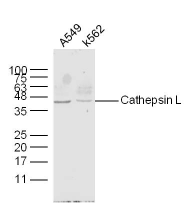
Figure 1. Sample: A549 Cell (Human) Lysate at 30 ug K562 Cell (Human) Lysate at 30 ug Primary: Anti- Cathepsin L (SL1508R) at 1/300 dilution Secondary: IRDye800CW Goat Anti-Rabbit IgG at 1/20000 dilution Predicted band size: 19/30/37 kD Observed band size: 40 kD
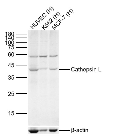
Figure 2. Sample: Lane 1: Human HUVEC cell Lysates Lane 2: Human K562 cell Lysates Lane 3: Human MCF-7 cell Lysates Primary: Anti-Cathepsin L (SL1508R) at 1/300 dilution Secondary: IRDye800CW Goat Anti-Rabbit IgG at 1/20000 dilution Predicted band size: 19/30/37kDa Observed band size: 40kDa
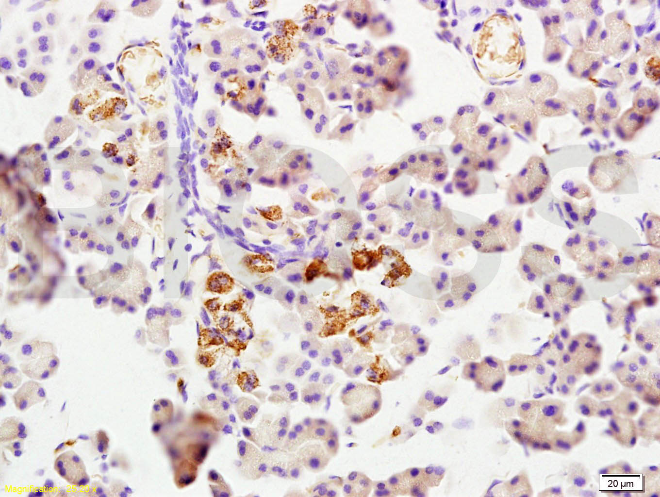
Figure 3. Tissue/cell: rat pancreas tissue; 4% Paraformaldehyde-fixed and paraffin-embedded; Antigen retrieval: citrate buffer ( 0.01M, pH 6.0 ), Boiling bathing for 15min; Block endogenous peroxidase by 3% Hydrogen peroxide for 30min; Blocking buffer (normal goat serum,C-0005) at 37℃ for 20 min; Incubation: Anti-Cathepsin L Polyclonal Antibody, Unconjugated(SL1508R) 1:200, overnight at 4°C, followed by conjugation to the secondary antibody(SP-0023) and DAB(C-0010) staining
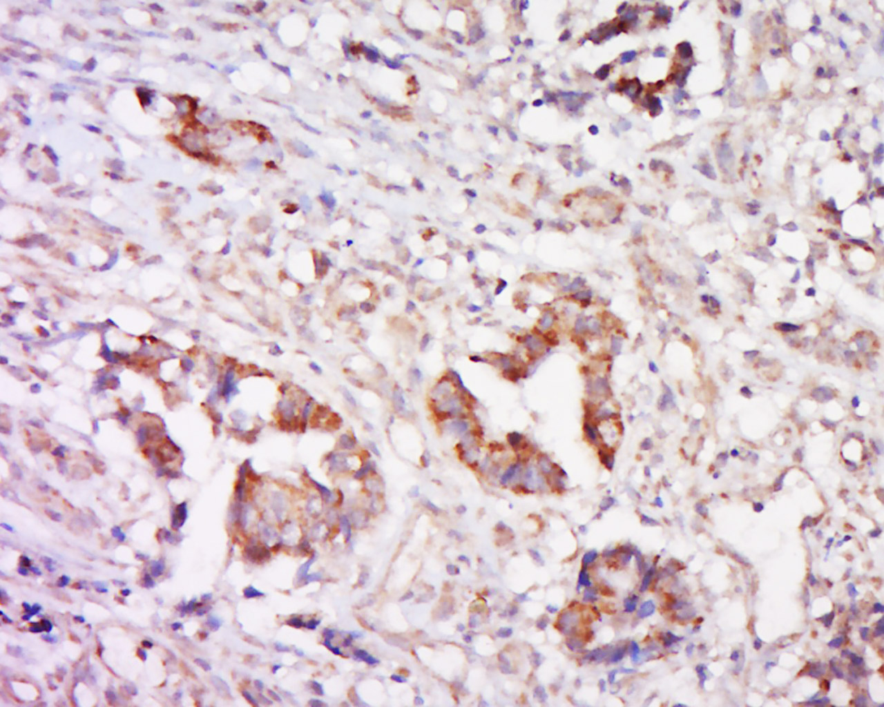
Figure 4. Tissue/cell: human cervical carcinoma; 4% Paraformaldehyde-fixed and paraffin-embedded; Antigen retrieval: citrate buffer ( 0.01M, pH 6.0 ), Boiling bathing for 15min; Block endogenous peroxidase by 3% Hydrogen peroxide for 30min; Blocking buffer (normal goat serum,C-0005) at 37℃ for 20 min; Incubation: Anti-Cathepsin Polyclonal Antibody, Unconjugated(SL1508R) 1:500, overnight at 4°C, followed by conjugation to the secondary antibody(SP-0023) and DAB(C-0010) staining
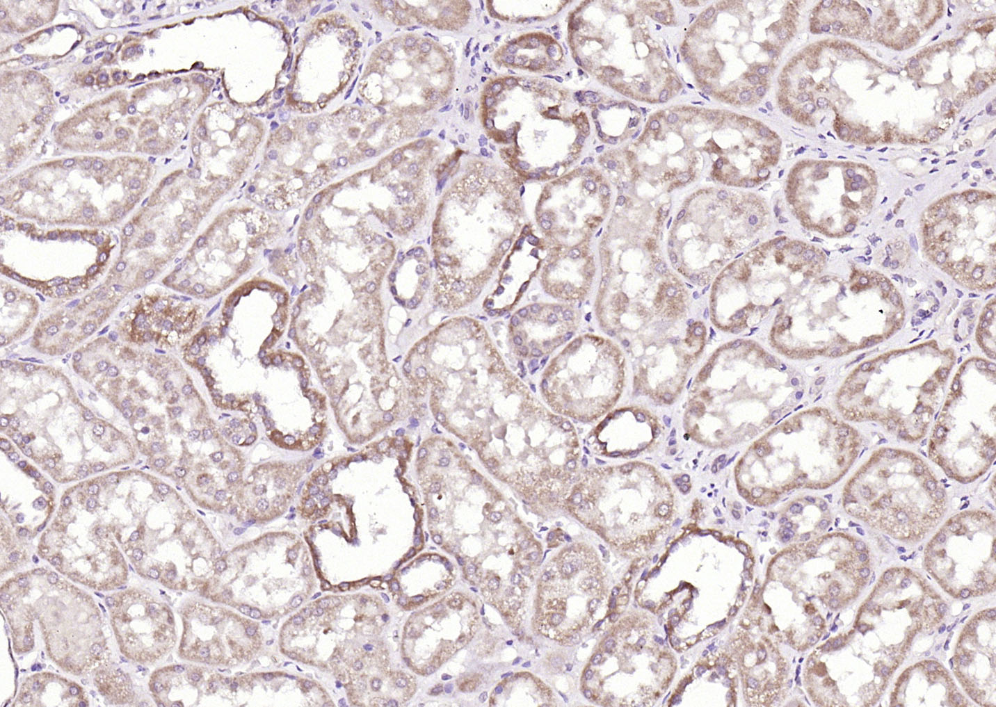
Figure 5. Paraformaldehyde-fixed, paraffin embedded (Human kidney); Antigen retrieval by boiling in sodium citrate buffer (pH6.0) for 15min; Block endogenous peroxidase by 3% hydrogen peroxide for 20 minutes; Blocking buffer (normal goat serum) at 37°C for 30min; Antibody incubation with (Cathepsin L) Polyclonal Antibody, Unconjugated (SL1508R) at 1:200 overnight at 4°C, followed by operating according to SP Kit(Rabbit) (sp-0023) instructionsand DAB staining
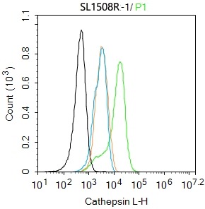
Figure 6. Blank control:A549. Primary Antibody (green line): Rabbit Anti-Cathepsin L antibody (SL1508R) Dilution: 1ug/Test; Secondary Antibody: Goat anti-rabbit IgG-FITC Dilution: 0.5ug/Test. Protocol The cells were fixed with 4% PFA (10min at room temperature)and then permeabilized with 0.1% PBST for 20 min at room temperature.The cells were then incubated in 5%BSA to block non-specific protein-protein interactions for 30 min at room temperature.Cells stained with Primary Antibody for 30 min at room temperature. The secondary antibody used for 40 min at room temperature. Acquisition of 20,000 events was performed
Ordering & shipping
- Minimum order: 1 unit
- Lead time: varies with order quantity and specifications. Please consult your local distributor or our sales team for confirmation
- Payment: T/T 100% in advance; PayPal; Alibaba Trade Assurance
- Shipping: FedEx / DHL / UPS express; cold-chain packing available on request
- Price: USD, per unit (50ul / 100ul)
Partial purchase records(bought amounts latest5)
| Username | Quantity | bought time |
| Os*** | 1 | 2026-08-13 |
| Ca*** | 1 | 2026-07-01 |
| Ge*** | 1 | 2026-06-27 |
| Um*** | 1 | 2026-03-21 |
| Qu*** | 1 | 2026-02-03 |
User Comment(Total0User Comment Num)
- No comment


 +86 571 56623320

 SUNLONG BIOTECH


