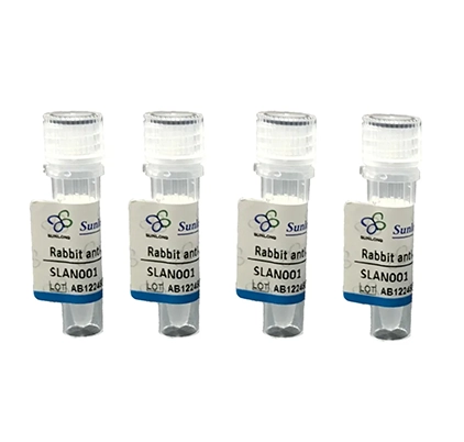
Details
Product Name HCP1 Chinese Name 血红素Transporter1抗体 Alias G21; HCP 1; Heme Carrier Protein 1; MGC9564; PCFT; PCFT/HCP1; PCFT_HUMAN; PDE7A; Proton coupled folate transporter; Proton-coupled folate transporter; SLC46A1; Solute carrier family 46 member 1; Research Area Cardiovascular Cell biology immunology Signal transduction The new supersedes the old Immunogen Species Rabbit Clonality Polyclonal React Species Human, Mouse, (predicted: Rat, Chicken, Cow, ) Applications WB=1:500-2000,IHC-P=1:100-500,IHC-F=1:100-500,ICC/IF=1:100-500,IF=1:100-500,Flow-Cyt=2ug/Test (Paraffin sections need antigen repair)
not yet tested in other applications.
optimal dilutions/concentrations should be determined by the end user.Theoretical molecular weight 50kDa Cellular localization The cell membrane Form Liquid Concentration 1mg/ml immunogen KLH conjugated synthetic peptide derived from human HCP1: 341-459/459 Lsotype IgG Purification affinity purified by Protein A Buffer Solution 1M TBS(pH7.4) with 1% BSA, 3% Proclin300 and 50% Glycerol. Storage Shipped at 4℃. Store at -20 °C for one year. Avoid repeated freeze/thaw cycles. Attention This product as supplied is intended for research use only, not for use in human, therapeutic or diagnostic applications. PubMed PubMed Product Detail This gene encodes a transmembrane proton-coupled folate transporter protein that facilitates the movement of folate and antifolate substrates across cell membranes, optimally in acidic pH environments. This protein is also expressed in the brain and choroid plexus where it transports folates into the central nervous system. This protein further functions as a heme transporter in duodenal enterocytes, and potentially in other tissues like liver and kidney. Its localization to the apical membrane or cytoplasm of intestinal cells is modulated by dietary iron levels. Mutations in this gene are associated with autosomal recessive hereditary folate malabsorption disease. Alternatively spliced transcript variants encoding different isoforms have been described for this gene. [provided by RefSeq, Aug 2013]
Function:
Has been shown to act both as an intestinal proton-coupled high-affinity folate transporter and as an intestinal heme transporter which mediates heme uptake from the gut lumen into duodenal epithelial cells. The iron is then released from heme and may be transported into the bloodstream. Dietary heme iron is an important nutritional source of iron. Shows a higher affinity for folate than heme.
Subcellular Location:
Apical cell membrane; Multi-pass membrane protein. Cytoplasm. Note=Localizes to the apical membrane of intestinal cells in iron-deficient cells, while it resides in the cytoplasm in iron-replete cells.
Tissue Specificity:
Expressed in kidney, liver, placenta, small intestine, spleen, retina and retinal pigment epithelium. Lower levels found in colon and testis. Very low levels in brain, lung, stomach, heart and muscle. In intestine, expressed in duodenum with lower levels in jejunum, ileum, cecum, rectum and segments of the colon.
DISEASE:
Hereditary folate malabsorption (HFM) [MIM:229050]: Rare autosomal recessive disorder characterized by impaired intestinal folate absorption with folate deficiency resulting in anemia, hypoimmunoglobulinemia with recurrent infections, and recurrent or chronic diarrhea. In many patients, neurological abnormalities such as seizures or mental retardation become apparent during early childhood, attributed to impaired transport of folates into the central nervous system. When diagnosed early, the disorder can be treated by administration of folate. If untreated, it can be fatal and, if treatment is delayed, the neurological defects can become permanent. Note=The disease is caused by mutations affecting the gene represented in this entry.
Similarity:
Belongs to the major facilitator superfamily. SLC46A family.
SWISS:
Q96NT5
Gene ID:
113235
Database links:Entrez Gene: 113235 Human
Entrez Gene: 52466 Mouse
Omim: 611672 Human
SwissProt: Q96NT5 Human
SwissProt: Q6PEM8 Mouse
Unigene: 131618 Mouse
Unigene: 153975 Rat
Product Picture
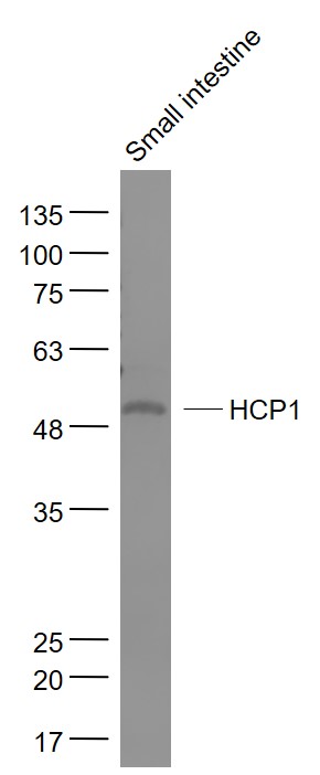
Figure 1. Sample: Small intestine (Mouse) Lysate at 40 ug Primary: Anti- HCP1 (SL15428R) at 1/1000 dilution Secondary: IRDye800CW Goat Anti-Rabbit IgG at 1/20000 dilution Predicted band size: 50 kD Observed band size: 50 kD
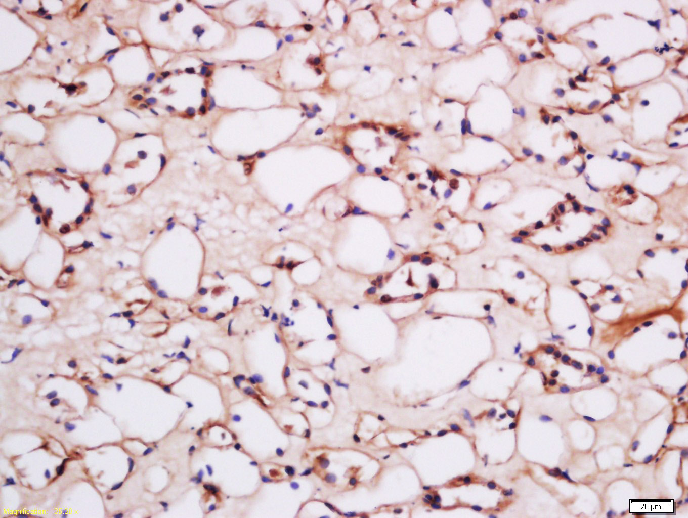
Figure 2. Tissue/cell: human kidney tissue; 4% Paraformaldehyde-fixed and paraffin-embedded; Antigen retrieval: citrate buffer ( 0.01M, pH 6.0 ), Boiling bathing for 15min; Block endogenous peroxidase by 3% Hydrogen peroxide for 30min; Blocking buffer (normal goat serum,C-0005) at 37℃ for 20 min; Incubation: Anti-HCP1 Polyclonal Antibody, Unconjugated(SL15428R) 1:200, overnight at 4°C, followed by conjugation to the secondary antibody(SP-0023) and DAB(C-0010) staining
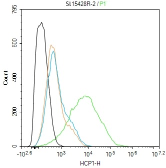
Figure 3. Blank control:Jurkat. Primary Antibody (green line): Rabbit Anti-HCP1 antibody (SL15428R) Dilution: 2ug/Test; Secondary Antibody (white blue line): Goat anti-rabbit IgG-FITC Dilution: 0.5ug/Test. Isotype control (orange line) : Normal Rabbit IgG Protocol The cells were incubated in 5%BSA to block non-specific protein-protein interactions for 30 min at room temperature.Cells stained with Primary Antibody for 30 min at room temperature. The secondary antibody used for 40 min at room temperature. Acquisition of 20,000 events was performed
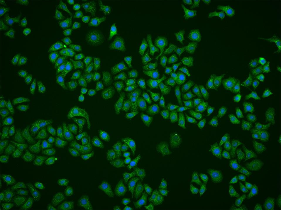
Figure 4. HepG2 cell; 4% Paraformaldehyde-fixed; Triton X-100 at room temperature for 20 min; Blocking buffer (normal goat serum, C-0005) at 37°C for 20 min; Antibody incubation with (HCP1) polyclonal Antibody, Unconjugated (SL15428R) 1:100, 90 minutes at 37°C; followed by a conjugated Goat Anti-Rabbit IgG antibody at 37°C for 90 minutes, DAPI (blue, C02-04002) was used to stain the cell nuclei
Ordering & shipping
- Minimum order: 1 unit
- Lead time: varies with order quantity and specifications. Please consult your local distributor or our sales team for confirmation
- Payment: T/T 100% in advance; PayPal; Alibaba Trade Assurance
- Shipping: FedEx / DHL / UPS express; cold-chain packing available on request
- Price: USD, per unit (50ul / 100ul)
Partial purchase records(bought amounts latest1)
| Username | Quantity | bought time |
| Le*** | 1 | 2026-07-28 |
User Comment(Total0User Comment Num)
- No comment


 +86 571 56623320

 SUNLONG BIOTECH


