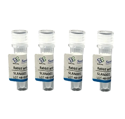
Details
Product Name ERVFRD-1 Chinese Name ERVFRD-1抗体 Alias SYCY2_HUMAN; EFRD1_HUMAN; Endogenous retrovirus group FRD member 1; Envelope polyprotein; HERV-FRD; HERV-FRD_6p24.1 provirus ancestral Env polyprotein; TM; SU; ERVFRD-1; ERVFRDE1; UNQ6191/PRO20218; literatures Research Area Bacteria and viruses Immunogen Species Rabbit Clonality Polyclonal React Species Human Applications WB (1:500-2000), IP (1:50-200)
Not yet tested in other applications.
Optimal working dilutions must be determined by the end user.Theoretical molecular weight 21/37/58kDa Cellular localization The cell membrane Form Liquid Concentration 1mg/ml immunogen KLH conjugated synthetic peptide derived from human HERV-FRD : 331-430/538 Lsotype IgG Purification affinity purified by Protein A Buffer Solution 1M TBS(pH7.4) with 1% BSA, 3% Proclin300 and 50% Glycerol. Storage Shipped at 4℃. Store at -20 °C for one year. Avoid repeated freeze/thaw cycles. Attention This product as supplied is intended for research use only, not for use in human, therapeutic or diagnostic applications. PubMed PubMed Product Detail Retroviral envelope proteins mediate receptor recognition and membrane fusion during early infection. Endogenous envelope proteins may have kept, lost or modified their original function during evolution. This endogenous envelope protein has retained its original fusogenic properties. Can make pseudotypes with MLV, HIV-1 or SIV-1 virions and confer infectivity.
SU mediates receptor recognition (By similarity).
TM anchors the envelope heterodimer to the viral membrane through one transmembrane domain. The other hydrophobic domain, called fusion peptide, mediates fusion of the viral membrane with the target cell membrane (By similarity).
Subunit:
The surface (SU) and transmembrane (TM) proteins form a heterodimer. SU and TM are attached by noncovalent interactions or by a labile interchain disulfide bond (By similarity).
Subcellular Location:
Virion.
Transmembrane protein: Cell membrane; Single-pass membrane protein (Potential).
Tissue Specificity:
Expressed at higher level in placenta. Expressed at lower level in adrenal, bone marrow, brain, breast, colon, kidney, lung, ovary, peripheral blood lymphocytes, prostate, skin, spleen, testis, thymus, thyroid, trachea.
Post-translational modifications:
Specific enzymatic cleavages in vivo yield the mature SU and TM proteins (By similarity).
The CXXC motif is highly conserved across a broad range of retroviral envelope proteins. It is thought to participate in the formation of a labile disulfide bond possibly with the CX6CC motif present in the transmembrane protein. Isomerization of the intersubunit disulfide bond to an SU intrachain disulfide bond is thought to occur upon receptor recognition in order to allow membrane fusion (By similarity).
Similarity:
Belongs to the gamma type-C retroviral envelope protein family. HERV class-I FRD env subfamily.
SWISS:
P60508
Gene ID:
2086
Database links:Entrez Gene: 2086 Human
SwissProt: P60508 Human
Product Picture
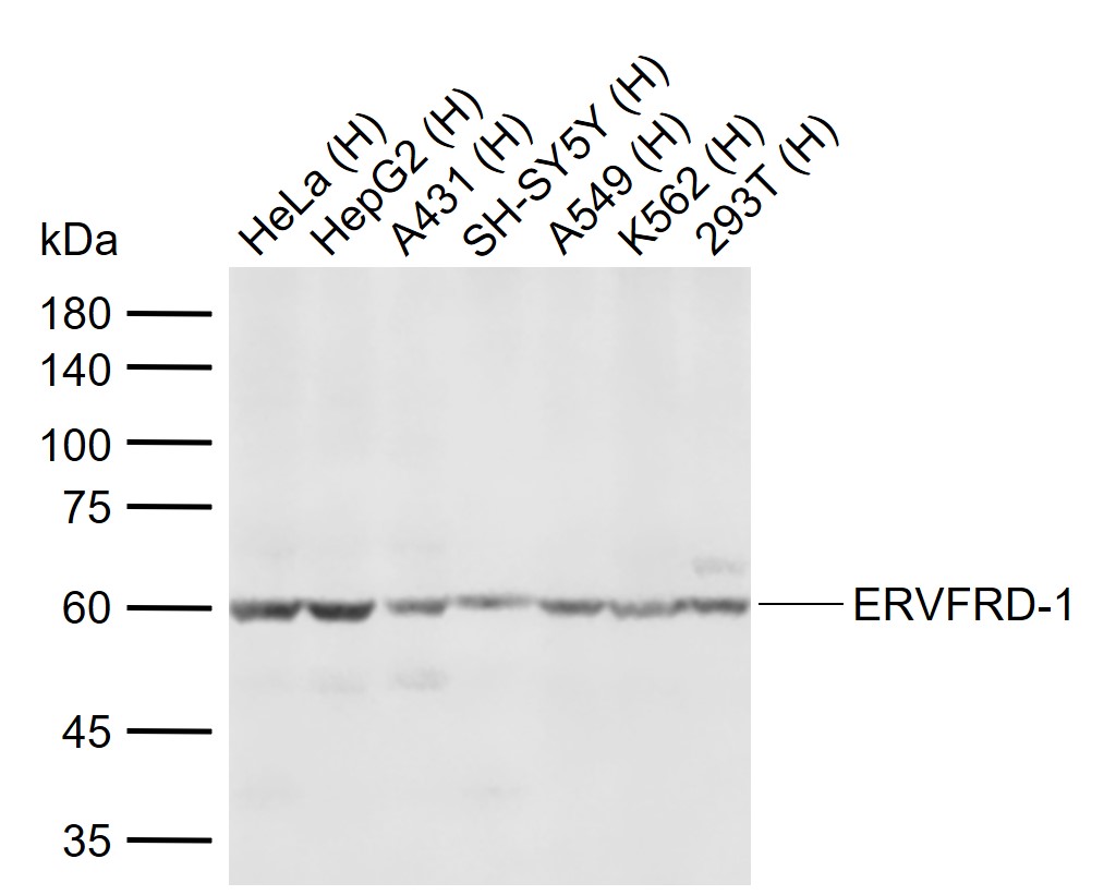
Figure 1. Sample: Lane 1: Human HeLa cell lysates Lane 2: Human HepG2 cell lysates Lane 3: Human A431 cell lysates Lane 4: Human SH-SY5Y cell lysates Lane 5: Human A549 cell lysates Lane 6: Human K562 cell lysates Lane 7: Human 293T cell lysates Primary: Anti-ERVFRD-1 (SL15466R) at 1/1000 dilution Secondary: IRDye800CW Goat Anti-Rabbit IgG at 1/20000 dilution Predicted band size: 21/37/58 kDa Observed band size: 60 kDa
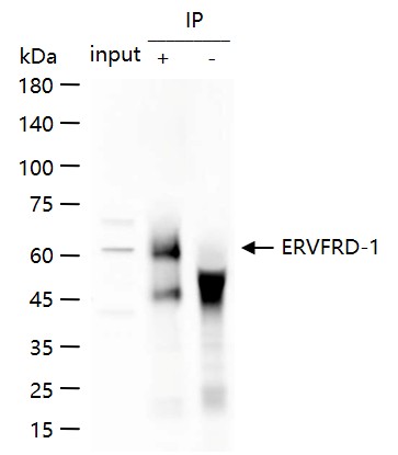
Figure 2. ERVFRD-1 was immunoprecipitated from HeLa cell lysate with SL15466R at 1/100 dilution. Western blot was performed from the immunoprecipitate using protein A/G beads. HRP conjugated Goat Anti-Rabbit IgG was used as secondary antibody. Lane 1: HeLa cell lysate (Input). Lane 2: b SL15466R IP in HeLa cell lysate. Lane 3: native Rabbit IgG IP in HeLa cell lysate (negative control)
Ordering & shipping
- Minimum order: 1 unit
- Lead time: varies with order quantity and specifications. Please consult your local distributor or our sales team for confirmation
- Payment: T/T 100% in advance; PayPal; Alibaba Trade Assurance
- Shipping: FedEx / DHL / UPS express; cold-chain packing available on request
- Price: USD, per unit (50ul / 100ul)
Partial purchase records(bought amounts latest5)
| Username | Quantity | bought time |
| Ya*** | 1 | 2026-07-29 |
| Ha*** | 1 | 2026-07-09 |
| Um*** | 1 | 2026-05-21 |
| Gr*** | 1 | 2026-03-26 |
| Zo*** | 1 | 2026-02-22 |
User Comment(Total0User Comment Num)
- No comment


 +86 571 56623320

 SUNLONG BIOTECH


