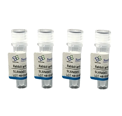
Rabbit Anti-RhoA antibody
RhoA protein; Aplysia ras related homolog 12; ARH 12; ARH12; ARHA; H 12; Oncogene RHO H12; Ras homolog gene family member A; RHO 12; Rho A; RHO H12; RHO12; RHOH 12; RHOH12; Small GTP binding protein Rho A; Small GTP binding protein RhoA; Transforming prot
All categories
View History [Clear]
Details
Product Name RhoA Chinese Name RhoA抗体 Alias RhoA protein; Aplysia ras related homolog 12; ARH 12; ARH12; ARHA; H 12; Oncogene RHO H12; Ras homolog gene family member A; RHO 12; Rho A; RHO H12; RHO12; RHOH 12; RHOH12; Small GTP binding protein Rho A; Small GTP binding protein RhoA; Transforming protein Rho A; Transforming protein RhoA. Research Area Tumour Signal transduction G protein signal Immunogen Species Rabbit Clonality Polyclonal React Species Human(predicted:Mouse,Rat,Dog,Pig,Cow,Horse,Rabbit,Sheep) Applications IHC-P=1:100-500,IHC-F=1:100-500,IF=1:100-500 (Paraffin sections need antigen repair)
not yet tested in other applications.
optimal dilutions/concentrations should be determined by the end user.Theoretical molecular weight 21kDa Cellular localization cytoplasmic The cell membrane Form Liquid Concentration 1mg/ml immunogen KLH conjugated synthetic peptide derived from human RhoA: 101-180/193 Lsotype IgG Purification affinity purified by Protein A Buffer Solution 1M TBS(pH7.4) with 1% BSA, 3% Proclin300 and 50% Glycerol. Storage Shipped at 4℃. Store at -20 °C for one year. Avoid repeated freeze/thaw cycles. Attention This product as supplied is intended for research use only, not for use in human, therapeutic or diagnostic applications. PubMed PubMed Product Detail This gene encodes a member of the Rho family of small GTPases, which cycle between inactive GDP-bound and active GTP-bound states and function as molecular switches in signal transduction cascades. Rho proteins promote reorganization of the actin cytoskeleton and regulate cell shape, attachment, and motility. The protein encoded by this gene is prenylated at its C-terminus, and localizes to the cytoplasm and plasma membrane. It is thought to be important in cell locomotion. Overexpression of this gene is associated with tumor cell proliferation and metastasis. Multiple alternatively spliced variants, encoding the same protein, have been identified.
Function:
Regulates a signal transduction pathway linking plasma membrane receptors to the assembly of focal adhesions and actin stress fibers. Involved in a microtubule-dependent signal that is required for the myosin contractile ring formation during cell cycle cytokinesis. Plays an essential role in cleavage furrow formation. Required for the apical junction formation of keratinocyte cell-cell adhesion. Serves as a target for the yopT cysteine peptidase from Yersinia pestis, vector of the plague, and Yersinia pseudotuberculosis, which causes gastrointestinal disorders. Stimulates PKN2 kinase activity. May be an activator of PLCE1. Activated by ARHGEF2, which promotes the exchange of GDP for GTP. Essential for the SPATA13-mediated regulation of cell migration and adhesion assembly and disassembly. The MEMO1-RHOA-DIAPH1 signaling pathway plays an important role in ERBB2-dependent stabilization of microtubules at the cell cortex. It controls the localization of APC and CLASP2 to the cell membrane, via the regulation of GSK3B activity. In turn, membrane-bound APC allows the localization of the MACF1 to the cell membrane, which is required for microtubule capture and stabilization.
Subunit:
Interacts with ARHGEF28. Binds PRKCL1, ROCK1 and ROCK2. Interacts with ARHGEF2, ARHGEF3, NET1 and RTKN. Interacts with PLCE1 and AKAP13. Interacts (in the constitutively activated, GTP-bound form) with DGKQ. Interacts with human respiratory syncytial virus (HRSV) protein F; this interaction facilitates virus-induced syncytium formation. Interacts with GNB2L1/RACK1; enhances RHOA activation. Interacts with PKP4; the interaction is detected at the midbody. Interacts (GTP-bound form preferentially) with PKN2; the interaction stimulates autophosphorylation and phosphorylation of PKN2.
Subcellular Location:
Cell membrane; Lipid-anchor; Cytoplasmic side. Cytoplasm, cytoskeleton. Cleavage furrow. Cytoplasm, cell cortex. Midbody. Note=Localized to cell-cell contacts in calcium-treated keratinocytes. Translocates to the equatorial region before furrow formation in a ECT2-dependent manner. Localizes to the equatorial cell cortex (at the site of the presumptive furrow) in early anaphase in a activated form and in a myosin- and actin-independent manner.
Post-translational modifications:
Substrate for botulinum ADP-ribosyltransferase.
Cleaved by yopT protease when the cell is infected by some Yersinia pathogens. This removes the lipid attachment, and leads to its displacement from plasma membrane and to subsequent cytoskeleton cleavage.
AMPylation at Tyr-34 and Thr-37 are mediated by bacterial enzymes in case of infection by H.somnus and V.parahaemolyticus, respectively. AMPylation occurs in the effector region and leads to inactivation of the GTPase activity by preventing the interaction with downstream effectors, thereby inhibiting actin assembly in infected cells. It is unclear whether some human enzyme mediates AMPylation; FICD has such ability in vitro but additional experiments remain to be done to confirm results in vivo.
Phosphorylation by PRKG1 at Ser-188 inactivates RHOA signaling.
Ubiquitinated by the BCR(BACURD1) and BCR(BACURD2) E3 ubiquitin ligase complexes, leading to its degradation by the proteasome, thereby regulating the actin cytoskeleton and cell migration.
Similarity:
Belongs to the small GTPase superfamily. Rho family.
SWISS:
P61586
Gene ID:
387
Database links:Entrez Gene: 395442 Chicken
Entrez Gene: 387 Huma
Entrez Gene: 11848 Mouse
Omim: 165390 Human
SwissProt: P61586 Human
SwissProt: Q9QUI0 Mouse
Unigene: 247077 Human
Unigene: 757 Mouse
Unigene: 107401 Rat
RhoA protein is mainly distributed in the cytoplasm and The cell membrane. The RhoA protein family is a key Signal transduction component in cellular processes such as controlling Cytoskeleton protein assembly and regulating gene expression. Typically, RhoA acts by activating its downstream Rho associated kinase (ROK). During the regulation of Cytoskeleton assembly, both RhoA and its targets are located in the cytoplasm.Product Picture 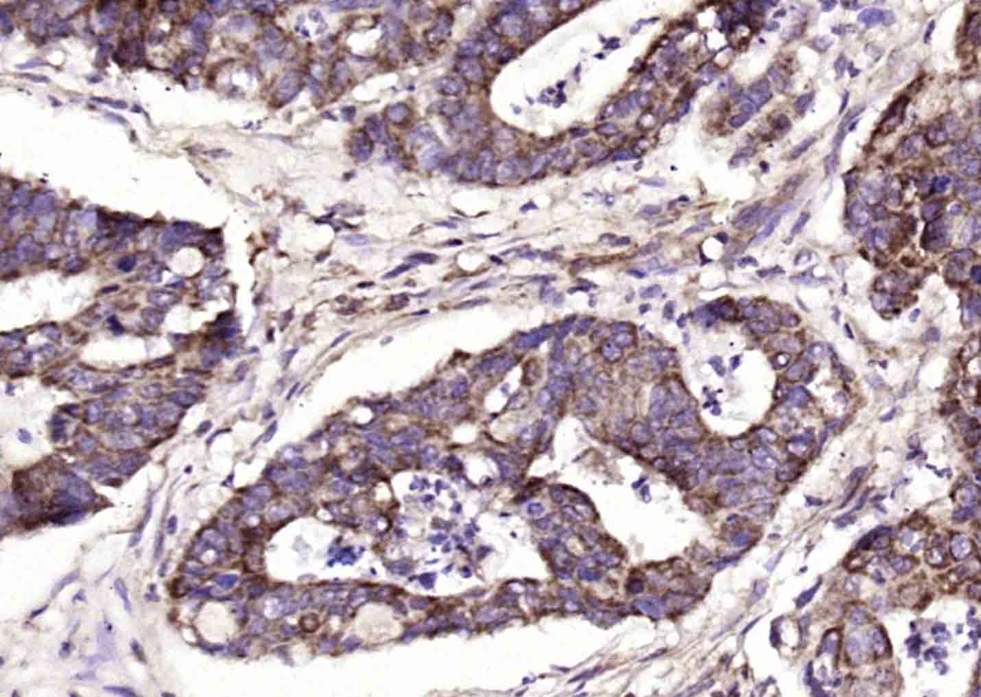 Paraformaldehyde-fixed, paraffin embedded (human colon carcinoma); Antigen retrieval by boiling in sodium citrate buffer (pH6.0) for 15min; Block endogenous peroxidase by 3% hydrogen peroxide for 20 minutes; Blocking buffer (normal goat serum) at 37°C for 30min; Antibody incubation with (RhoA) Polyclonal Antibody, Unconjugated (SL22249R) at 1:200 overnight at 4°C, followed by operating according to SP Kit(Rabbit) (sp-0023) instructionsand DAB staining.
Ordering & shipping
- Minimum order: 1 unit
- Lead time: varies with order quantity and specifications. Please consult your local distributor or our sales team for confirmation
- Payment: T/T 100% in advance; PayPal; Alibaba Trade Assurance
- Shipping: FedEx / DHL / UPS express; cold-chain packing available on request
- Price: USD, per unit (50ul / 100ul)
Partial purchase records(bought amounts latest4)
| Username | Quantity | bought time |
| Fi*** | 1 | 2026-07-26 |
| Vi*** | 1 | 2026-06-17 |
| Ge*** | 1 | 2026-05-31 |
| Re*** | 1 | 2026-04-21 |
User Comment(Total0User Comment Num)
- No comment


 +86 571 56623320

 SUNLONG BIOTECH


